

Loading, please wait...
Loading, please wait...

Post-cardiac arrest brain injury represents the primary cause of mortality in patients who achieve sustained return of spontaneous circulation. The pathophysiological cascade begins with immediate global cerebral ischemia during circulatory arrest. Furthermore, secondary reperfusion injury accelerates cellular damage once circulation returns. Reperfusion triggers microvascular dysfunction, oxidative stress, and persistent neuroinflammation. Consequently, severe neuronal destruction can lead to persistent coma or brain death. However, early neurological recovery pathways remain heterogeneous among intensive care admissions. Clinicians frequently encounter diagnostic dilemmas when determining irreversible brain injury versus potentially survivable metabolic depression. Therefore, modern critical care protocols prioritize multimodal neuroprognostication to navigate these complex recovery patterns. Premature pessimism often leads to inappropriate withdrawal of life-sustaining treatment in patients who might otherwise achieve meaningful functional recovery. Conversely, indefinite aggressive therapy in nonsurvivable injury causes emotional distress for families and misallocates critical care resources. The 2025 European Resuscitation Council guidelines establish a standardized pathway that integrates clinical and diagnostic findings. By adopting this rigorous framework, medical teams systematically evaluate cortical integrity while avoiding subjective assumptions. Ultimately, structured post-resuscitation care ensures that intensivists deliver patient-centered decisions grounded in robust neurophysiological evidence.
Modern intensive care strictly discourages relying on any single prognostic parameter to predict poor neurological outcome. Instead, clinicians must apply multimodal neuroprognostication through a synchronized, stepwise algorithm. This multifaceted paradigm synthesizes bedside physical examinations, continuous electroencephalography, evoked potentials, serum biomarkers, and neuroimaging modalities. Moreover, each modality evaluates distinct anatomical regions and cellular functions across the central nervous system. For instance, somatosensory evoked potentials test subcortical pathways, whereas biochemical markers quantify active neuronal injury. Consequently, combining multiple concordant indicators dramatically reduces the false-positive rate. In fact, clinical trials demonstrate that relying on isolated findings can lead to a false-positive prediction rate approaching thirty percent. When critical care specialists synthesize multiple concordant parameters, however, the false-positive rate falls near zero. Additionally, timing remains crucial when executing this diagnostic protocol. Intensivists must delay formal prognostication until at least seventy-two hours after the return of spontaneous circulation. Evaluating comatose patients before this seventy-two-hour benchmark risks inaccurate assessments due to transient post-ischemic stunning. Furthermore, active targeted temperature management alters neurological responsiveness during initial resuscitation. Therefore, postponing definitive evaluations allows the central nervous system adequate opportunity to recover physiological stability.
A meticulous neurological examination serves as the bedrock of post-resuscitation prognostication. Specifically, clinicians must assess brainstem reflexes after patient rewarming and metabolic stabilization. The bilateral absence of pupillary light reflexes at seventy-two hours represents a highly specific marker of devastating brain injury. Similarly, the bilateral loss of corneal reflexes indicates severe pontine and midbrain dysfunction. However, practitioners must ensure that ocular trauma or systemic anticholinergic medications do not confound these cranial nerve findings. In addition to cranial reflexes, intensivists systematically evaluate the best motor response on the Glasgow Coma Scale. Extensor posturing or complete flaccidity in response to painful stimuli indicates widespread hemispheric disruption. Nevertheless, motor scoring alone lacks sufficient specificity to dictate treatment withdrawal independently. Clinicians must also identify and document post-arrest myoclonus. Status myoclonus presenting within the first seventy-two hours historically signaled an unfavorable outcome. However, recent evidence reveals that some patients with early myoclonus still recover awareness, particularly when epileptiform activity responds to antiseizure drugs. Therefore, intensivists must not consider early myoclonic twitches as an absolute indicator of hopelessness. Instead, clinical examination must guide subsequent neurophysiological testing to confirm structural destruction.
Electrophysiological studies provide objective insight into cortical viability and somatosensory connectivity. In particular, continuous electroencephalography monitors real-time cerebral electrical activity throughout the critical care admission. Highly malignant electroencephalographic patterns include persistent background suppression, burst-suppression with identical bursts, and generalized periodic discharges. When these malignant patterns persist beyond twenty-four to forty-eight hours without sedatives, they strongly correlate with irreversible hypoxic insult. Conversely, continuous background reactivity and sleep spindles suggest intact cortical architecture and potential for functional recovery. Furthermore, short-latency somatosensory evoked potentials evaluate thalamocortical pathway conduction. The bilateral absence of the N20 cortical potential reliably predicts poor outcome with exceptional specificity. In addition to electrophysiology, circulating biochemical markers provide quantifiable measurements of neuronal necrosis. Neuron-specific enolase serves as the most widely validated serum biomarker in clinical practice. Peak levels exceeding sixty micrograms per liter at forty-eight or seventy-two hours indicate extensive cellular death. Moreover, serial sampling provides greater prognostic value than isolated values, as rising trends demonstrate ongoing secondary necrosis. Consequently, neurofilament light chain has emerged as an ultra-sensitive biomarker that reinforces neuron-specific enolase data during clinical evaluations.
Neuroimaging offers direct structural confirmation of global hypoxic-ischemic brain damage. Computed tomography performed within the first seventy-two hours can demonstrate diffuse cerebral edema. Specifically, marked attenuation of the gray-white matter ratio signifies severe cytotoxic edema and tissue necrosis. In addition, brain magnetic resonance imaging provides remarkable sensitivity for ischemic injury between two and five days post-arrest. Diffusion-weighted imaging reveals widespread cortical and basal ganglia diffusion restriction that correlates with permanent cell injury. However, neuroimaging interpretations must align with clinical findings rather than standing alone. Furthermore, intensivists must rigorously eliminate clinical confounders before finalizing any prognostic determination. Residual sedative infusions and neuromuscular blockade represent the most common confounding factors in the intensive care unit. For example, renal or hepatic impairment significantly delays drug clearance, prolonging drug-induced coma. Similarly, severe metabolic disturbances, including profound hyponatremia and severe hypoxemia, alter mental status. Physicians must rewarm hypothermic patients to normal body temperature before assessing brainstem reflexes. In addition, critical care teams should utilize therapeutic drug monitoring and short-acting sedatives to facilitate reliable neurological checks. Ultimately, eliminating these reversible confounders prevents tragic diagnostic errors and protects vulnerable patients during recovery.
Ethical decision-making in post-cardiac arrest care demands transparent communication among intensivists, neurologists, nurses, and family surrogates. Neurological prognostication should never function as an isolated academic exercise. Instead, it provides the clinical foundation for establishing realistic goals of care. Critical care clinicians must share diagnostic uncertainties openly with family members during scheduled conferences. Moreover, physicians must explain that neuroprognostication evaluates the statistical probability of functional recovery rather than offering absolute certainty. When multimodal assessments demonstrate concordant evidence of irreversible, devastating neurological injury, clinicians can compassionately recommend shifting from curative interventions to comfort-focused palliative care. Consequently, this shared decision-making model prevents prolonged, nonbeneficial intensive care while honoring patient dignity and autonomy. Conversely, if diagnostic tests yield discordant or indeterminate results, the medical team must extend the observation period. Many patients with delayed awakening gradually demonstrate neurological improvement when given sufficient time and physiological support. Furthermore, modern rehabilitation specialists should assess surviving patients early to identify physical, cognitive, and emotional rehabilitation needs. Ultimately, combining precise prognostication with empathetic family communication ensures ethical, compassionate care throughout the intensive care unit journey.
Evaluating comatose patients prior to seventy-two hours post-resuscitation carries significant clinical risk because ischemic brain tissue requires adequate time to recover from acute metabolic stunning. In addition, lingering sedative agents, analgesics, and targeted temperature management frequently depress physical responses during early intensive care. Waiting seventy-two hours allows drug clearance and physiological stabilization. Consequently, delaying formal prognostication prevents premature withdrawal of life-sustaining treatment and ensures that clinical predictions remain accurate and reliable.
Guidelines identify several robust indicators that reliably predict an unfavorable neurological recovery when present together. Specifically, bilateral absence of pupillary light reflexes and absent corneal reflexes at seventy-two hours strongly suggest irreversible damage. Furthermore, bilateral absence of N20 responses during somatosensory evoked potential testing and persistently suppressed electroencephalogram backgrounds indicate catastrophic cortical death. High serum neuron-specific enolase levels exceeding sixty micrograms per liter at forty-eight hours corroborate extensive neuronal necrosis across multiple testing modalities.
Targeted temperature management significantly alters drug pharmacokinetics by impairing hepatic clearance and reducing renal excretion of common sedatives and paralytics. Moreover, hypothermia itself blunts brainstem reflexes and alters electroencephalographic waveforms during active cooling. Therefore, intensivists must successfully rewarm patients to normothermia and maintain controlled temperatures below thirty-seven and a half degrees Celsius before prognosticating. Clinicians must wait until sedative medications fully metabolize to avoid mistaking transient pharmacological depression for irreversible post-anoxic encephalopathy.
Disclaimer: This content is for informational and educational purposes only... Refer to the latest local and national guidelines for clinical practice.
References

Read summarized clinical updates, watch expert medical content, and earn CME certifications right from your smartphone.
Open your smartphone camera to scan and install.





The 2025 resuscitation guidelines highlight multimodal neuroprognostication for post-cardiac arrest brain injury. By integrating clinical exams, EEG, imaging, and serum biomarkers after resolving confounders, critical care teams avoid premature treatment withdrawal and improve post-resuscitation patient outcomes.
Today

Assam reduced maternal sepsis deaths from 21% to 8% through rigorous infection prevention protocols and digital monitoring. Explore the clinical strategies, sterilization quality checks, early recognition pathways, and antimicrobial stewardship lessons essential for obstetric teams across India.
Today

At the Cardiological Society of India Kerala conference, specialists called for a calibrated review of DGHS curbs on reusing single-use items. Regulated reprocessing of catheters and guidewires could bridge the gap between rising cardiac intervention costs and government health insurance reimbursement limits.
Today
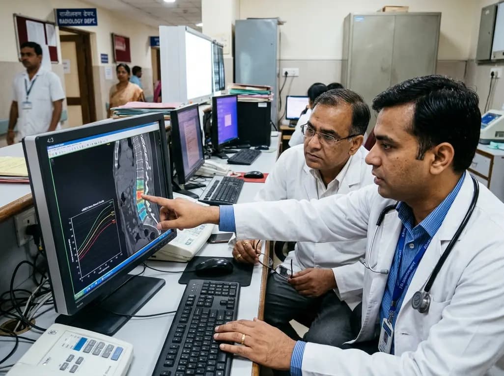
CT-derived volumetric bone mineral density centile curves offer an accurate method for opportunistic osteoporosis screening. This approach identifies high-risk patients who missed routine DXA scans and reliably predicts major osteoporotic fractures, addressing significant diagnostic gaps in clinical practice.
Today

A nationwide real-world cohort study reveals that sequential antiresorptive therapy following teriparatide reduces hip fracture risk by over 50%. Immediate initiation within 90 days and high treatment adherence are critical to preserve skeletal gains in severe osteoporosis.
Today
