

Loading, please wait...
Loading, please wait...

Osteoporosis remains a major silent epidemic that clinicians frequently fail to identify before debilitating skeletal injuries occur. Dual-energy X-ray absorptiometry continues to serve as the established clinical standard for evaluating skeletal fragility, but diagnostic access and patient adherence remain persistently limited across many healthcare settings. Fortunately, opportunistic osteoporosis screening using routinely acquired clinical computed tomography scans presents an exceptional pathway to address this care gap without adding medical expenses or extra radiation exposure.
Fragility fractures generate devastating morbidity, extended hospitalizations, and considerable mortality in aging populations. Dual-energy X-ray absorptiometry undeniably provides accurate areal bone mineral density measurements. However, real-world registry studies show that less than twenty percent of eligible individuals receive regular preventative examinations. Consequently, primary care physicians and specialists miss critical opportunities to prescribe bone-protective pharmacotherapy before catastrophic fractures happen. Meanwhile, millions of older patients undergo routine computed tomography of the chest, abdomen, and spine for general medical indications. Radiologists and treating clinicians can readily leverage these existing volumetric examinations. By evaluating trabecular bone attenuation on routine imaging, doctors capture hidden skeletal deterioration early. Therefore, implementing proactive opportunistic osteoporosis screening creates a practical bridge between daily diagnostic imaging and timely fracture prevention.
Historically, clinicians utilized simple single-slice region of interest attenuation values measured in Hounsfield units to estimate bone health. While practical, this basic technique exhibits significant variability across distinct scanner manufacturers and contrast enhancement protocols. To solve this clinical dilemma, researchers recently investigated automated volumetric bone mineral density reference curves. The study examined a retrospective cohort of 4,418 clinical computed tomography scans, linking a substantial subset of 1,088 examinations with dual-energy X-ray absorptiometry results performed within twelve months. Deep learning segmentation models automatically extracted trabecular compartments of the first lumbar vertebra and the femoral neck. Subsequently, investigators utilized internal tissue calibration to compute standardized volumetric measurements. Furthermore, they constructed sex-specific centile curves using generalized additive models for location, scale, and shape. This sophisticated pipeline produced reliable percentile distributions that directly reflect age-related physiological bone deterioration in real clinical populations.
A central challenge in quantitative computed tomography lies in defining clear, clinically relevant thresholds that correspond to standard diagnostic definitions. In this landmark validation, researchers applied receiver operating characteristic curves and the Youden index to anchor computed tomography percentiles directly against established dual-energy X-ray absorptiometry classifications. The derived volumetric bone mineral density cut-points demonstrated robust diagnostic concordance for identifying skeletal degradation. Interestingly, the investigation revealed that among individuals who subsequently sustained a major osteoporotic fracture, 66.3% of women and 41.9% of men had vertebral volumetric values falling directly below the computed tomography osteoporosis threshold. Most alarmingly, 44.8% of these fracture-positive women and 76.9% of the fracture-positive men had never received formal bone densitometry. Consequently, automated opportunistic identification successfully flags individuals who slip completely through conventional preventative health safety nets.
The ultimate test of any bone health metric resides in its prospective fracture prediction ability. Investigators linked clinical records to longitudinal administrative databases to follow incident major osteoporotic fracture events over prolonged follow-up periods. Using multivariable Cox proportional hazards models, researchers evaluated time to incident skeletal fracture across all cohorts. The results demonstrated that individuals classified as osteoporotic by volumetric computed tomography thresholds faced up to a 2.86-fold higher risk of sustaining an incident major osteoporotic fracture compared to individuals with normal volumetric bone values. Notably, this elevated risk profile remained consistent across both sexes and across diverse anatomical vertebral sites. These outcomes confirm that volumetric thresholds do not merely reflect mathematical correlations with absorptiometry. Instead, they capture biologically meaningful bone structural fragility that directly predicts imminent skeletal collapse and traumatic admissions.
Integrating opportunistic analysis directly into clinical workflows offers an actionable opportunity to optimize fracture liaison services and preventive medical care. When patients undergo abdominal or spinal computed tomography for unrelated indications, artificial intelligence algorithms can silently segment trabecular bone and calculate volumetric density. When automated values breach established thresholds, the radiology system generates an opportunistic diagnostic flag. Hospital systems can immediately route this information to primary care physicians, geriatricians, and rheumatologists. Consequently, clinicians can initiate dietary interventions, vitamin D supplementation, and antiresorptive or osteoanabolic medications before fractures happen. In resource-constrained settings where specialized bone densitometry equipment remains sparse, this pragmatic paradigm extracts immense clinical value from existing radiological investigations. By transforming routine diagnostic scans into opportunistic preventive tools, modern medicine can substantially curb the escalating global burden of fragility fractures.
Opportunistic screening evaluates bone mineral density from clinical computed tomography scans ordered for unrelated medical reasons, such as abdominal pain or oncology follow-up. By measuring trabecular bone attenuation without requiring additional radiation or dedicated appointments, clinicians detect undiagnosed skeletal fragility early. This pragmatic approach reliably directs high-risk patients toward formal bone evaluations and timely preventative therapies.
Dual-energy X-ray absorptiometry measures areal bone mineral density in two dimensions, which can be falsely elevated by spinal osteoarthritis, aortic calcifications, and patient positioning artifacts. In contrast, computed tomography-derived volumetric bone mineral density evaluates true three-dimensional trabecular bone compartments. This precise structural differentiation provides accurate assessments of metabolic bone degradation unaffected by surrounding degenerative sclerosis.
When an opportunistic computed tomography report indicates low volumetric bone mineral density, physicians should interpret the result as a validated diagnostic flag. Clinicians must conduct a comprehensive fracture risk assessment, review secondary causes of bone loss, and evaluate falls risk. When clinically indicated, doctors can initiate antiresorptive or osteoanabolic treatment regimens promptly to avoid future fractures.
Disclaimer: This content is for informational and educational purposes only. It is not intended to provide medical advice, diagnosis, or treatment. Always seek the advice of your physician or other qualified health provider with any questions you may have regarding a medical condition. Refer to the latest local and national guidelines for clinical practice.
References

Read summarized clinical updates, watch expert medical content, and earn CME certifications right from your smartphone.
Open your smartphone camera to scan and install.





CT-derived volumetric bone mineral density centile curves offer an accurate method for opportunistic osteoporosis screening. This approach identifies high-risk patients who missed routine DXA scans and reliably predicts major osteoporotic fractures, addressing significant diagnostic gaps in clinical practice.
Today

Assam reduced maternal sepsis deaths from 21% to 8% through rigorous infection prevention protocols and digital monitoring. Explore the clinical strategies, sterilization quality checks, early recognition pathways, and antimicrobial stewardship lessons essential for obstetric teams across India.
Today
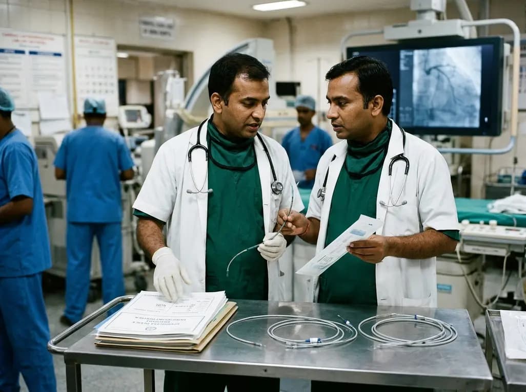
At the Cardiological Society of India Kerala conference, specialists called for a calibrated review of DGHS curbs on reusing single-use items. Regulated reprocessing of catheters and guidewires could bridge the gap between rising cardiac intervention costs and government health insurance reimbursement limits.
Today
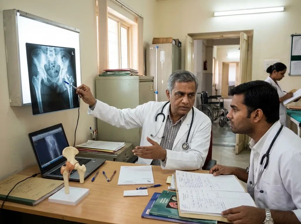
A nationwide real-world cohort study reveals that sequential antiresorptive therapy following teriparatide reduces hip fracture risk by over 50%. Immediate initiation within 90 days and high treatment adherence are critical to preserve skeletal gains in severe osteoporosis.
Today
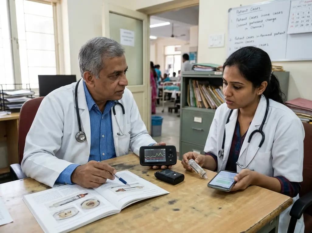
A case report details how reusing single-use insulin cartridges caused device failure and diabetic ketoacidosis in a type 1 diabetes patient using an advanced hybrid closed-loop system. The report highlights technological blindspots, mechanical degradation, and safety protocols during supply shortages.
Today
