.webp&w=384&q=75)

Loading, please wait...
Loading, please wait...

Preoperative bone evaluation remains fundamental when planning spinal instrumentation. Spine surgeons often encounter difficulties when assessing bone health in older adults, because conventional dual-energy X-ray absorptiometry frequently overestimates bone mineral density. Degenerative joint disease, vascular calcifications, and osteophytes artificially elevate planar density readings. Consequently, clinicians increasingly utilize advanced cross-sectional imaging to quantify skeletal strength. Finite element analysis provides an objective computational method that simulates structural failure under axial loading, directly determining vertebral compression strength. However, clinicians continually debate which routine preoperative imaging parameters offer the greatest diagnostic accuracy. While computed tomography measures volumetric trabecular density, magnetic resonance imaging provides scores for bone quality and paraspinal musculature. Furthermore, plain radiographs reveal abdominal aortic calcification. Understanding the distinct predictive value of each parameter is therefore crucial for surgical planning. A pivotal study by Anna-Maria Mielke and colleagues investigated these multimodal biomarkers to determine their relative contribution to vertebral mechanical strength.
Spinal constructs require robust vertebral purchase to prevent cage subsidence, pedicle screw loosening, and proximal junctional failure. Finite element analysis models reproduce physiological loading conditions by transforming three-dimensional computed tomography data into detailed computational meshes. Through this mathematical approach, biomechanical engineers reliably quantify vertebral compression strength without destructive cadaveric testing. Standard planar imaging captures only two-dimensional areal density, which obscures localized trabecular microarchitectural deterioration. In contrast, computational simulations replicate axial compressive forces distributed across the endplates. Additionally, finite element modeling incorporates heterogeneous trabecular patterns, cortical shell thickness, and localized material properties. Biomechanical investigations demonstrate that simulated failure loads correlate strongly with direct experimental testing. Consequently, finite element analysis represents an established benchmark for non-invasive skeletal assessment. However, full computational simulations require specialized software and extensive processing time. These technical demands prevent routine implementation in busy clinics. Surgeons therefore require reliable imaging proxies that closely reflect these computational results to optimize risk stratification.
Quantitative computed tomography provides an accurate assessment of trabecular architecture without artifact interference from degenerative osteophytes. In their cross-sectional analysis of seventy-six lumbar fusion patients, Mielke and colleagues evaluated volumetric bone mineral density at the first and second lumbar vertebrae using asynchronous protocols. The study revealed that volumetric bone mineral density was the dominant independent predictor of vertebral failure load across all multivariable models. Specifically, each increase of one milligram per cubic centimeter in trabecular density corresponded to a failure load increase between sixty-five and sixty-seven Newtons. Furthermore, this association demonstrated remarkable statistical significance in every analytical model. Trabecular bone absorbs substantial axial compressive stress within the lumbar spine. When osteoporosis degrades internal trabecular connectivity, the vertebral centrum loses compressive resistance rapidly. Unlike dual-energy absorptiometry, quantitative computed tomography isolates trabecular bone voxels from cortical sclerosis. Consequently, volumetric density measurements provide direct insight into mechanical load tolerance, allowing surgeons to identify occult osteopenia before performing spinal reconstruction.
Clinicians frequently explore magnetic resonance imaging as an opportunistic alternative for skeletal assessment. The vertebral bone quality score evaluates non-contrast T1-weighted sequences by measuring vertebral signal intensity relative to cerebrospinal fluid. Because osteopenia accelerates adipocyte conversion within bone marrow, elevated signal ratios typically suggest reduced skeletal health. Similarly, clinicians often consider abdominal aortic calcification an indirect marker of systemic vascular disease and altered calcium metabolism. Vascular rigidification may restrict local microvascular perfusion to the vertebral endplates, accelerating osseous degeneration. Nevertheless, multivariable regression demonstrated that neither magnetic resonance scores nor aortic calcification grades independently predicted vertebral compression strength. Although these parameters reflect biological aging, they fail to enhance mechanical strength prediction once models incorporate volumetric computed tomography. Magnetic resonance signals primarily reflect marrow fat infiltration rather than trabecular mineral content. Consequently, spine surgeons should exercise caution when evaluating skeletal integrity solely through magnetic resonance imaging or abdominal radiographs.
Paraspinal musculature provides dynamic stability and distributes mechanical loads across the lumbar motion segment. Chronic back pain, sedentary lifestyles, and lumbar degeneration often cause multifidus muscle atrophy, resulting in fibrous replacement and fatty infiltration. In the study, investigators measured multifidus fatty infiltration at the fourth lumbar level using axial T2-weighted magnetic resonance imaging. Interestingly, the regression analysis demonstrated a positive trend between multifidus fatty infiltration and vertebral compression strength, although this relationship did not reach statistical significance. Additionally, incorporating muscle fat measurements modestly increased the overall predictive fit of the model. This unexpected association likely reflects adaptive load redistribution within the degenerated spine. When stabilizing paraspinal musculature weakens, the anterior vertebral column bears greater static axial loading. Over time, persistent compressive forces can stimulate compensatory trabecular remodeling or sclerotic bone formation. Therefore, while paraspinal muscle fatty infiltration provides valuable contextual clinical insight, clinicians must not interpret muscle preservation as a guarantee of mechanical bone strength.
Preoperative bone quality identification directly influences surgical strategy and reduces instrumentation failure rates. In osteoporotic patients, inadequate vertebral strength substantially elevates the danger of pedicle screw pullout and proximal junctional kyphosis. By establishing volumetric density as the primary imaging determinant of vertebral strength, this study supports refined preoperative algorithms. When computed tomography reveals compromised bone strength, surgeons can implement proactive protective measures. For example, surgical teams may choose fenestrated cement-augmented pedicle screws, bicortical purchase, or expandable interbody devices to enhance construct stability. Additionally, identifying skeletal fragility enables targeted medical optimization before elective procedures. Clinicians can prescribe osteoanabolic agents, such as teriparatide, several weeks prior to surgery to stimulate trabecular bone formation. Conversely, surgical teams should not rely exclusively on qualitative magnetic resonance interpretation when assessing structural integrity. Combining opportunistic computed tomography evaluation with individualized surgical techniques significantly improves long-term patient outcomes.
Quantitative computed tomography measures three-dimensional trabecular bone density inside the vertebral centrum. Because trabecular architecture supports the majority of axial compressive loads, volumetric density correlates strongly with mechanical failure thresholds. Unlike planar radiographs, volumetric computed tomography eliminates confounding artifacts caused by aortic calcifications, scoliosis, and degenerative facet hypertrophy.
Magnetic resonance vertebral bone quality scores assess fatty bone marrow infiltration rather than mineralized osseous architecture. Although magnetic resonance provides valuable opportunistic insight into bone health, multivariable analyses demonstrate that it does not independently predict mechanical vertebral strength. Therefore, quantitative computed tomography remains the essential imaging tool for evaluating biomechanical bone integrity.
Multifidus fatty infiltration indicates progressive muscle degeneration and decreased dynamic spinal stability. When posterior muscular support declines, the anterior column bears greater static mechanical stress. This sustained axial loading can stimulate compensatory trabecular remodeling and sclerotic bone adaptation within the vertebral body, creating a positive statistical trend with compressive strength despite muscular deterioration.
Disclaimer: This content is for informational and educational purposes only... Refer to the latest local and national guidelines for clinical practice.
References

Read summarized clinical updates, watch expert medical content, and earn CME certifications right from your smartphone.
Open your smartphone camera to scan and install.





A new Spine study demonstrates that QCT-derived volumetric BMD is the primary independent predictor of finite element lumbar vertebral compression strength. MRI-based bone quality and aortic calcification showed no independent value, while multifidus fatty infiltration displayed an exploratory trend.
Today
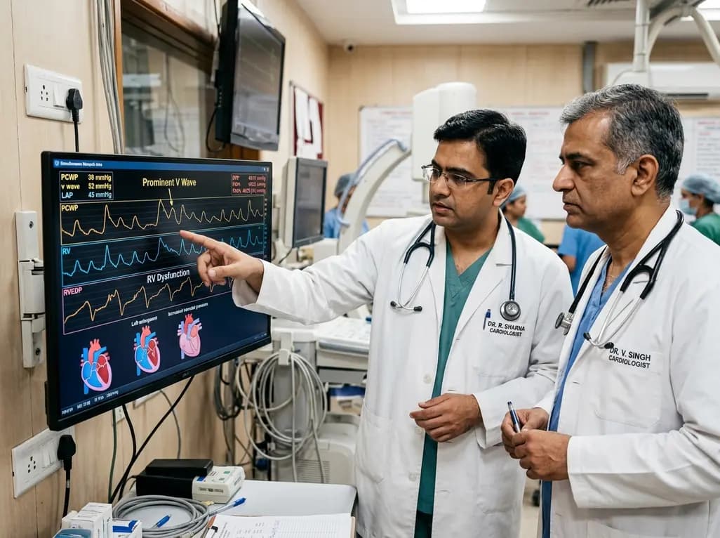
Left atrial stiffening generates prominent V waves that pressurize the pulmonary circulation and augment right ventricular afterload independently of intrinsic pulmonary vascular disease in patients with heart failure with preserved ejection fraction.
Today

A meta-analysis demonstrates that secondhand smoke exposure significantly elevates fetal loss risk by 21% among nonsmoking pregnant women. Clinicians must implement household screening and family-centered cessation interventions to prevent avoidable miscarriages and stillbirths.
Today

Preclinical validation demonstrates that a novel contact force-integrated pulsed field quantization index accurately predicts lesion depth in real time, achieving 100% durable pulmonary vein isolation without collateral injury.
Today
A new cross-sectional evaluation reveals how regional lean mass and bone mineral content interact in resistance-trained men, showing that whole-body size and allometric scaling explain shared musculoskeletal phenotypes and strength relationships.
Today
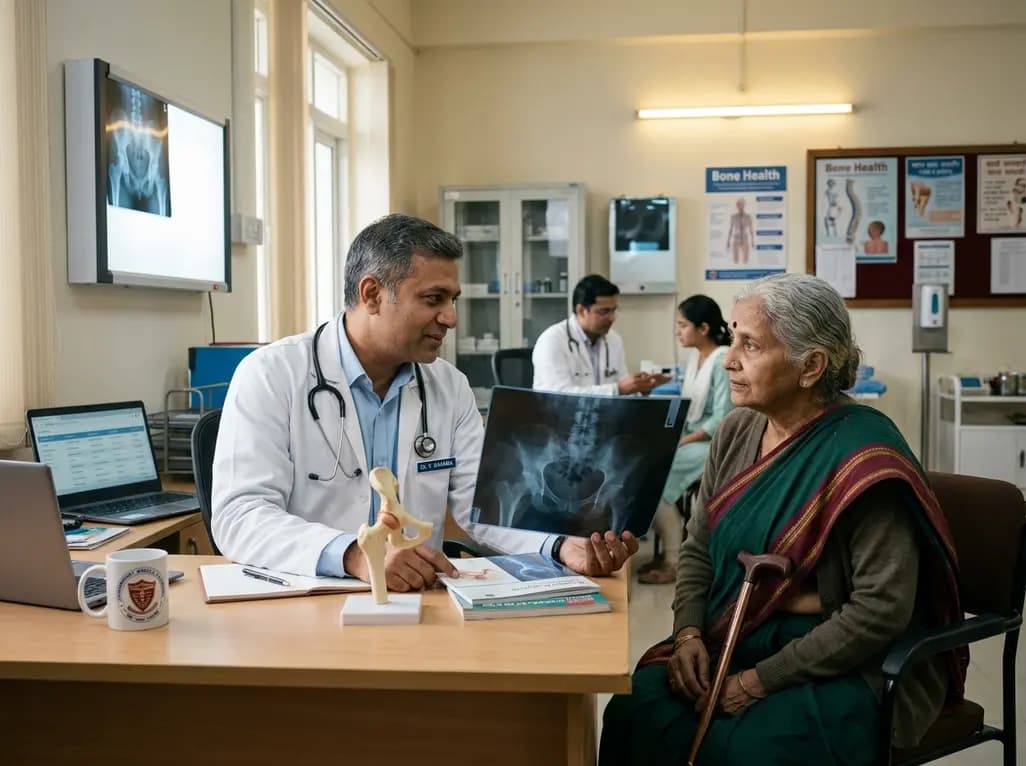
Explore the emerging role of glucose metabolic reprogramming in exercise-attenuated osteoporosis, examining how physical activity influences osteoblast glycolysis, osteoclast bioenergetics, and skeletal remodeling pathways to improve bone mineral density.
Today
