.webp&w=384&q=75)

Loading, please wait...
Loading, please wait...

Assessment of regional lean mass and bone density provides sports physicians with critical data regarding athlete performance and skeletal durability. Clinicians recognize that muscular development correlates with bone mineral accumulation in active young populations. However, healthcare professionals often debate whether these observed correlations reflect direct structural dependence or simply shared total body dimensions. A cross-sectional analysis in resistance-trained males evaluates this question by examining regional dual-energy X-ray absorptiometry measures alongside functional leg press strength.
Resistance exercise places powerful tensile and compressive mechanical loads on skeletal tissue. Consequently, muscular hypertrophy and osteogenesis frequently progress in tandem across training cycles. Dual-energy X-ray absorptiometry offers precise segment-specific quantification of soft tissue and bone parameters. In a cohort of 44 resistance-trained men, researchers analyzed broad upper and lower body lean mass alongside regional bone mineral content, projected bone area, and areal bone mineral density. Initially, regional lean mass displayed strong positive correlations with bone mineral content across multiple anatomical regions when models adjusted for age and height. Specifically, arm lean mass showed a standardized correlation of 0.605 with arm bone mineral content. Similarly, rib bone mineral content reached a value of 0.608, while leg bone mineral content demonstrated an association of 0.429. Additionally, pelvic bone mineral content exhibited an association of 0.550. These standard adjustments suggest substantial coupling between muscularity and local mineralization. Nevertheless, sports medicine physicians must interpret these standard associations with caution, because height and age adjustments do not fully account for total mass volume.
When clinicians examine musculoskeletal variables, body size functions as a major confounding variable. Therefore, investigators applied allometric scaling, total body mass adjustments, and projected bone area corrections to the baseline dual-energy X-ray absorptiometry dataset. As a result, several previously robust correlations decreased in magnitude or lost statistical precision. For example, larger individuals naturally possess heavier skeletal frames, larger projected bone areas, and greater absolute muscle bulk. Consequently, unadjusted statistical models often amplify apparent biological couplings that simply stem from overall body volume. The study prioritized Benjamini-Hochberg corrections across 18 adjusted cross-sectional tests to restrict false discovery rates. This statistical rigor confirmed that apparent regional bone adaptations frequently diminish once models control for total body mass. Orthopedic specialists and exercise physiologists should therefore recognize that skeletal robusticity primarily reflects generalized somatotype and overall body frame size rather than isolated regional muscular loading alone.
Functional lower-extremity strength forms a cornerstone of athletic power and musculoskeletal rehabilitation. The investigators evaluated dynamic muscular performance using estimated leg press one-repetition maximum tests. Interestingly, pelvic bone mineral content remained conditionally associated with estimated leg press one-repetition maximum after controlling for age, height, and lower body lean mass. The standardized coefficient reached 0.501, achieving significance with a Benjamini-Hochberg corrected q-value of 0.001. Although this statistical link appears robust, the researchers appropriately emphasize that this metric represents an exploratory conditional finding. Specifically, the association cannot isolate the individual contributions of pelvic architecture, skeletal proportions, specific resistance training history, or unmeasured connective tissue factors. Clinicians must not assume that increasing pelvic mineral mass directly boosts lower limb force generation. Instead, physicians should view pelvic mineralization and leg press capacity as coordinated components of a broader, high-load musculoskeletal phenotype.
For clinicians managing athletes, these findings provide essential guidance for interpreting body composition scans. Sports physicians frequently order dual-energy X-ray absorptiometry scans to track rehabilitation, assess injury risk, or optimize competitive performance. However, misinterpreting regional mineral metrics as standalone determinants of physical performance can lead to flawed training guidance. Instead, clinicians should recognize that high regional lean mass, sturdy bone structure, and high force production co-develop within an integrated bodily framework. Furthermore, clinicians treating patients in physical therapy should focus on holistic structural loading rather than attempting to isolate bone density changes at specific functional joints. Comprehensive compound resistance exercises, such as multi-joint squats and leg presses, naturally provide concurrent anabolic stimuli across lower-body muscle groups and pelvic structures. Therefore, therapeutic interventions should consistently emphasize progressive overload across whole kinematic chains to build systemic musculoskeletal resilience.
Translating these athletic observations into everyday medical practice benefits diverse clinical cohorts, including individuals with low bone density. Doctors frequently encounter patients who present with osteopenia or sarcopenia. Because regional lean mass and bone mineral content correlate tightly with overall body frame size, clinicians should prioritize whole-body strength protocols to stimulate both tissues concurrently. In addition, practitioners must avoid viewing bone mineral density in isolation from muscular capacity. When evaluating fracture risk or functional decline, measuring both muscle mass and physical functional capability provides far greater predictive utility than bone densitometry alone. Resistance training serves as a non-pharmacological pillar that simultaneously preserves bone geometry, expands lean tissue, and enhances joint stability. Consequently, integrating progressive resistance training into routine preventive care promotes enduring physical independence and skeletal longevity across the lifespan.
Regional lean mass generates direct tensile force on adjacent skeletal structures during muscular contraction. This mechanical strain stimulates osteoblast activity, bone remodeling, and local mineral deposition. However, overall body size and bone surface area strongly confound this relationship. Therefore, clinicians must interpret regional lean mass and bone associations alongside total body mass to distinguish true localized tissue adaptations from general somatic dimensions.
Pelvic bone mineral content connects conditionally to leg press strength because the pelvic girdle acts as the central mechanical foundation during bilateral compound pressing. Athletes who consistently lift heavy loads develop robust pelvic architecture, broad skeletal dimensions, and substantial neuromuscular drive. Thus, this statistical association reflects a coordinated high-force musculoskeletal phenotype rather than an isolated skeletal driver of force output.
Physicians should use allometric scaling to adjust physiological variables for non-linear differences in body geometry and size. Standard ratio scaling often creates statistical artifacts in taller or heavier individuals. By utilizing allometric models and projected bone area corrections, clinicians can accurately isolate genuine tissue adaptations, monitor therapeutic progress, and assess structural integrity without body mass distortion.
Disclaimer: This content is for informational and educational purposes only. It is not intended to substitute for professional medical advice, diagnosis, or treatment. Clinicians must apply independent medical judgment when evaluating individual patients. Refer to the latest local and national guidelines for clinical practice.
References
Bagheri R et al. Regional lean mass, bone mineral measures, and estimated leg press 1-RM in resistance-trained males. J Int Soc Sports Nutr. 2026 Dec 30. doi: 10.1080/15502783.2026.2746457. PMID: 42860013.
Duplanty AA, Levitt DE, Hill DW, McFarlin BK, DiMarco NM, Vingren JL. Resistance training is associated with higher bone mineral density among young adult male distance runners independent of physiological factors. J Strength Cond Res. 2018;32(6):1594-1600.
Pinheiro MB, Oliveira J, Bauman A, Fairhall N, Kwok W, Sherrington C. Evidence on physical activity and osteoporosis prevention for healthcare providers. Br J Sports Med. 2020;54(23):1392-1399.

Read summarized clinical updates, watch expert medical content, and earn CME certifications right from your smartphone.
Open your smartphone camera to scan and install.





A new cross-sectional evaluation reveals how regional lean mass and bone mineral content interact in resistance-trained men, showing that whole-body size and allometric scaling explain shared musculoskeletal phenotypes and strength relationships.
Today
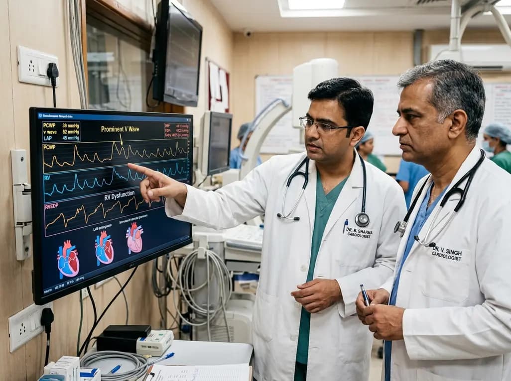
Left atrial stiffening generates prominent V waves that pressurize the pulmonary circulation and augment right ventricular afterload independently of intrinsic pulmonary vascular disease in patients with heart failure with preserved ejection fraction.
Today
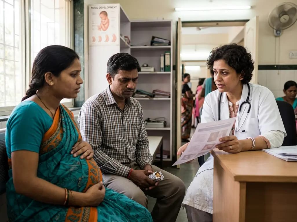
A meta-analysis demonstrates that secondhand smoke exposure significantly elevates fetal loss risk by 21% among nonsmoking pregnant women. Clinicians must implement household screening and family-centered cessation interventions to prevent avoidable miscarriages and stillbirths.
Today
Preclinical validation demonstrates that a novel contact force-integrated pulsed field quantization index accurately predicts lesion depth in real time, achieving 100% durable pulmonary vein isolation without collateral injury.
Today
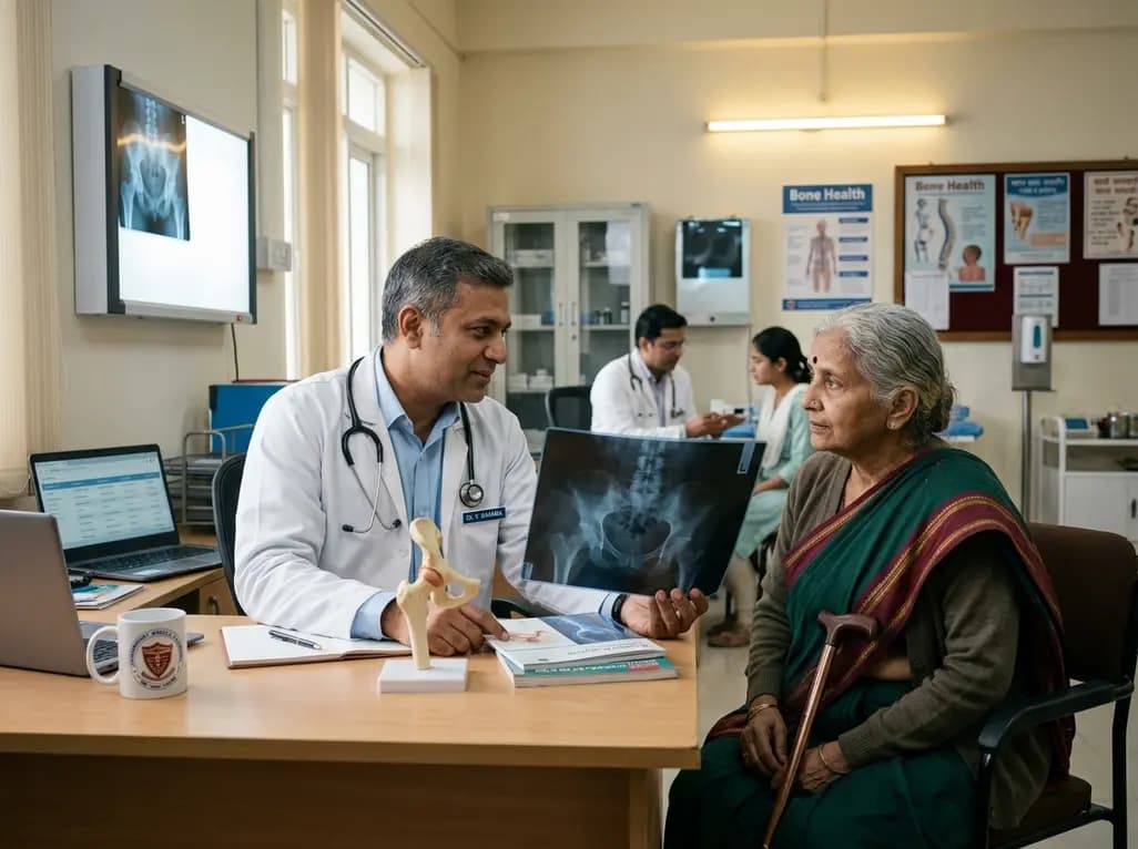
Explore the emerging role of glucose metabolic reprogramming in exercise-attenuated osteoporosis, examining how physical activity influences osteoblast glycolysis, osteoclast bioenergetics, and skeletal remodeling pathways to improve bone mineral density.
Today
