

Loading, please wait...
Loading, please wait...

Oral squamous cell carcinoma continues to cause substantial morbidity and mortality globally, primarily due to delayed detection and pronounced biological heterogeneity. Traditional screening methods frequently fail to distinguish early malignant transitions from benign reactive conditions. However, the emergence of multimodal AI in oral cancer offers a transformative strategy to overcome these longstanding diagnostic hurdles. By integrating histopathological whole slide images, cross-sectional radiology, genomic profiles, and patient clinical variables, computational models provide comprehensive oncological assessments. Recent evidence confirms that deep learning systems that synthesize disparate diagnostic modalities offer superior predictive accuracy compared to isolated single-stream evaluations.
Oral malignancies frequently evolve from oral potentially malignant disorders, presenting clinicians with ambiguous tissue manifestations that hinder definitive staging. Consequently, subjective visual screening and localized biopsies often miss aggressive sub-clonal populations. Clinicians require holistic decision support systems that synthesize surface mucosal patterns with underlying deep tissue architecture and molecular alterations. Integrating multimodal inputs directly addresses this diagnostic gap by capturing complex biological dynamics across macroscopic and microscopic scales.
Furthermore, early diagnosis drastically alters five-year survival metrics, elevating rates from under forty percent in regional disease to over eighty percent in localized lesions. Machine learning architectures process clinical photographs alongside high-resolution histopathology and patient demographic risk factors to identify malignant transformations early. Therefore, this computational synthesis provides oncologists and oral surgeons with actionable insights during initial evaluations. By reducing inter-observer variability among pathologists, these automated algorithms accelerate definitive surgical and therapeutic planning. Ultimately, applying multi-source data architectures establishes a reliable foundation for individualized patient risk stratification.
Developing effective computational models requires sophisticated algorithms capable of extracting complementary features from vastly different biomedical data types. Convolutional neural networks routinely handle pixel-level patterns within radiological scans and digital histopathology slides. Meanwhile, deep transformers process high-dimensional genomic sequences and unstructured clinical notes with remarkable contextual awareness. In addition, graph neural networks effectively model biological networks and cellular spatial arrangements across the tumor microenvironment.
However, simple algorithmic stacking rarely yields optimal clinical results. Modern frameworks employ intermediate and late fusion strategies governed by cross-modal attention mechanisms. These attention layers dynamically weigh the clinical importance of each stream, ensuring that uninformative or noisy inputs do not compromise overall model performance. For instance, when radiographic tumor boundaries appear poorly defined, the network prioritizes clear cellular atypia from digitized tissue biopsies. Similarly, graph embedding models preserve spatial relationships between tumor-infiltrating lymphocytes and malignant epithelial cells. Consequently, these integrated deep learning frameworks reconstruct a coherent, multi-scale picture of patient pathology that single-modality networks cannot achieve.
Comprehensive evaluations demonstrate that multimodal architectures systematically achieve higher areas under the receiver operating characteristic curve than single-modality algorithms. Unimodal models that rely solely on clinical photographs or radiographic slices often struggle with overlapping visual phenotypes. In contrast, multi-tiered platforms incorporate molecular markers alongside cross-sectional imaging, significantly minimizing false-positive classifications. This multi-layered approach reliably differentiates benign keratosis from high-grade dysplasia and invasive oral carcinoma.
Moreover, multimodal frameworks show exceptional performance in predicting overall survival, local recurrence, and occult cervical lymph node metastasis. By correlating depth of invasion from radiological scans with gene expression profiles and surgical margins, algorithms reliably identify patients requiring elective neck dissection. In addition, predictive modeling forecasts treatment responses to induction chemotherapy and targeted radiation protocols. As a result, multidisciplinary tumor boards gain access to calibrated risk probabilities that support evidence-based treatment de-escalation or intensification. These diagnostic improvements underscore the tremendous potential of integrated computational workflows in routine oncology practice.
Despite impressive experimental metrics, several technical and structural barriers impede the seamless clinical translation of computational diagnostic systems. Most published studies rely on retrospective cohorts derived from centralized public repositories, which frequently introduce institutional sampling bias. Consequently, algorithms often experience substantial performance drops when applied to independent patient populations across diverse geographical regions. Differences in biopsy preparation, staining protocols, and imaging equipment contribute to domain shift problems that challenge algorithmic reliability.
Furthermore, clinical real-world deployment requires strict standardization of biomedical data formats and robust imputation methods for missing variables. In routine hospital settings, patients rarely possess complete genomic profiles, advanced radiographic scans, and standardized clinical notes simultaneously. Therefore, developers must design resilient models that maintain diagnostic efficacy even when specific data modalities are absent. Establishing vendor-neutral data pipelines and open-access multi-institutional datasets will prove vital in overcoming these data inconsistencies. Only through rigorous multi-center prospective validation can researchers verify algorithmic generalizability and build clinical trust across diverse healthcare delivery systems.
Integrating artificial intelligence into oncological decision-making raises critical ethical, legal, and operational considerations that regulatory bodies must address. Black-box deep learning architectures create hesitation among practicing clinicians who require explainable rationales before altering surgical or chemotherapeutic plans. Therefore, future research must incorporate explainable artificial intelligence techniques, such as attention maps and feature importance rankings, to justify model outputs clearly. Transparent algorithmic reasoning empowers clinicians to validate computational suggestions against standard clinical guidelines.
Additionally, algorithmic governance must protect sensitive patient health information through federated learning protocols and differential privacy techniques. These distributed training methodologies allow computational models to learn from multi-center international registries without centralizing confidential patient records. Moreover, software developers and clinical researchers must actively mitigate demographic biases within training sets to ensure equitable diagnostic performance across all socio-economic groups. Ultimately, successful clinical implementation depends on designing intuitive decision-support interfaces that seamlessly integrate into existing hospital workflows. As interdisciplinary collaboration deepens, advanced computational oncology will transform early detection protocols and personalized treatment paradigms worldwide.
Multimodal systems combine diverse diagnostic streams, such as histopathology, radiological imaging, genomics, and clinical histories, to construct a complete profile of tumor biology. While single-modality models frequently struggle with ambiguous tissue appearances, multimodal architectures leverage complementary features across macroscopic and molecular levels. Consequently, this integrated approach significantly reduces false-positive interpretations, improves overall diagnostic accuracy, and delivers more dependable prognostic assessments for individual cancer patients.
Computational models integrate digitized hematoxylin and eosin histopathology slides, computed tomography, and magnetic resonance imaging scans. Furthermore, these platforms incorporate high-throughput genomic data, including somatic mutations and RNA expression levels, alongside clinical records containing patient demographics, tobacco exposure, and staging information. Cross-modal attention mechanisms dynamically fuse these complementary inputs to evaluate tumor margins, guide treatment selection, and assess metastatic risk accurately.
Primary translational obstacles include limited model generalizability across diverse populations, retrospective dataset biases, and absent clinical validation studies. Additionally, real-world clinics often lack standardized data pipelines, leading to frequent missing modalities such as genomic profiling. Clinicians also demand transparent, explainable decision rationales before adopting automated tools. Overcoming these hurdles requires prospective multi-center trials, robust data imputation methods, and strict regulatory frameworks.
Disclaimer: This content is for informational and educational purposes only and does not constitute medical advice or establish a doctor-patient relationship. Healthcare professionals must exercise independent clinical judgment and verify diagnostic criteria independently. Refer to the latest local and national guidelines for clinical practice.
References
Deka B et al. Multimodal AI in Oral Cancer: A PRISMA-Guided Systematic Review. Healthc Technol Lett. 2026 undefined undefined. doi: 10.1049/htl2.70105. PMID: 42999821.
Hajare D, Patil SV. Next-Generation Multimodal AI for Early Oral Cancer Prediction and Patient-Specific Treatment. Smart Trends in Computing and Communications. Springer; 2026:145-158.
Kumar P, Singh R, Sharma A. Multimodal AI in early detection of oral potentially malignant disorders and oral cancer: Integrating imaging, histopathology, and genomics. J Orofac Health Sci. 2026;14(2):89-102.

Read summarized clinical updates, watch expert medical content, and earn CME certifications right from your smartphone.
Open your smartphone camera to scan and install.





A PRISMA-guided review of 82 studies shows that multimodal AI in oral cancer outperforms unimodal tools by fusing histopathology, radiology, genomics, and clinical records. This integrated framework boosts diagnostic precision and survival prediction while paving the way for targeted clinical oncology.
Today
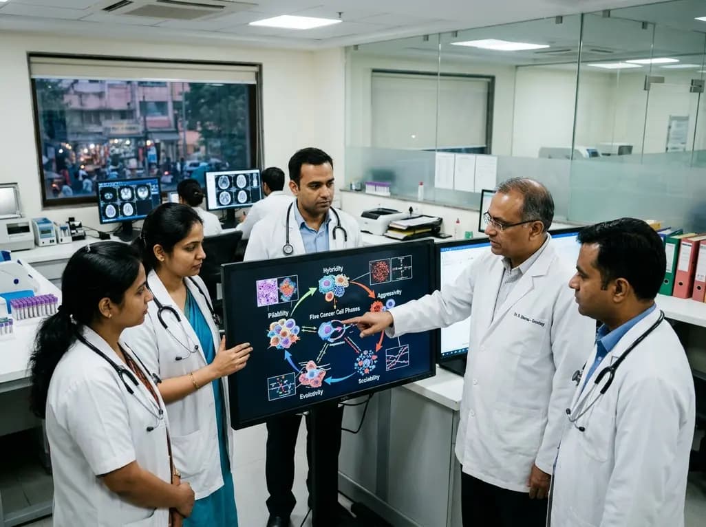
The PHASE framework conceptualizes cancer biology through five interacting processes: Pliability, Hybridity, Aggressivity, Sociability, and Evolutivity. This regulatory circuit offers a systems-level perspective on tumor heterogeneity, disease progression, and interaction-guided therapeutic strategies.
Today
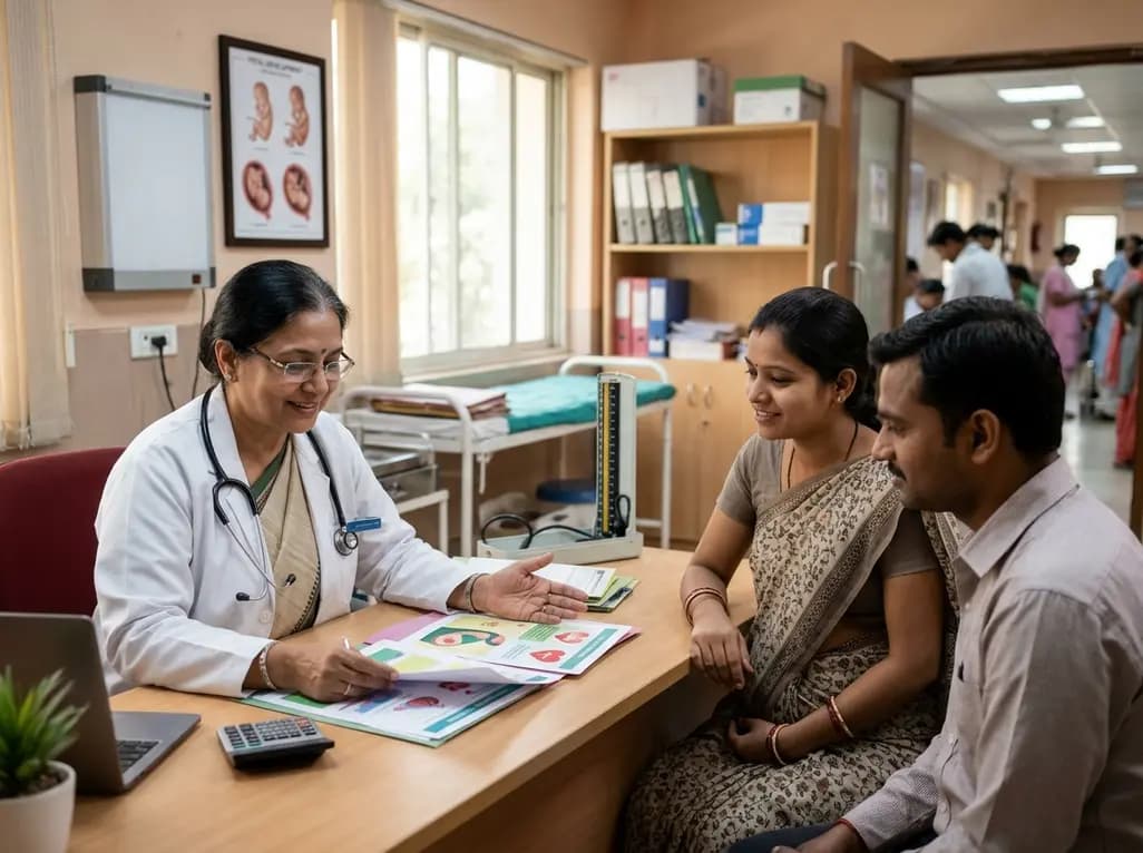
Maternal cardiovascular risk factors such as hypertension, obesity, and diabetes significantly elevate offspring cardiovascular disease risk through placental dysfunction and fetal programming. Enhancing preconception screening and antenatal care offers clinicians a powerful strategy to protect long-term heart health.
Today
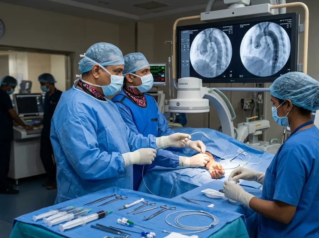
The novel CAST technique utilizes the indwelling Safari XS guidewire as a tracking rail to reposition a radial pigtail catheter into the descending aorta during minimalist TAVI, overcoming challenging vascular anatomy without requiring secondary femoral access or specialized equipment.
Today
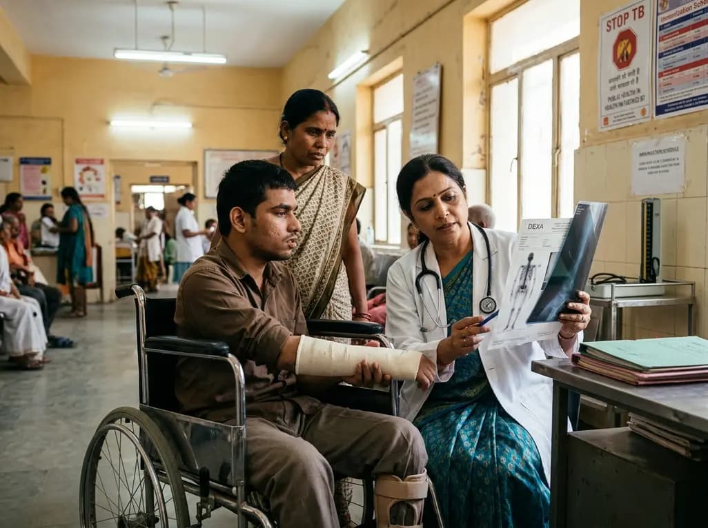
A retrospective cohort study reveals that lumbar spine bone mineral density does not distinguish extremity long bone fracture status in adults with severe motor and intellectual disabilities. Because age drove fracture risk rather than spinal density, clinicians must adopt comprehensive assessment tools.
Yesterday
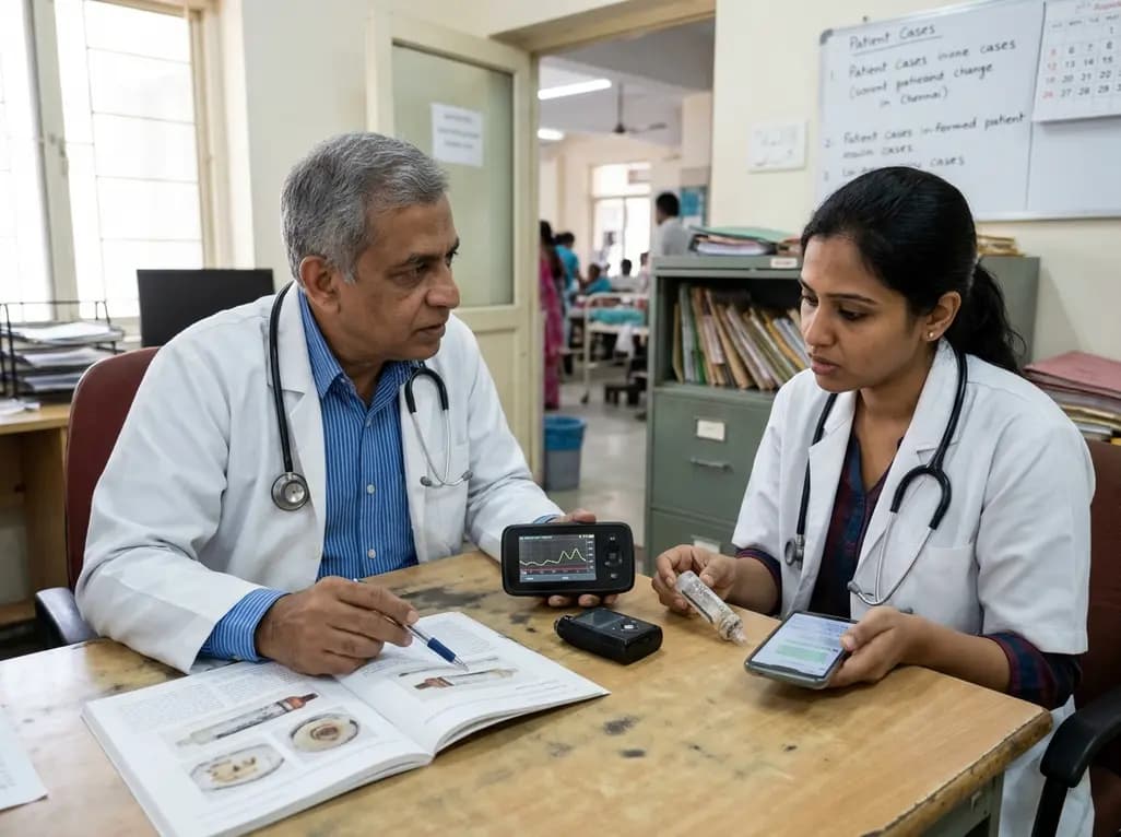
A case report details how reusing single-use insulin cartridges caused device failure and diabetic ketoacidosis in a type 1 diabetes patient using an advanced hybrid closed-loop system. The report highlights technological blindspots, mechanical degradation, and safety protocols during supply shortages.
Today
