

Loading, please wait...
Loading, please wait...

Adults with severe motor and intellectual disabilities face substantial risks of fragility fractures, particularly across extremity long bones. Clinicians frequently utilize dual-energy X-ray absorptiometry to quantify lumbar spine BMD in these vulnerable individuals. However, routine spinal measurements often fail to reflect the true structural fragility of non-weight-bearing limbs. Patients with profound neuromotor impairments frequently remain non-ambulatory throughout their lives. Consequently, their peripheral skeleton experiences severe mechanical unloading, which dramatically accelerates localized bone resorption. In addition, chronic anticonvulsant therapy, poor nutritional intake, and limited sunlight exposure further degrade bone microarchitecture. Healthcare providers often expect axial bone density scans to mirror peripheral skeletal integrity. Nevertheless, recent clinical evidence demonstrates notable discordance between spinal readings and actual fracture events. Clinicians frequently observe low spinal mineral scores across fractured and non-fractured cohorts alike. Therefore, relying exclusively on axial densitometry creates significant blind spots when determining individual peripheral fracture risk. Medical teams require a comprehensive diagnostic framework to safeguard bone health in non-ambulatory populations.
A recent retrospective cohort study evaluated 86 adults with severe motor and intellectual disabilities to determine whether lumbar spine BMD distinguishes extremity long bone fractures. Researchers analyzed adult patients who underwent dual-energy X-ray absorptiometry between 2023 and 2024. The investigators compared 23 patients with documented extremity fractures against 63 individuals without peripheral fractures over a 10-year observational period. Interestingly, lumbar densitometry demonstrated exceptional technical reproducibility, with an intraclass correlation coefficient of 0.939. Mean lumbar spine bone density was modestly lower in the fracture group compared to the non-fracture cohort. However, this difference failed to achieve statistical significance. Furthermore, both groups displayed markedly depressed mean Z-scores well below negative two standard deviations. When investigators executed multivariable logistic regression, advancing age strongly predicted extremity long bone fractures. Conversely, lumbar bone mineral density showed no meaningful statistical association with fracture occurrence. These crucial findings confirm that spinal densitometry alone cannot reliably stratify peripheral skeletal fragility in disabled adults.
Several distinct physiological mechanisms explain why central densitometry fails to capture extremity fragility in disabled adults. First, mechanical loading directly governs skeletal adaptation through bone mechanotransduction pathways. Because non-ambulatory individuals spend prolonged periods seated or recumbent, extremity long bones experience severe disuse osteopenia. In contrast, the axial skeleton still supports trunk weight during assisted sitting, preserving relatively higher trabecular mass in the spine. Second, anatomical positioning artifacts frequently distort spinal dual-energy absorptiometry results. Severe scoliosis, pelvic obliquity, and muscular contractures alter vertebral alignment in neuromuscular disorders. Furthermore, degenerative sclerosis, aortic calcifications, and prior surgical instrumentation artificially elevate measured lumbar density values. As a result, the lumbar spine appears falsely preserved despite profound generalized skeletal deterioration. In addition, extremity cortical bone undergoes selective thinning and endocortical resorption that planar spinal scans cannot evaluate. Cortical porosity in the femur and tibia severely compromises bone strength. Consequently, gentle caregiver transfers readily trigger long bone fractures despite unremarkable spinal scans.
Because spinal bone measurements provide incomplete clinical information, clinicians must evaluate broader risk profiles to forecast fractures effectively. Multivariable analysis confirms that advancing chronological age independently increases peripheral fracture susceptibility. Over decades, persistent immobility progressively compounds skeletal fragility. In addition, prolonged therapy with hepatic enzyme-inducing antiepileptic medications accelerates vitamin D catabolism, triggering chronic secondary hyperparathyroidism. Patients with spastic quadriparesis also experience recurring involuntary muscle spasms. These abrupt, forceful contractions exert violent bending forces across structurally brittle femoral and humeral shafts. Furthermore, nutritional compromise represents another critical risk factor in adults with severe motor impairments. Severe dysphagia frequently restricts protein and micronutrient intake, impairing bone matrix synthesis. Similarly, low body mass index directly correlates with elevated fracture vulnerability due to diminished soft-tissue cushioning during accidental trauma. Therefore, medical teams cannot evaluate skeletal vulnerability through isolated radiological scores. Instead, clinicians must evaluate patient age, seizure activity, nutritional status, and handling techniques collectively to gauge genuine fragility.
Effective fracture prevention in adults with severe disabilities requires a proactive strategy that transcends conventional osteoporosis protocols. Healthcare professionals should consider peripheral skeletal assessments, including distal femur evaluations where specialized software and positioning allow. In addition, physiotherapists must train caregivers in gentle transfer protocols to avoid twisting forces during daily hygiene and repositioning. Minimizing rotational stress significantly lowers the incidence of accidental spiral fractures in brittle long bones. Furthermore, physicians must optimize nutritional support, ensuring adequate calcium intake and maintaining 25-hydroxyvitamin D concentrations within target therapeutic ranges. When patients exhibit severe generalized bone fragility or prior low-impact trauma, clinicians should consider anti-resorptive pharmacotherapy. Bisphosphonates, including intravenous zoledronic acid, effectively reduce bone turnover markers and stabilize skeletal density in disabled populations. However, healthcare teams must monitor renal function and serum calcium diligently before and after infusion. By combining careful environmental modifications with tailored pharmacological therapy, clinicians can effectively prevent disabling skeletal injuries.
Adults with severe disabilities experience pronounced mechanical unloading in their extremities due to immobility, causing severe regional cortical thinning. In contrast, the lumbar spine bears partial body weight during supported sitting, preserving central trabecular mass. Furthermore, scoliosis, vertebral contractures, and aortic calcifications artificially elevate spinal density readings. Consequently, lumbar measurements obscure profound peripheral fragility, leaving clinicians unable to differentiate fracture status through spinal densitometry alone.
Clinicians can utilize distal femur dual-energy X-ray absorptiometry to assess regional skeletal mass in non-ambulatory individuals directly. Additionally, high-resolution peripheral quantitative computed tomography accurately measures volumetric bone density and cortical porosity without positional distortion. Emerging bedside technologies like radiofrequency echographic multi-spectrometry also provide reliable bone quality evaluations. These specialized peripheral modalities capture structural microarchitecture changes far more effectively than traditional planar spinal scans.
Healthcare providers should implement a comprehensive assessment protocol rather than relying solely on axial densitometry scores. Clinicians must evaluate advancing age, nutritional adequacy, serum vitamin D levels, and long-term antiepileptic drug exposure. Furthermore, care teams should assess spasticity severity, transfer techniques, and prior low-trauma fracture events. Integrating these multidimensional clinical factors allows physicians to identify high-risk individuals early and initiate preventive antiresorptive therapies appropriately.
Disclaimer: This content is for informational and educational purposes only and is not intended to serve as medical advice, diagnosis, or treatment. Healthcare professionals must evaluate each clinical situation individually and consult standard medical textbooks, peer-reviewed literature, and relevant professional guidelines. Patients should seek guidance from a qualified physician regarding any medical condition or treatment decision. The author and publisher disclaim any liability for adverse outcomes resulting from reliance on the information contained herein. Refer to the latest local and national guidelines for clinical practice.
References

Read summarized clinical updates, watch expert medical content, and earn CME certifications right from your smartphone.
Open your smartphone camera to scan and install.





A retrospective cohort study reveals that lumbar spine bone mineral density does not distinguish extremity long bone fracture status in adults with severe motor and intellectual disabilities. Because age drove fracture risk rather than spinal density, clinicians must adopt comprehensive assessment tools.
Today

A landmark study reveals that an annual weight loss of 5% or more and an FVC decline of 10% or more independently worsen survival in idiopathic pulmonary fibrosis. When both occur, mortality hazard increases more than fourfold, highlighting the critical necessity of routine nutritional monitoring in clinic.
Today
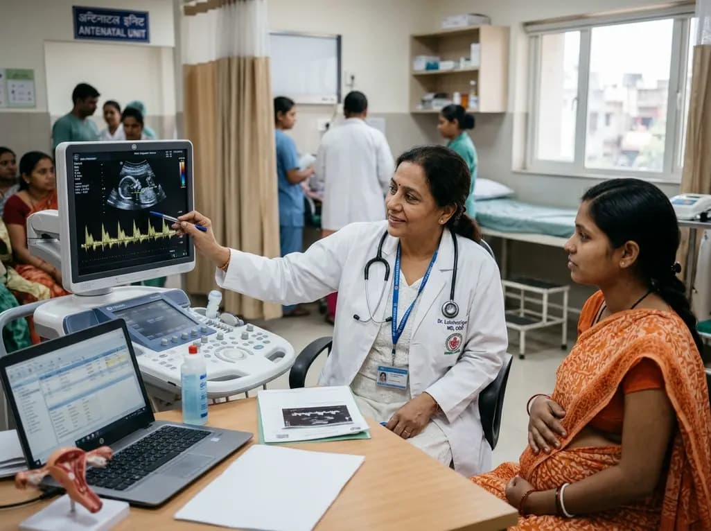
Non-RhD alloimmunization poses an emerging challenge in modern obstetric care as RhIG reduces RhD cases. This review highlights key non-RhD antigens, Doppler monitoring, and neonatal management strategies.
Today

New phase I data evaluate the impact of food and beverages on enlicitide, the first oral PCSK9 inhibitor. While food reduces peak absorption, model simulations demonstrate robust LDL-C lowering of approximately 60%, confirming meaningful pharmacokinetic forgiveness and flexible real-world administration.
Today

A landmark UK Biobank study by Karolinska Institutet researchers reveals that optimal rest mitigates neurodegeneration in cardiometabolic illness. Patients maintaining high sleep quality demonstrated superior cognitive scores and larger brain volumes, highlighting sleep optimization as an actionable clinical strategy.
Today
