

Loading, please wait...
Loading, please wait...

Modern neuro-oncology relies heavily on molecular profiling to classify diffuse brain tumors accurately. Clinicians frequently encounter multicentric IDH-mutant gliomas during baseline neuroimaging evaluations. Historically, medical teams assumed that anatomically separated intracranial lesions shared identical histopathological and genetic characteristics. Clinicians often presumed that secondary foci arose through simple metastatic dissemination from a primary index tumor. However, recent evidence challenges this long-standing clinical dogma. Researchers have uncovered substantial molecular discordance across distinct anatomic tumor sites in the same patient. Isocitrate dehydrogenase mutations represent early driver events in gliomagenesis. Nevertheless, branching clonal evolution can produce distinct neoplastic lineages across separated cerebral hemispheres. Consequently, neurosurgeons and oncologists must reconsider their diagnostic workflows. When clinicians assume biological identity without sampling each lesion, they risk serious diagnostic misclassification. Therefore, understanding intracranial heterogeneity has become vital for optimal neuro-oncological management. Evaluating each anatomical focus individually helps clinicians avoid inaccurate prognostic assessments.
Recent investigative findings provide striking evidence regarding divergent tumour trajectories within multicentric presentations. In a pivotal clinical series, investigators analyzed patients presenting with radiographically distinct multifocal disease where at least one focus harbored an IDH mutation. Surprisingly, multi-site tissue sampling revealed substantial histo-molecular discordance among synchronous lesions. In several examined patients, distinct intracranial foci exhibited completely mismatched foundational driver mutations. Moreover, other cases showed divergent clonal pathways, such as discordant 1p/19q codeletion status between separate lesions. Additionally, some synchronous lesions demonstrated asynchronous malignant progression, where one tumor displayed low-grade histology while another had already transformed into high-grade disease. These findings demonstrate that radiographic congruence does not guarantee biological identity. Instead, independent oncogenic transformations or early divergent clonal branching can generate distinct tumors simultaneously. Therefore, treating clinicians cannot rely on a single tissue biopsy to represent the entire intracranial tumor burden. Consequently, neuro-oncologists must recognize that separate lesions may follow entirely independent biological timelines and clinical trajectories.
The biological mechanisms underlying multicentric IDH-mutant gliomas involve complex evolutionary processes. Gliomagenesis typically begins with truncal alterations in metabolic enzymes like IDH1 or IDH2. However, secondary genetic events frequently diverge as cells migrate or develop independently within distant brain parenchyma. For instance, the acquisition or loss of 1p/19q co-deletion defines entirely separate WHO diagnostic entities, distinguishing oligodendrogliomas from astrocytomas. If one lesion retains 1p/19q integrity while another loses both arms, the patient simultaneously harbors two distinct glioma subtypes. Furthermore, differences in CDKN2A/B homozygous deletion or ATRX expression can drive asynchronous malignant progression. As a result, one anatomical focus may behave indolently, whereas another focus rapidly acquires aggressive features. In addition, metachronous lesions, which appear months or years after the initial presentation, frequently display heightened clonal divergence. Thus, advanced molecular testing, including next-generation sequencing, must interrogate all accessible lesions. Without detailed spatial profiling, clinicians cannot grasp the true genomic architecture of multicentric disease.
Pathological discordance between distinct intracranial lesions directly alters therapeutic strategies. In clinical practice, therapeutic plans depend heavily on precise histopathological grading and molecular markers. For example, discovering an astrocytoma in one hemisphere and an oligodendroglioma in another necessitates distinct chemotherapy regimens and radiation planning. When multifocal sampling occurred in clinical studies, the discovered pathological differences directly changed management in half of synchronous cases. Specifically, treating physicians modified systemic regimens, escalated radiation targets, or initiated targeted therapies based on specific focal mutations. Conversely, presumptive diagnosis often leads to undertreating aggressive secondary lesions or overtreating indolent foci. Furthermore, emerging targeted treatments, such as brain-penetrant IDH inhibitors, demonstrate variable efficacy depending on co-occurring molecular alterations. If an unsampled lesion lacks the sensitizing profile of the primary tumor, targeted monotherapy will fail. Consequently, clinicians must recognize that accurate tissue acquisition protects patients from inappropriate management strategies.
Historically, neurosurgeons avoided biopsying multiple lesions to minimize procedural morbidity and surgical risks. In review cohorts, surgeons frequently deferred additional biopsies because they presumed histological concordance with the primary resected mass. In fact, nearly thirty percent of unsampled secondary lesions were technically accessible for stereotactic biopsy, yet surgeons left them unsampled due to traditional dogma. However, modern stereotactic navigation, robotic guidance, and minimally invasive techniques have significantly reduced surgical morbidity. Consequently, the threshold for multi-site tissue acquisition should shift substantially in contemporary practice. Whenever secondary intracranial lesions are surgically accessible without high neurological risk, teams should obtain dedicated tissue samples. Furthermore, neuropathologists must apply modern integrated CNS WHO diagnostics, combining morphology, immunohistochemistry, and next-generation sequencing for every specimen. If stereotactic biopsy is unsafe due to deep anatomical location or eloquent cortex involvement, clinicians must document diagnostic uncertainty. In such cases, advanced physiological neuroimaging, such as MR spectroscopy or amino acid PET, can provide non-invasive biological clues.
Managing patients with multicentric gliomas requires proactive multidisciplinary coordination among neurosurgeons, neuropathologists, neuroradiologists, and oncologists. First, diagnostic radiologists must thoroughly document the spatial distribution, diffusion characteristics, and contrast enhancement of every separate intracranial lesion. Second, neurosurgical teams should critically evaluate the feasibility of dual-target sampling during initial operative planning. Performing concurrent stereotactic biopsies or staged resections often provides critical diagnostic clarity without adding substantial morbidity. Third, pathology laboratories must process all sampled specimens with comprehensive next-generation sequencing rather than relying solely on rapid frozen sections. Furthermore, clinicians must integrate findings through molecular tumor boards to formulate individualized treatment sequences. In cases where discordant grades exist, therapy must target the highest-grade component while monitoring the lower-grade lesion. Additionally, clinicians should remain vigilant during long-term surveillance because metachronous recurrence may harbor novel genomic alterations. By implementing these systematic protocols, neuro-oncology teams can successfully navigate tumor heterogeneity. Ultimately, personalized neuro-oncology demands rigorous molecular verification across all anatomically separated lesions.
Multifocal gliomas typically spread through established white matter pathways or cerebrospinal fluid channels from a single origin. In contrast, multicentric gliomas represent separate anatomical lesions without connecting pathways. They can arise from independent oncogenic events or early branching clonal evolution, frequently producing discordant histological grades and distinct molecular driver profiles across lesions.
Biopsying multiple lesions prevents diagnostic misclassification caused by assuming pathological concordance. Studies confirm that separate foci can harbor mismatched molecular drivers or different tumor grades. Acquiring tissue from accessible secondary lesions directly influences chemotherapy selection, radiation field design, and targeted therapy indications, ensuring clinicians deliver personalized and effective oncological care.
Neuropathologists should evaluate all resected or biopsied specimens using integrated CNS WHO criteria. Testing requires routine histopathology, immunohistochemistry for IDH1 R132H and ATRX, and next-generation sequencing panels. Additionally, assessing 1p/19q codeletion and CDKN2A/B homozygous deletion is critical to distinguish oligodendroglioma from astrocytoma and identify high-grade molecular features accurately.
Disclaimer: This content is for informational and educational purposes only... Refer to the latest local and national guidelines for clinical practice.
References

Read summarized clinical updates, watch expert medical content, and earn CME certifications right from your smartphone.
Open your smartphone camera to scan and install.





Multicentric IDH-mutant gliomas demonstrate significant spatial and temporal molecular discordance across distinct lesions. Presuming histological uniformity can lead to clinical misclassification, highlighting the essential need for multifocal tissue sampling and integrated next-generation molecular profiling.
Today

A new study demonstrates how explainable AI guides transparent fall risk prediction models in older adults. By pairing random survival forests and SHAP with Cox regression, researchers achieved superior accuracy and eliminated unneeded variables, enhancing clinical decision-making and patient safety.
Today

Ascertainment bias in cancer case-control studies inflates odds ratios and penetrance estimates when cases are drawn from high-risk clinics. A landmark modeling study demonstrates substantial bias ranging up to 23.72, highlighting the critical need for ascertainment adjustment in genetic risk counseling.
Today
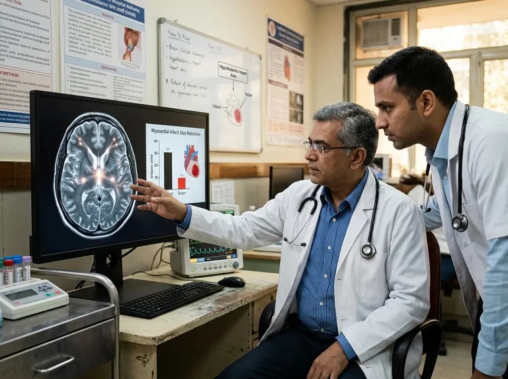
Researchers demonstrate that central induction of synthetic torpor in non-hibernating rats provides striking cardioprotection, reducing myocardial infarct size by ~40% during ischaemia-reperfusion injury through endogenous survival signaling, independent of hypothermia.
Today
A prospective study evaluates DeepSeek for knee osteoarthritis outcomes. While the model excels at baseline diagnosis and clinical scoring, its six-month prognostic predictions fall short of clinicians and simple persistence models, demonstrating high run-to-run variability.
Today
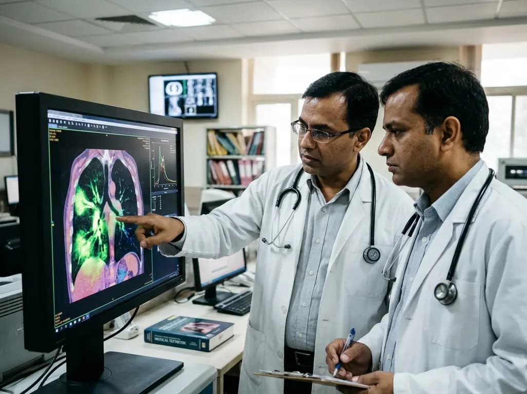
A novel fluorine-functionalized near-infrared copper fluorescent probe selectively illuminates lung tissue, revealing critical links between hyperglycemia, ER stress, and pulmonary malignancy for improved early diagnosis.
Today
