

Loading, please wait...
Loading, please wait...

Managing patients with multicentric IDH-mutant gliomas represents one of the most formidable challenges in modern neuro-oncology. Historically, clinicians viewed multicentric lower-grade gliomas as uniformly disseminated diseases with dismal prognoses and limited surgical options. However, emerging longitudinal evidence reveals that spatially separated lesions exhibit profound biological and clinical divergence. A landmark study published by Gordon and colleagues in the Journal of Neuro-Oncology investigates these intricate relationships across nearly eight years of clinical follow-up. By evaluating patient trajectories managed through awake functional-based resection, the investigators demonstrated that aggressive surgical management remains feasible and remarkably safe. Consequently, this evolving paradigm challenges conventional therapeutic dogmas, offering renewed hope for preserving survival and neurocognitive function.
Multicentric lower-grade gliomas differ fundamentally from multifocal gliomas because they lack microscopic or radiological continuity along established white matter tracts. Therefore, neuro-oncologists must understand whether these separate foci share an identical molecular identity or follow divergent evolutionary paths. In their cohort analysis, Gordon and colleagues evaluated nine patients presenting with eighteen distinct tumor sites. The team classified molecular profiles by assessing isocitrate dehydrogenase mutation status and 1p/19q-codeletion patterns. Interestingly, defining molecular markers were concordant in five patients, discordant in three patients, and unresolved in one individual. This finding proves that spatially separated lesions within the same patient do not necessarily harbor identical diagnostic profiles. Furthermore, molecular concordance did not guarantee parallel histopathological behavior or uniform cellular differentiation over time. For instance, an astrocytic lesion in one hemisphere may display low mitotic activity, whereas a contralateral oligodendroglial lesion accelerates into higher histological malignancy. Clinicians must recognize that a single biopsy or resection from one nodule cannot reliably represent the overall intracranial disease burden. Accordingly, comprehensive molecular characterization across all accessible lesions provides indispensable insights that guide appropriate risk stratification and individualized therapeutic selection.
Beyond baseline genetic differences, spatial lesions demonstrate remarkably divergent temporal trajectories during prolonged follow-up. Specifically, the investigators observed asynchronous progression patterns in six patients, whereas only three individuals exhibited synchronous disease evolution. These growth kinetics indicate that distinct tumor sites follow autonomous clocks rather than coordinated systemic escalation. Consequently, one anatomical lesion may remain quiescent for years while an alternate site undergoes abrupt volumetric enlargement or anaplastic transformation. Furthermore, systemic medical interventions reveal equally complex inter-lesional disparities. In this cohort, six patients received systemic alkylating chemotherapy with temozolomide. Half of these treated patients experienced discordant therapeutic responses between their separate tumor sites. In several cases, one lesion demonstrated objective volumetric regression while a concurrent lesion demonstrated relentless progression or therapeutic resistance. Therefore, neuro-oncologists cannot assume that systemic cytotoxic chemotherapy provides uniform cytoreduction across all intracranial disease sites. Instead, clinicians must evaluate each anatomical lesion as an independent entity with unique microenvironmental and genetic determinants. When clinicians observe radiological recurrence at a single site, they should not automatically abandon effective systemic regimens if other lesions remain controlled. Instead, surgeons can deliver targeted local interventions while preserving effective systemic therapy.
Given the pronounced spatial and temporal heterogeneity of multicentric disease, surgical sequencing demands meticulous personalization. The surgical team performed seventeen awake functional-based resections across twenty-two site-level surgical targets. Rather than pursuing simultaneous, highly disruptive bilateral craniotomies, the neurosurgeons tailored single-stage or staged interventions based on individualized functional priorities. Crucially, the surgeons achieved an extent of resection reaching or exceeding ninety percent in over eighty-one percent of resected sites. Moreover, the median extent of resection across all interventions reached a remarkable one hundred percent. To achieve such extensive cytoreduction without causing permanent neurological deficits, surgeons employed intraoperative direct electrical stimulation mapping. This awake mapping technique identifies critical cortical and subcortical pathways supporting language, motor execution, visuospatial awareness, and executive cognition. Consequently, the surgical resection continues until surgeons identify the functional boundary of eloquent white matter tracts, rather than halting at arbitrary anatomical borders. In patients with bilateral or distant multicentric lesions, staged surgical interventions allow the brain to recover and reorganize functional networks through neuroplasticity. Therefore, surgeons can successfully resect a dominant hemispheric lesion and subsequently address a contralateral lesion months or years later without compounding postoperative disability.
The paramount goal of modern neuro-oncological surgery remains the optimization of the oncofunctional balance. Prolonging overall survival offers little clinical value if aggressive cytoreduction leaves the patient functionally incapacitated. In this study, the longitudinal functional outcomes were remarkably encouraging. Among the eight surviving patients evaluated at a median follow-up of 7.8 years, the median Karnofsky Performance Status score was ninety, ranging between eighty and one hundred. In addition, eight of the nine patients maintained or resumed their professional or academic careers during at least part of their disease course. The remaining patient successfully preserved daily personal autonomy and engaged in active social participation. Although detailed neuropsychological assessments revealed heterogeneous and descriptive patterns, patients generally maintained their baseline cognitive competence. These findings confirm that aggressive, repeated surgical interventions do not inevitably precipitate cognitive decline or functional dependency. Instead, awake intraoperative cognitive mapping actively shields complex neurocognitive networks, including executive functioning and working memory. Consequently, patients sustain meaningful social and vocational lives while harboring a chronic, controlled multicentric brain neoplasm. Multimodal rehabilitation and personalized cognitive therapy further reinforce these favorable functional trajectories over prolonged observation periods.
The biological and clinical insights from this investigation demand a profound shift in clinical tumor board discussions. Traditionally, multidisciplinary teams often classified multicentric low-grade gliomas as inoperable, immediately relegating patients to definitive chemoradiation or watchful waiting. However, these new findings demonstrate that clinicians should not treat multicentric gliomas as uniform, untreatable entities. Because lesions often progress asynchronously and respond discordantly to medical therapies, teams must continuously re-evaluate each tumor focus independently. For example, when an individual lesion shows active growth while other lesions remain stable, clinicians can deploy targeted surgical resection to that specific site. Furthermore, reserving extensive brain radiotherapy prevents premature neurocognitive deterioration in younger patients who may survive for decades. By integrating repeated awake functional resections with deferred, lesion-specific therapies, clinicians preserve critical cognitive reserve. Multidisciplinary teams should include experienced awake neurosurgeons, neuro-oncologists, neuropsychologists, and neuroradiologists who understand dynamic tumor connectomics. Additionally, regular advanced neuroimaging, including perfusion protocols and metabolic spectroscopy, aids in identifying local dedifferentiation early. Ultimately, embracing an individualized longitudinal management framework transforms multicentric lower-grade glioma from a hopeless prognosis into a manageable chronic disease.
Implementing an oncofunctional paradigm requires standardized protocols that unify molecular testing, neurosurgical precision, and functional surveillance. First, neurosurgical centers must adopt comprehensive molecular sequencing for every surgically removed tissue specimen. Because spatial lesions frequently exhibit discordant 1p/19q or IDH profiles, testing each distinct surgical sample prevents misdirected adjuvant therapy. Second, surgical teams must develop structured intraoperative awake testing protocols tailored to specific lesion locations. Speech therapists and neuropsychologists should collaborate closely in the operating room to test multimodal cognitive domains during resection. Moreover, clinicians must establish rigorous postoperative rehabilitation pathways that support swift occupational reintegration. Routine post-surgical evaluations should monitor not only gross motor function but also subtle executive and psychological changes. Finally, longitudinal clinical trials must incorporate multicentric glioma cohorts into prospective registries. Expanding multicenter collaborative databases will refine surgical timing, optimize adjuvant sequencing, and establish evidence-based guidelines. Through this integrated approach, surgical neuro-oncology continues to elevate survival benchmarks while protecting the fundamental human capacities of every patient.
Multicentric IDH-mutant gliomas can arise from separate ancestral clones or diverge early during tumor evolution. Although initial oncogenic hits such as IDH1 mutations may coincide, subsequent genomic alterations, including 1p/19q codeletion and TP53 mutations, diverge across anatomical sites. Consequently, spatial niche variations and microenvironmental pressures drive distinct molecular and histopathological pathways.
Awake functional mapping identifies essential cortical and subcortical pathways in real time. Therefore, neurosurgeons can maximize the extent of resection while protecting speech, executive, and motor networks. This functional-guided strategy prevents permanent postoperative deficits, enabling patients to maintain high functional independence, preserve cognitive health, and return to their regular occupational activities.
No, systemic chemotherapy cannot reliably replace surgery for secondary lesions. The study revealed discordant inter-lesional responses to temozolomide in 50% of treated patients, meaning one lesion shrank while another progressed. Therefore, clinicians must combine systemic therapies with targeted, functional-based resections rather than relying solely on medical therapy for multicentric control.
Disclaimer: This content is for informational and educational purposes only... Refer to the latest local and national guidelines for clinical practice.
References

Read summarized clinical updates, watch expert medical content, and earn CME certifications right from your smartphone.
Open your smartphone camera to scan and install.





A study on multicentric IDH-mutant gliomas reveals marked inter-lesional molecular and progression heterogeneity. Awake functional-based resection achieved a 100% median resection extent across sites while preserving long-term performance status and professional autonomy in surviving patients.
Today

A Bayesian component network meta-analysis of 130 RCTs reveals that self-monitoring and gamification provide the clearest positive effects in mHealth physical activity interventions, while social comparison may have adverse psychological trade-offs.
Today

The GESS-AEEM consensus establishes a clear clinical implementation framework for testosterone therapy in postmenopausal women with hypoactive sexual desire disorder, focusing on transdermal dosing, precise laboratory monitoring, and safety communication.
Today

A multicenter observational study evaluated an 8-week digital health monitoring platform using wearable sensors in ambulatory heart failure. While participants achieved high daily wear-time of 11.7 hours and 80% completion, initial recruitment revealed significant technology-related onboarding barriers.
Today
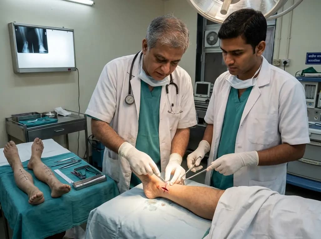
A paired cadaveric study compared triple and quintuple percutaneous Achilles tendon lengthening. While quintuple hemisection showed numerically greater elongation, differences were not statistically significant.
Yesterday

A clinical decision-support framework using health checkup data from 231,683 adults predicts incident obesity with high discrimination (AUROC 0.918). The system integrates temporal survival analysis and risk-factor explanations to prioritize individualized preventive interventions.
Today
