

Loading, please wait...
Loading, please wait...

Managing acute neurovascular crises requires prompt therapeutic decision-making. Clinicians frequently encounter severe diagnostic and therapeutic dilemmas when handling Moyamoya acute ischemic stroke in emergency rooms. Moyamoya disease presents progressive, non-atherosclerotic stenosis of the terminal internal carotid arteries. This occlusion leads to an unstable network of fragile parenchymal collaterals. Consequently, when acute vessel occlusion occurs, standard emergency interventions carry distinct hemodynamic risks. Physicians must weigh tissue salvage against hemorrhagic conversion. Recent multi-center registry analyses provide valuable real-world evidence. These data clarify how reperfusion therapies perform in this rare condition.
Moyamoya disease represents a unique structural arteriopathy that fundamentally alters cerebral hemodynamics. The underlying pathology involves bilateral chronic steno-occlusion of the supraclinoid internal carotid arteries and circle of Willis branches. In response, cerebral microvasculature forms a hazy, smoke-like collateral network. Therefore, ischemic events in these patients do not always follow typical thromboembolic pathways. In many instances, strokes arise from progressive hemodynamic insufficiency, low perfusion pressure, or microthrombi originating in unstable collateral vessels.
Additionally, the clinical presentation often differs from routine large vessel occlusions. Patients may exhibit fluctuating neurologic symptoms, recurrent transient ischemic attacks, or sudden catastrophic deficits. Younger demographics, particularly children and young adults, show significant vulnerability. Furthermore, clinicians often detect severe baseline hypoperfusion across extensive vascular territories. Standard stroke protocols rely on rapid recanalization of large anatomical conduits. However, in Moyamoya vasculopathy, the primary trunk may already be chronically narrowed or occluded. Consequently, aggressive interventions can endanger the delicate collateral pathways that sustain penumbral tissue. Physicians must exercise caution, because misinterpreting these fragile vessels as acute thrombi can trigger inappropriate clinical maneuvers. Neurologists must promptly identify the underlying arteriopathy on urgent vascular imaging to prevent improper acute management.
A nationwide retrospective analysis of over 1.7 million stroke admissions demonstrated that Moyamoya disease accounts for approximately 0.2 percent of acute ischemic stroke hospitalizations. Interestingly, clinicians utilize reperfusion modalities far less frequently in this population than in conventional stroke cohorts. For instance, the adjusted odds of receiving endovascular thrombectomy decrease significantly for patients with Moyamoya disease. Similarly, systemic intravenous thrombolysis administration rates remain markedly lower.
Several technical and diagnostic barriers explain this reduced utilization. First, emergency physicians often hesitate to administer thrombolysis due to the historical fear of provoking intracranial hemorrhage from fragile collateral vessels. Second, interventionalists face formidable anatomical challenges when performing catheter angiography. The marked tapering and stenosis of carotid arteries complicate microcatheter navigation. Furthermore, distinguishing acute embolic occlusions from chronic in-situ steno-occlusive lesions remains exceptionally difficult on non-invasive angiograms. Consequently, intervention teams frequently hesitate during initial therapeutic windows. Additionally, unstandardized institutional protocols often discourage interventionists from treating complex intracranial arteriopathies under time pressure. Therefore, multidisciplinary teams must assess physiological perfusion, baseline functional status, and vascular anatomy collaboratively before initiating acute recanalization procedures.
When an acute embolic occlusion superimposes on Moyamoya vasculopathy, endovascular thrombectomy offers potential tissue rescue. However, interventionalists must approach these procedures with meticulous care. Recent real-world data indicate that mechanical thrombectomy can achieve successful recanalization in carefully selected candidates. Nevertheless, procedural complication rates require vigilant attention. Navigating stent retrievers or aspiration catheters through chronically narrowed vessels risks arterial dissection, vasospasm, and vessel rupture.
Furthermore, hemodynamic swings during general anesthesia or conscious sedation can critically compromise collateral flow. As a result, maintaining optimal mean arterial pressure during the intervention is paramount. Successful recanalization rapidly restores cerebral blood volume. However, sudden reperfusion into dysplastic capillary beds carries an elevated risk of reperfusion injury and cerebral hyperperfusion syndrome. Post-procedural management requires rigorous blood pressure control in specialized intensive care units. In addition, neurointerventional teams should favor gentle aspiration techniques or smaller profile devices to minimize shear stress against diseased endothelial linings. When executed properly, endovascular recanalization prevents extensive infarction without excessively escalating fatal hemorrhagic transformations. Skilled operators must constantly balance ischemic territory salvage against iatrogenic vessel injury.
Intravenous thrombolysis remains the primary pharmacological therapy for acute ischemic stroke presenting within narrow time windows. Nonetheless, its safety profile in Moyamoya disease historically generated substantial debate. The presence of abnormal, thinned, and microaneurysmal lenticulostriate collaterals creates persistent concern for catastrophic hemorrhagic conversion. Despite these theoretical hazards, observational data show that selected patients receive intravenous thrombolysis without prohibitive rates of symptomatic intracerebral hemorrhage.
Moreover, multivariable analyses reveal comparable safety outcomes between Moyamoya cohorts and general stroke cohorts treated with thrombolytic agents. Consequently, clinicians should not view Moyamoya disease as an absolute contraindication to standard intravenous thrombolysis when acute non-hemorrhagic deficits occur within eligible windows. However, strict adherence to blood pressure protocols remains essential before and after drug infusion. Clinicians must maintain systolic blood pressure below 180 millimeters of mercury to protect delicate collateral pathways. Additionally, advanced multimodal imaging, such as magnetic resonance perfusion and susceptibility-weighted sequences, should evaluate microbleeds and core-penumbra mismatches. Thus, careful baseline screening enables physicians to administer intravenous alteplase or tenecteplase safely. Systematic post-thrombolysis neurological checks guarantee early detection of any emerging hemorrhagic complications.
In India and South Asia, Moyamoya disease presents unique diagnostic challenges across diverse healthcare settings. Although historically regarded as an East Asian disease, Moyamoya disease represents an important cause of young stroke in Indian neurological practice. Many patients present with atypical ischemic manifestations, recurrent transient ischemic attacks, or early-onset stroke without traditional cardiovascular risk factors. Unfortunately, lack of early access to digital subtraction angiography or advanced magnetic resonance imaging frequently delays definitive diagnosis.
Therefore, emergency departments across tertiary Indian hospitals must incorporate rapid intracranial vessel imaging into routine stroke algorithms. When treating Moyamoya acute ischemic stroke, physicians must integrate acute hemodynamic stabilization with long-term revascularization planning. Direct or indirect surgical revascularization, such as superficial temporal artery to middle cerebral artery bypass or encephaloduroarteriosynangiosis, provides proven secondary stroke prevention. In addition, acute teams must coordinate closely with experienced neurosurgeons during the index admission. Standardizing multidisciplinary pathways ensures timely reperfusion, minimizes procedural complications, and facilitates seamless transition to surgical collateral augmentation. Ultimately, heightened clinical awareness enables Indian clinicians to preserve functional independence in vulnerable young patients.
Endovascular thrombectomy can be safe and effective for select Moyamoya patients suffering acute large vessel occlusions. However, procedural risks are considerably higher than in standard stroke cases. Diseased vessel walls, tortuous collateral vessels, and chronic arterial stenosis elevate the risk of arterial rupture, dissection, and hyperperfusion hemorrhage. Consequently, neurointerventionalists must carefully evaluate vascular anatomy, choose low-profile retrieval devices, and maintain stable cerebral perfusion pressure throughout the entire procedure.
Clinicians can administer intravenous thrombolysis to eligible Moyamoya patients presenting within the standard acute therapeutic window. Although fragile collateral vessels raise theoretical hemorrhage concerns, real-world registry studies show acceptable safety profiles without excessive symptomatic intracerebral bleeding. Nevertheless, physicians must rigorously exclude existing hemorrhage via baseline neuroimaging and maintain strict blood pressure targets below established guideline thresholds before, during, and after pharmacological infusion.
Surgical revascularization represents the definitive gold standard for secondary stroke prevention in patients with Moyamoya disease. Once clinicians stabilize the acute ischemic event, elective revascularization procedures significantly reduce recurrent ischemic and hemorrhagic episodes. Neurosurgeons perform direct extra-intracranial arterial bypass, indirect encephaloduroarteriosynangiosis, or combined techniques. These surgeries establish durable collateral flow, successfully augmenting regional brain perfusion and relieving hemodynamic stress on vulnerable parenchymal networks.
Disclaimer: This content is for informational and educational purposes only. It is not intended to be a substitute for professional medical advice, diagnosis, or treatment. Always seek the advice of your physician or other qualified health provider with any questions you may have regarding a medical condition. Never disregard professional medical advice or delay in seeking it because of something you have read here. Refer to the latest local and national guidelines for clinical practice.
References

Read summarized clinical updates, watch expert medical content, and earn CME certifications right from your smartphone.
Open your smartphone camera to scan and install.





A nationwide analysis evaluates the safety and efficacy of endovascular thrombectomy and intravenous thrombolysis in Moyamoya disease patients presenting with acute ischemic stroke, offering vital insights for emergency revascularization protocols.
Today
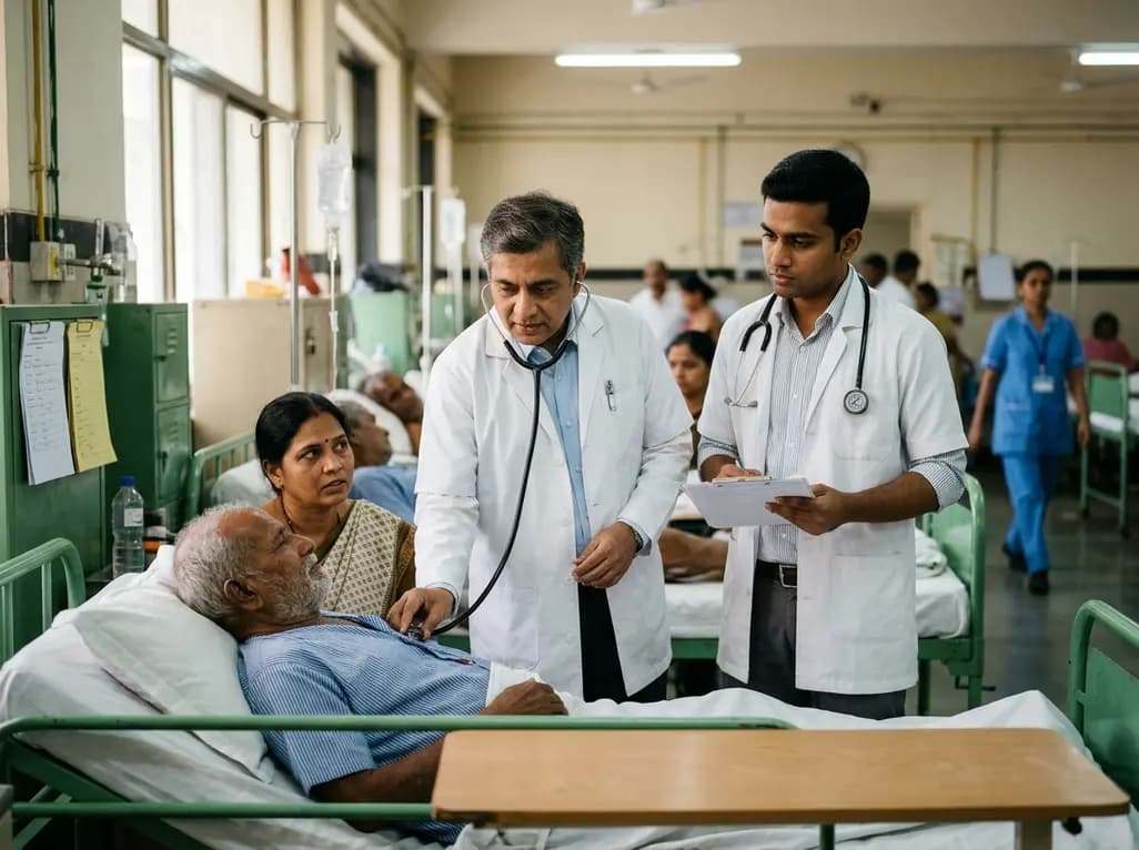
A qualitative study investigates whether traditional bedside clinical examinations still mirror contemporary physician practice. Findings support retaining long cases while reforming short cases toward hypothesis-driven assessments.
Today
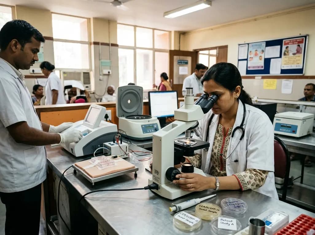
A pilot study evaluated WearDOXXN, a capillary microfluidic sweat patch, for noninvasive breast cancer monitoring by comparing sweat and serum CEA and PIP/GCDFP-15 levels.
Today
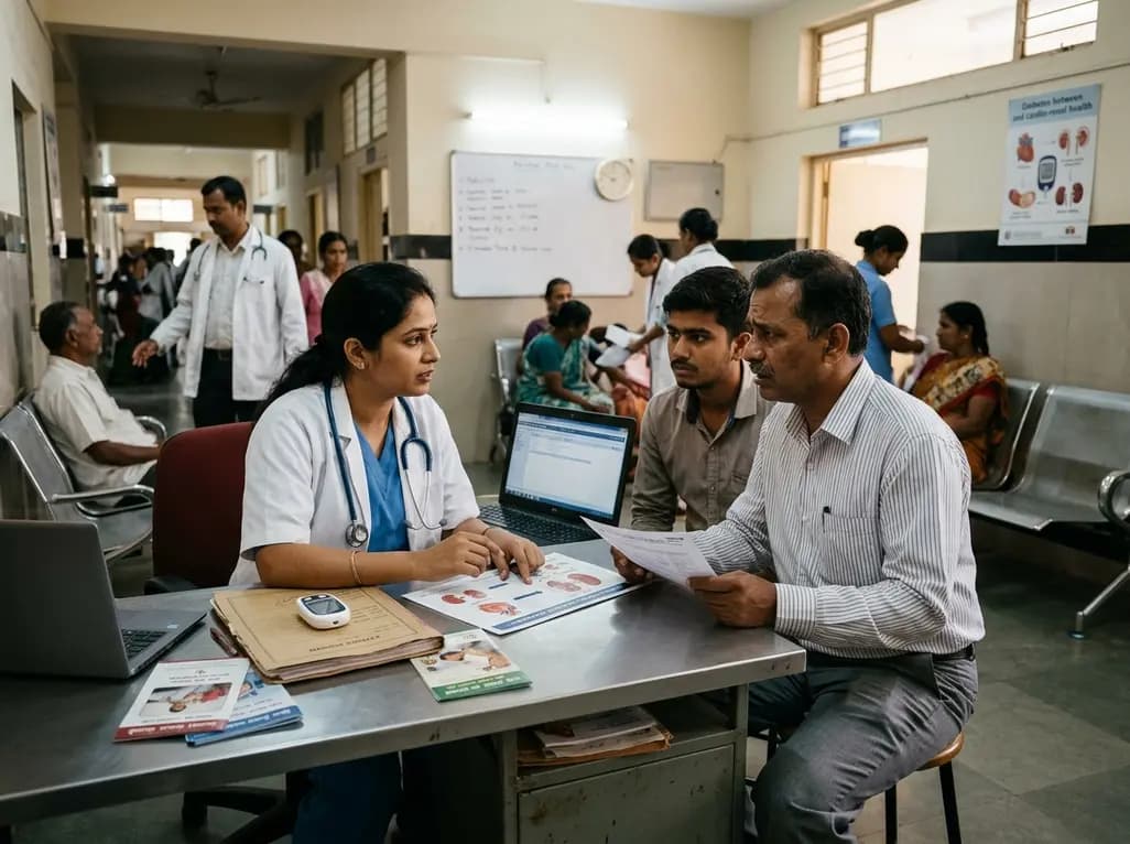
India faces a massive diabetes burden with over 89 million adults affected. Managing blood sugar in isolation fails to prevent severe cardiorenal complications. Clinicians must adopt an integrated metabolic ecosystem that prioritizes early detection, multifactorial risk assessment, and personalized therapies.
Today
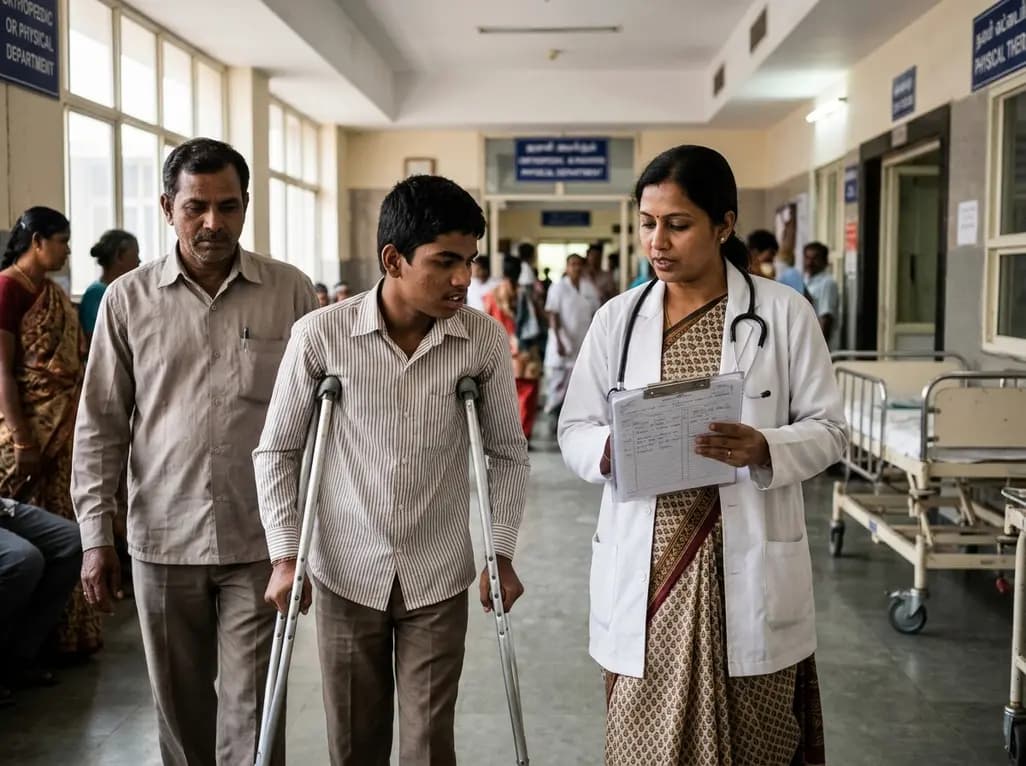
The Gross Motor Function Classification System (GMFCS) stops its validated age bands at 18. Recent evidence emphasizes that evaluating adult cerebral palsy requires purpose-sensitive documentation combining adolescent ratings with dynamic profiles of mobility, fatigue, pain, and community participation.
Today
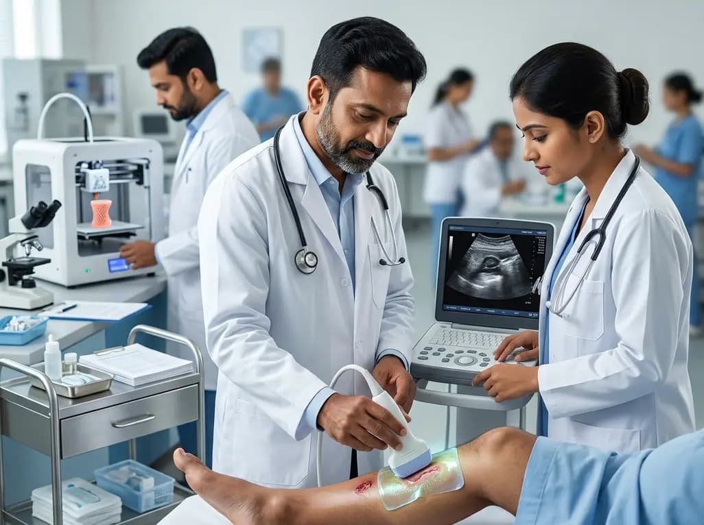
A breakthrough 3D-printed piezoelectric collagen hydrogel activated by ultrasound accelerates diabetic wound repair, demonstrating 98.5% wound closure, robust antibacterial action, and enhanced vascularization without toxic chemical crosslinkers.
Today
