.webp&w=384&q=75)

Loading, please wait...
Loading, please wait...
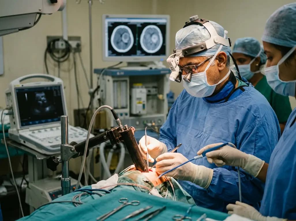
Surgical access to subcortical and intraventricular lesions poses substantial technical demands for operating teams. Deep lesions require delicate navigation through sensitive functional tracts. The modified tubular retractor offers a transformative solution for neurosurgeons operating in low-resource healthcare environments. Historically, traditional blade retractors applied focal pressure on vulnerable cerebral parenchyma. This mechanical pressure frequently induced localized ischemia, cerebral edema, and secondary neurological deficits. Although commercial tubular retractors distribute radial pressure evenly, their high acquisition cost restricts widespread adoption. Furthermore, standard tubular retractors often present narrow viewing corridors that hinder surgical manoeuvrability for lesions measuring 3 cm or larger. To resolve these persistent operative bottlenecks, neurosurgeons developed an innovative, low-cost tubular retractor. This indigenously engineered system incorporates an expanded optical window and an ergonomic trajectory guide. Consequently, surgical teams can achieve superior visualization without purchasing expensive proprietary systems. Recent clinical trials highlight how this device balances affordability with high surgical efficacy.
Accessing deep intracranial pathologies has traditionally relied on self-retaining blade retractors, such as Leyla spatulas. However, continuous blade retraction exerts asymmetric shear stress across cortical margins. Consequently, patients often experience postoperative brain contusions, venous infarction, and prolonged cerebral edema. In contrast, cylindrical retractor systems disperse mechanical forces radially over a broader contact surface. This cylindrical geometry preserves white matter tracts during transsulcal entry corridors.
Nevertheless, existing conventional tubular retractor models have notable limitations. For instance, repurposed syringe barrels often restrict illumination and surgical freedom. These makeshift systems frequently cause instrument crowding when surgeons deploy ultrasonic aspirators alongside bipolar forceps and suction tubes. Moreover, commercial stereotactic channel devices remain cost-prohibitive for many public hospitals across developing nations. When lesions exceed 3 cm, standard narrow-lumen tubes severely constrain angular visualization, preventing complete tumor removal. Therefore, surgeons operating in resource-limited operating suites require an adaptable, inexpensive tool. An optimal device must deliver superior illumination, wide instrument angles, and reliable structural stability without escalating procedural costs.
To address these technical barriers, neurosurgical researchers introduced a customized device. The modified tubular retractor integrates several critical structural enhancements tailored for challenging intracranial exposures. First, the cannula features a transparent outer wall with a distinctive 2.5 cm distal slanted opening. This beveled window substantially expands the deep optical field, allowing dynamic visual tracking of tumor margins. Second, the system features a flared proximal collar that prevents instrument clashing during microscopic or endoscopic procedures.
Additionally, the retractor utilizes an atraumatic tapered obturator with an integrated neuronavigation lumen. This channel permits real-time, image-guided transsulcal cannulation along predefined subcortical white matter pathways. Surgeons can securely anchor the tube using standard Leyla retractor arms, ensuring robust intraoperative stability. Importantly, manufacturing this indigenously modified device costs a fraction of imported commercial alternatives. By combining transparent materials, angular viewing ports, and navigation compatibility, the design dramatically enhances surgical dexterity. Consequently, neurosurgical teams can maneuver ultrasonic aspirators and micro-instruments smoothly without compromising surrounding healthy neural tissue.
Clinical efficacy was demonstrated in a prospective comparative cohort study of 105 patients with deep brain tumors measuring 3 cm or larger. Investigators stratified participants into three distinct cohorts based on the retraction technique employed. Group 1 underwent traditional Leyla spatula retraction, Group 2 utilized conventional syringe-derived tubular retractors, and Group 3 received surgery using the modified tubular device.
The clinical findings demonstrated striking differences in surgical precision and resection completeness. Specifically, gross total resection reached 91.4% in the modified retractor group. In comparison, surgeons achieved gross total resection in only 78.9% of the Leyla spatula cohort and 28.9% of the conventional tubular cohort. Logistic regression modeling confirmed that retractor design served as an independent predictor of residual tumor (Odds Ratio: 2.59, p = 0.035). Furthermore, intraoperative visualization quality independently influenced surgical clearance (Odds Ratio: 2.01, p = 0.036). The modified design provided an expansive operative corridor that permitted meticulous peripheral tumor dissection. As a result, operating teams excised deep-seated lesions comprehensively while preserving adjacent neurovascular structures and critical functional boundaries.
Beyond achieving higher resection rates, the modified retractor system yielded superior clinical recovery profiles. Patients undergoing surgery with the novel tubular design experienced significantly lower rates of retraction-related cortical injury and hemorrhage. In addition, the reduced surgical trauma directly translated into shorter stays in intensive care units.
Long-term functional evaluations further reinforced these therapeutic advantages. At three months post-surgery, 91% of patients in the modified retractor group achieved favorable functional recovery, defined as a Glasgow Outcome Score of 4 or 5. Conversely, patients treated with conventional blade or basic tubular retractors experienced higher rates of persistent neurological deficits and prolonged rehabilitation needs. The gentle, radial tissue displacement maintained microvascular perfusion in surrounding parenchymal tissues. Therefore, postoperative edema subsided rapidly, enabling early mobilization and faster overall recovery. These robust functional outcomes emphasize that structural modifications in retractor hardware directly improve long-term patient quality of life.
The development of this indigenous surgical device offers profound implications for neurosurgical practice in low- and middle-income countries. Advanced minimally invasive neurosurgery often remains inaccessible due to expensive proprietary consumables and specialized instrumentation. However, this low-cost innovation democratizes safe subcortical surgery without compromising patient safety or oncological principles.
Furthermore, this surgical approach seamlessly integrates with standard operating room equipment, including conventional microscopes, endoscopes, and standard neuronavigation platforms. Surgical trainees can readily acquire the necessary procedural skills, flattening the learning curve for minimally invasive channel-based surgery. While these prospective findings are highly promising, clinicians must note study limitations. The non-randomized cohort design and lack of intraoperative fluorescence mapping require careful consideration. Therefore, future multicentric trials should evaluate long-term oncological survival across diverse tumor histologies. Nevertheless, this modified retractor establishes a practical, scalable paradigm for delivering high-quality neurosurgical care in resource-limited operating rooms.
A modified tubular retractor is an indigenously redesigned cylindrical surgical cannula used in neurosurgery. It features a transparent body, a slanted distal window, and an obturator compatible with neuronavigation. This design enables minimally invasive access to deep intracranial tumors while protecting surrounding brain parenchyma.
The device provides a wider operative field through its slanted distal aperture and flared proximal collar. These ergonomic improvements eliminate instrument crowding, enhance micro-instrument manoeuvrability, and provide superior illumination. Consequently, surgeons achieve significantly higher rates of gross total tumor resection compared with conventional syringe-based or blade retractors.
This indigenous device costs significantly less than commercial proprietary retractors, making minimally invasive brain surgery affordable. Additionally, it integrates easily with standard Leyla arms and standard neuronavigation systems. Patients benefit from reduced brain retraction trauma, fewer surgical complications, shorter intensive care unit stays, and superior neurological recovery scores.
Disclaimer: This content is for informational and educational purposes only. It does not constitute medical advice or replace professional judgment. Refer to the latest local and national guidelines for clinical practice.
References
Jha VC et al. Modified tubular retractor for deep brain tumour resection: enhanced surgical outcomes in resource-constrained settings. Neurosurg Rev. 2026 May 28. doi: 10.1007/s10143-026-04338-2. PMID: 42204019.
Eichberg DG, Buttrick S, Snelling B, et al. Use of Tubular Retractor for Resection of Deep-Seated Cerebral Tumors and Colloid Cysts: Single Surgeon Experience and Review of the Literature. World Neurosurg. 2018;112:e50-e60. doi:10.1016/j.wneu.2017.12.023.
Mittelman L, Sistiaga IL, Seenarine N, et al. Connectome-guided resection of deep-seated brain tumors using tubular retractors: matched cohort outcomes and exploratory quantitative tractometry. J Neurooncol. 2026;177(3):481-492. doi:10.1007/s11060-026-05578-4.

Read summarized clinical updates, watch expert medical content, and earn CME certifications right from your smartphone.
Open your smartphone camera to scan and install.





A prospective study evaluates an innovative, low-cost modified tubular retractor for deep brain tumors ≥3 cm, demonstrating a 91.4% gross total resection rate, reduced complications, and improved functional outcomes in resource-constrained surgical settings.
last month

A comprehensive analysis of serum bile acid variability establishes robust reference ranges and reveals how sex, age, BMI, and smoking alter circulating profiles.
Today

A mixed-methods study evaluates a digital perinatal navigator app for high-risk pregnancies, demonstrating high usability, reduced distress, and improved awareness of vital healthcare services.
Today

A user-centered study demonstrates that AI-assisted carotid ultrasound enables nonexpert primary care staff to detect subclinical atherosclerosis. With real-time guidance and workflow optimization, this technology supports early cardiovascular risk communication and task-shifting in routine clinical practice.
Today

A hyaluronic acid-modified metal-polyphenol nanocomposite successfully eliminates reactive oxygen species in chondrocytes, halts cartilage breakdown, and promotes tissue anabolism, presenting a novel disease-modifying strategy for early osteoarthritis.
Today

A pilot randomized trial demonstrates that digital wellness applications and medically tailored meals significantly attenuate rapid weight regain following GLP-1 receptor agonist discontinuation, providing valuable transitional support for long-term obesity management.
Today
