.webp&w=384&q=75)

Loading, please wait...
Loading, please wait...

Atherosclerotic cardiovascular disease remains a leading cause of premature mortality worldwide, especially in populations undergoing rapid epidemiological transitions. Traditional risk calculators often fail to capture individual plaque vulnerability and vascular aging. Therefore, visualizing arterial pathology provides a powerful catalyst for timely lifestyle modification and medical intervention. Recent innovations in point-of-care devices have introduced AI-assisted carotid ultrasound into frontline settings. By leveraging automated image acquisition and real-time guidance, this approach allows nonexpert healthcare professionals to identify subclinical atherosclerosis. Consequently, routine cardiovascular risk stratification is undergoing a transformative shift toward accessible, community-level vascular assessment.
Cardiovascular disease frequently develops silently over several decades before presenting as myocardial infarction or acute ischemic stroke. In routine practice, clinicians estimate future risk using scoring algorithms based on systemic biomarkers such as blood pressure and cholesterol levels. However, these conventional risk tools frequently underestimate danger in intermediate-risk individuals who harbor significant arterial plaques. Consequently, patients often exhibit low compliance with preventive pharmacotherapy and lifestyle recommendations because asymptomatic figures feel abstract.
Visual risk communication directly overcomes this psychological disconnect by showing individuals their actual arterial anatomy. In landmark trials, providing pictorial reports of carotid plaque formation substantially improved patient engagement and accelerated lipid-lowering therapy initiation. Furthermore, witnessing visible arterial thickening prompts patients to adopt healthier dietary and exercise habits more consistently. Historically, obtaining these high-quality vascular images required expert sonographers and costly hospital-based equipment. This logistical bottleneck severely restricted widespread preventive screening, particularly in busy primary healthcare centers and underserved rural clinics. Therefore, decentralizing vascular imaging through task-shifting represents an urgent clinical priority to bridge this gap in preventive cardiology.
Point-of-care ultrasonography has historically demanded extensive operator training, fine motor skills, and specialized anatomical knowledge. For nonexpert clinicians, locating the carotid bifurcation and capturing an optimal longitudinal plane present substantial technical hurdles. Fortunately, modern advances in machine learning have transformed this landscape through AI-assisted carotid ultrasound systems designed specifically for novice users. These intelligent platforms integrate automated neural networks that process real-time video feeds during probe manipulation.
Specifically, the software provides dynamic visual cues and automated artery segmentation to guide the operator toward correct anatomical landmarks. When the probe aligns with the carotid lumen, the algorithm verifies image quality and automatically freezes optimal frames for analysis. Additionally, artificial intelligence tools can instantly quantify carotid intima-media thickness and identify distinct plaque morphology. Thus, technology effectively standardizes diagnostic image acquisition across varying skill levels. By removing the steep learning curve, intelligent handheld scanners permit task-shifting from scarce specialist physicians to frontline health workers. As a result, preventive screening moves directly into community practices where early detection yields the highest clinical benefit.
To evaluate how nonexpert healthcare staff handle intelligent ultrasound tools, researchers conducted a user-centered development study in northern Sweden. The investigation evaluated fourteen nurse assistants from four diverse primary care centers representing urban, rural, and remote settings. These nonexpert operators tested a specialized prototype incorporating real-time guidance and automated carotid segmentation directly within their daily clinic routines.
Through semi-structured interviews and pragmatic workflow observations, the investigators identified four fundamental themes governing clinical adoption. First, the diagnostic technology must integrate smoothly into the rigid time constraints of primary care visits. Second, robust real-time system guidance serves as an essential prerequisite for nonexperts to achieve procedural accuracy. Third, participants emphasized that practical, peer-supported training environments build enduring technical competence far better than theoretical tutorials alone. Fourth, operators required substantial confidence during patient encounters to explain findings clearly without creating unnecessary anxiety. Importantly, the qualitative analysis demonstrated that nonexpert healthcare workers could successfully capture diagnostic carotid images when software interfaces provided intuitive navigational prompts. Consequently, user-centered design emerged as the defining factor for converting technical innovation into frontline preventive action.
Deploying point-of-care devices in busy outpatient environments frequently reveals unforeseen practical barriers that laboratory testing overlooks. During prototype evaluations, observers documented recurring usability challenges involving probe ergonomics, physical cable management, and interface navigation. Moreover, operators experienced notable time pressure while attempting to balance rapid data entry with patient positioning. Maintaining steady transducer contact along the lateral neck also induced operator hand fatigue during extended scanning sessions.
In response to these pragmatic observations, developers introduced several iterative hardware and software refinements. For example, engineers redesigned the graphical interface to eliminate redundant menus and incorporated clear audio feedback to confirm optimal image capture. In addition, the team enhanced probe ergonomics to support stable grip postures and simplified patient demographic entry. Revised training protocols also incorporated hands-on peer practice modules to familiarize staff with transducer angles. These modifications significantly reduced procedural frustration and accelerated overall examination duration. Ultimately, addressing human factors ensured that nonexpert operators felt supported rather than burdened by algorithmic guidance. When medical technology respects clinical workflow realities, frontline healthcare providers embrace preventive diagnostic innovations with genuine enthusiasm.
The clinical insights from this user-centered trial offer profound implications for healthcare delivery across India. South Asian populations face an aggressive, premature onset of atherosclerotic coronary artery disease, often manifesting a decade earlier than Western cohorts. However, specialized imaging infrastructure and trained cardiologists remain heavily concentrated in tier-one urban tertiary hospitals. Rural primary health centres and overburdened community health centres rarely possess the resources to offer comprehensive cardiovascular screening.
Implementing intelligent, handheld point-of-care ultrasound could bridge this diagnostic disparity by enabling task-shifting to auxiliary nurse midwives and general practitioners. Primary care teams could screen asymptomatic individuals during regular wellness checks and hypertension clinics. When screening reveals occult carotid plaques, doctors can promptly initiate evidence-based statin therapy and intensive lifestyle counselling. Furthermore, displaying real-time vascular images helps overcome patient skepticism, significantly enhancing adherence to preventive prescriptions. Nevertheless, sustainable implementation in India will require structured vernacular training, robust ergonomic hardware, and clear clinical referral pathways. By uniting intelligent ultrasound guidance with decentralized primary care teams, health systems can democratize vascular disease prevention and curb catastrophic cardiovascular events.
Yes, nonexpert healthcare personnel can achieve reliable diagnostic imaging when supported by real-time artificial intelligence guidance. Recent implementation trials demonstrate that intuitive software interfaces, automated vessel segmentation, and audio feedback allow nonexperts to acquire high-quality carotid scans. However, successful execution requires structured, peer-supported hands-on training alongside ergonomic probe design. With appropriate workflow integration, trained frontline staff can effectively perform preliminary screening, identify subclinical atherosclerosis, and flag high-risk individuals for physician review.
Visual risk communication transforms abstract statistical estimates into tangible personal reality by showing patients their actual carotid artery images. When individuals visibly observe atherosclerotic plaque accumulation and arterial wall thickening, they comprehend their cardiovascular risk far more clearly. Consequently, this emotional and cognitive awareness prompts stronger motivation for behavioral modification. Clinical trials confirm that pictorial feedback significantly accelerates the initiation of lipid-lowering pharmacotherapy and fosters sustained adherence to healthy diet and exercise recommendations.
Clinics must overcome several critical operational challenges before implementing point-of-care vascular imaging. Specifically, primary care centers must accommodate the scanning procedure within strict consultation time limits without overloading staff. Additionally, administrators must resolve ergonomic issues, including probe handling strain, cable clutter, and cumbersome software navigation. Establishing standardized peer-supported training programs and clear referral protocols for positive scans is also essential. Addressing these practical factors ensures seamless adoption without disrupting everyday primary care productivity.
Disclaimer: This content is for informational and educational purposes only... Refer to the latest local and national guidelines for clinical practice.
References

Read summarized clinical updates, watch expert medical content, and earn CME certifications right from your smartphone.
Open your smartphone camera to scan and install.





A user-centered study demonstrates that AI-assisted carotid ultrasound enables nonexpert primary care staff to detect subclinical atherosclerosis. With real-time guidance and workflow optimization, this technology supports early cardiovascular risk communication and task-shifting in routine clinical practice.
Today

New evidence from Karolinska Institutet shows women vaccinated against HPV prior to conception have significantly lower rates of preterm birth and major perinatal complications. Clinicians in India can leverage these findings to strengthen adolescent immunisation and reproductive health advocacy.
Today
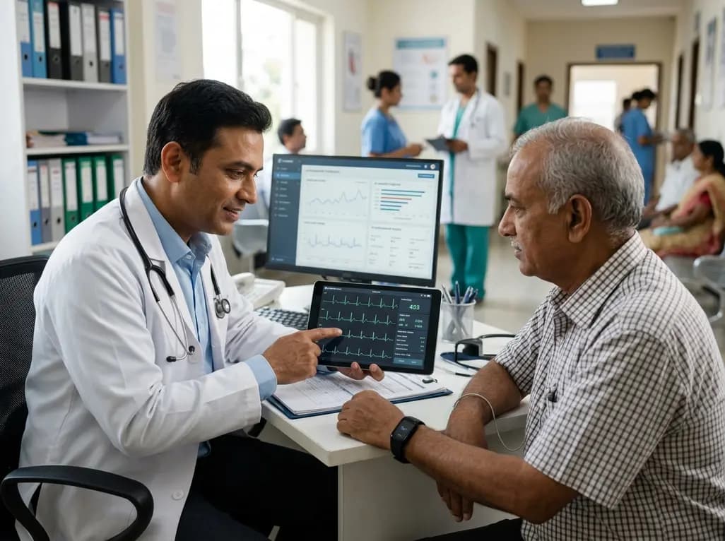
Wearable sensors and remote patient monitoring are shifting cardiovascular care from intermittent clinic visits to continuous physiological surveillance. Artificial intelligence and multimodal biosensors now empower earlier diagnosis and targeted interventions for heart disease.
Today
A hyaluronic acid-modified metal-polyphenol nanocomposite successfully eliminates reactive oxygen species in chondrocytes, halts cartilage breakdown, and promotes tissue anabolism, presenting a novel disease-modifying strategy for early osteoarthritis.
Today
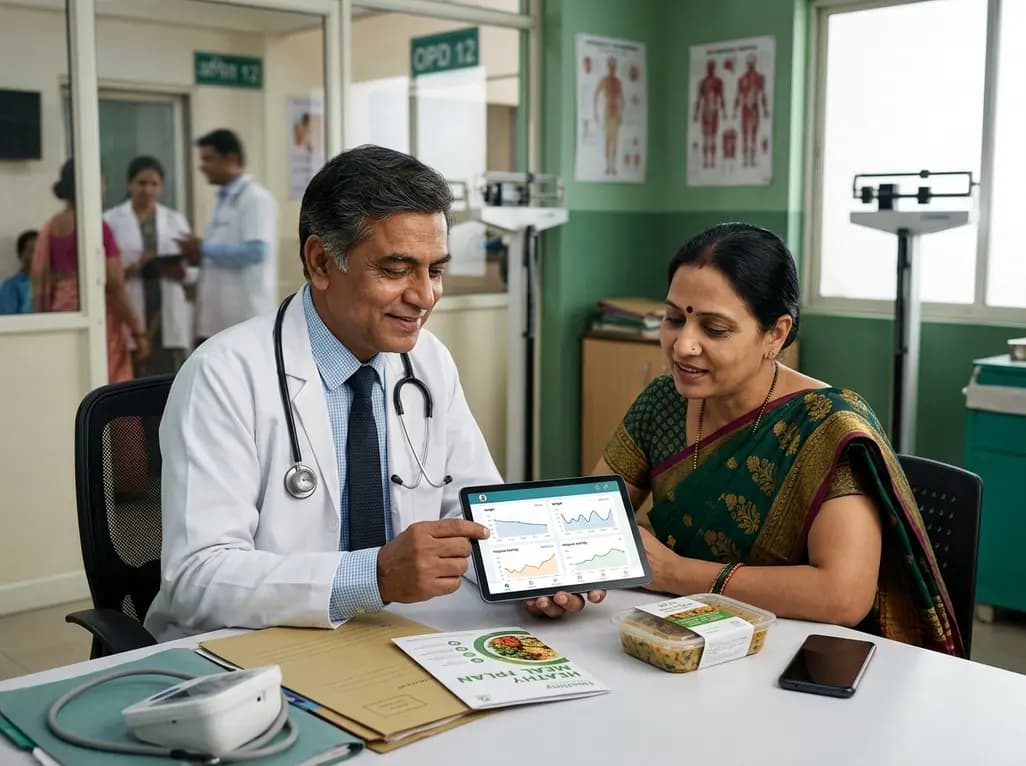
A pilot randomized trial demonstrates that digital wellness applications and medically tailored meals significantly attenuate rapid weight regain following GLP-1 receptor agonist discontinuation, providing valuable transitional support for long-term obesity management.
Today
