.webp&w=384&q=75)

Loading, please wait...
Loading, please wait...
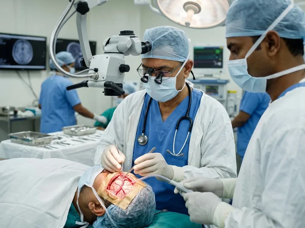
Middle meningeal artery (MMA) preservation is a cornerstone of successful revascularization in patients with moyamoya disease (MMD). Because these patients rely on external-to-internal carotid collaterals, any damage to the MMA during surgery can compromise cerebral blood flow. Recent research highlights the middle meningeal artery canal. This bony structure often encases the vessel, making it susceptible to injury during craniotomy.
Researchers analyzed 175 dry skulls and 200 CT scans. They found the frontal MMA branch traverses a bony canal in 91% of adults. However, this incidence varies significantly with age. Before fontanelle closure, the presence of a canal is rare. Conversely, the likelihood of finding a middle meningeal artery canal increases once children reach two years of age. For surgeons, understanding this developmental anatomy is essential to avoid inadvertent vessel transection when lifting the bone flap.
Notably, the study compared patients with MMD to those with atherosclerotic cerebrovascular disease (ACVD). While the prevalence of the frontal branch canal was similar between groups, MMD patients exhibited longer and wider canals. Specifically, the mean canal length in MMD patients was approximately 15.9 mm compared to 13.3 mm in the ACVD group. These findings suggest the MMA in moyamoya patients may be more extensively "buried" within the skull. This depth requires extra caution during the bone-drilling process.
To locate these structures, surgeons can look roughly one thumb's width posterior to the pterion landmark. Advanced preoperative imaging remains the best tool to map the exact entry and exit points of the canal. By identifying these landmarks, neurosurgeons can implement a vessel-sparing craniotomy, ensuring the integrity of the patient's critical collateral pathways.
In moyamoya disease, the MMA acts as a vital collateral pathway providing blood to the brain. If the artery is encased in a bony canal, it can be easily damaged during the bone-cutting phase of surgery, potentially causing a stroke.
The canal is almost non-existent in infants under two years old. Its prevalence increases significantly after fontanelle closure, appearing in nearly 48% of children over age two and over 91% of adults.
High-resolution cranial CT scans are the gold standard for identifying the location, length, and width of the bony canal, allowing for precise surgical planning and vessel preservation.
Disclaimer: This content is for informational and educational purposes only and does not constitute medical advice or a professional relationship. Always seek the advice of a physician or other qualified health provider with any questions you may have regarding a medical condition. Refer to the latest local and national guidelines for clinical practice.
References
Mertens R et al. Anatomy of the bony canal containing the middle meningeal artery for a vessel-sparing craniotomy in moyamoya disease. J Neurosurg. 2026 Jun 05. doi: 10.3171/2026.1.JNS252290. PMID: 42247704.
Hori S, Kashiwazaki D, Akioka N, et al. Surgical anatomy and preservation of the middle meningeal artery during bypass surgery for moyamoya disease. Acta Neurochir (Wien). 2015;157(1):29-36. doi: 10.1007/s00701-014-2282-7.
Eberlova L et al. Bony canal and grooves of the middle meningeal artery: mythic structures in anatomy and neurosurgery? Folia Morphol (Warsz). 2020;79(3):450-461. doi: 10.5603/FM.a2019.0098.

Read summarized clinical updates, watch expert medical content, and earn CME certifications right from your smartphone.
Open your smartphone camera to scan and install.





New research examines middle meningeal artery canal anatomy, providing crucial data for vessel-sparing craniotomy in Moyamoya disease patients....
4 months ago

Discover the crucial role of glymphatic dysfunction in stroke, where perivascular clearance failure and neuroinflammation exacerbate cerebral edema and post-stroke brain injury.
Today

A prenatal case highlights severe glutaric acidemia type II presenting as bilateral hyperechoic cystic kidneys at 18 weeks. Whole-exome sequencing revealed a novel homozygous ETFDH frameshift variant, demonstrating the power of rapid genetic testing in fetal renal anomalies.
Today

Review how tailored exercise programs benefit frail older patients with heart failure. Explore pathophysiological mechanisms, individualized training prescriptions, and strategies to overcome adherence barriers in clinical practice.
Yesterday

This comparative study evaluates posterior labral tears with and without glenohumeral instability, highlighting distinct demographic patterns, MRI underestimation in stable shoulders, and successful arthroscopic repair outcomes.
Yesterday

A clinical study demonstrates that early intervention with tocilizumab, either as monotherapy or bridged with corticosteroids, achieves significant proptosis reduction and resolves active inflammation in treatment-naive thyroid eye disease.
Yesterday
