.webp&w=384&q=75)

Loading, please wait...
Loading, please wait...
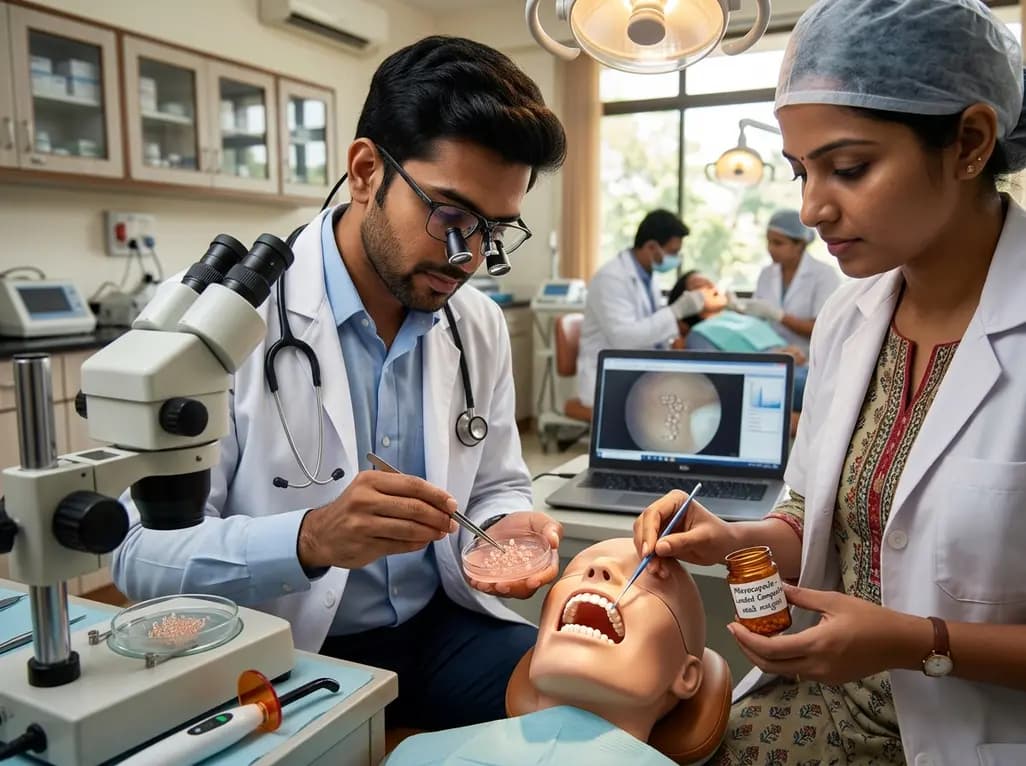
Resin-based dental materials have transformed modern restorative dentistry by combining outstanding esthetics with conservative cavity preparation. However, dynamic masticatory forces, thermal cycling, and enzymatic challenges frequently generate microscopic cracks within the resin matrix. When left unchecked, these microcracks propagate and inevitably cause catastrophic restoration fracture or marginal breakdown. Consequently, bioinspired self-healing dental composites have emerged as a promising strategy to autonomously intercept damage before clinical failure occurs. Understanding how microcapsule integration governs structural resilience remains essential for advancing these restorative materials into everyday clinical practice.
Modern restorative workflows depend heavily on dimethacrylate resins like Bis-GMA and TEGDMA. Although these polymeric networks offer remarkable initial hardness, they remain inherently susceptible to fatigue wear and volumetric shrinkage. Over time, repetitive chewing cycles induce localized stress concentrations at tooth-restoration interfaces. Consequently, subsurface microcracks develop deep within the composite bulk where dental clinicians cannot visually identify them. In addition, salivary fluids infiltrate these microscopic fissures and accelerate hydrolytic breakdown of polymer chains. Bacterial colonies then quickly colonize the expanding crevices, which promotes secondary caries and marginal discoloration. Therefore, conventional treatment requires invasive replacement of the compromised filling, sacrificing healthy dental hard tissue in the process. Such repeated restorative cycles shorten tooth survival and escalate dental treatment expenses for patients. Furthermore, traditional resin matrices lack intrinsic biological capacity to heal physical defects once microfractures initiate. To overcome these persistent clinical limitations, biomaterial scientists have turned toward autonomous repair systems. Integrating reactive chemical payloads directly into restorative formulations offers an innovative solution to combat mechanical fatigue. By sealing microcracks at their earliest onset, clinicians can potentially extend the clinical service life of tooth-colored restorations.
Autonomous repair systems mimic natural biological tissue by sealing damage without human intervention. In self-healing dental composites, extrinsic microcapsules function as microscopic reservoirs distributed uniformly throughout the resin matrix. Typically, engineers fabricate these microcapsules with poly(urea-formaldehyde) or functionalized silica shells. Inside these protective shells, liquid healing agents remain stable until physical stress triggers their therapeutic release. When an advancing crack ruptures the capsule wall, capillary action immediately draws the liquid monomer into the crack plane. For instance, formulations frequently utilize reactive triethylene glycol dimethacrylate combined with tertiary amine activators or dicyclopentadiene monomers. Subsequently, the released liquid contacts chemical catalysts embedded within the adjacent composite matrix. This contact immediately triggers localized polymerization, which hardens the monomer and binds the fractured surfaces together. Consequently, the newly formed polymer network arrests further crack propagation and restores structural continuity. Laboratory evaluations measure this recovery by comparing post-healing fracture toughness against pristine baseline values. Recent systematic analyses demonstrate that optimized systems can achieve fracture toughness recovery exceeding seventy percent. Thus, microencapsulation establishes a robust physical defense against progressive fatigue damage in restorative environments.
Achieving effective self-healing requires striking a delicate compromise between healing efficiency and mechanical stability. When manufacturers add high concentrations of microcapsules, crack repair capability logically rises due to abundant monomer availability. However, incorporating excessive microcapsule volumes inevitably displaces high-strength inorganic glass fillers from the resin composite. As a result, the baseline flexural strength, elastic modulus, and surface hardness decline significantly. Porous microcapsules can also act as structural stress concentrators if their shell walls rupture prematurely during chewing. Preclinical systematic review data indicate that microcapsule loading between 5 wt% and 15 wt% establishes an optimal therapeutic balance. Within this specific concentration window, composites achieve notable fracture toughness recovery between 60% and 75% without compromising essential baseline metrics. Conversely, loading levels exceeding 15 wt% frequently produce severe reductions in baseline strength that fail international restorative standards. On the other hand, concentrations below 5 wt% provide insufficient monomer volume to seal propagating fissures reliably. Therefore, meticulous control of capsule concentration, diameter distribution, and matrix adhesion ensures reliable crack interception without sacrificing restorative durability.
Biocompatibility represents a non-negotiable prerequisite for any biomaterial destined for prolonged intraoral placement. Because dental resins interact directly with dentinal tubules and adjacent gingival margins, chemical leaching causes justifiable clinical concern. Fortunately, preclinical cytotoxicity assays utilizing human gingival and pulp fibroblasts consistently show acceptable cytocompatibility for microcapsule formulations. Poly(urea-formaldehyde) shells demonstrate minimal residual formaldehyde release when researchers employ rigorous post-synthesis washing protocols. Moreover, silica-based microcapsules exhibit outstanding biological safety and bond intimately with silanated methacrylate networks. Beyond autonomic structural repair, innovative self-healing formulations offer valuable multifunctional advantages. For example, several experimental systems integrate antibacterial agents like quaternary ammonium compounds alongside healing microcapsules. This dual strategy actively suppresses Streptococcus mutans biofilms while simultaneously sealing internal microcracks. Additionally, incorporating calcium phosphate compounds or bioactive glass nanoparticles promotes ion release and local remineralization. Consequently, these multifunctional composites not only repair structural fractures but also actively inhibit marginal demineralization. Clinicians thereby gain a restorative material that targets both physical breakdown and recurrent biological pathology simultaneously.
Despite promising laboratory outcomes, several critical barriers impede immediate commercial translation of self-healing composites. First, substantial methodological heterogeneity across in vitro investigations limits direct performance comparisons between different chemical platforms. Researchers frequently utilize divergent fracture toughness protocols, specimen geometries, and aging regimens. Second, true autonomic healing requires prolonged chemical stability under rigorous hydrothermal conditions. Intraoral saliva, temperature shifts, and enzymatic saliva degradation may alter shell permeability over extended clinical timeframes. Furthermore, microcapsules provide only single-event healing at any specific crack site, because depleted capsules cannot replenish their monomer contents. To address this issue, researchers are currently designing vascularized microvascular networks and dynamic covalent reversible chemistries. In addition, comprehensive in vivo animal trials and long-term randomized clinical trials remain exceptionally sparse. Regulatory bodies require rigorous proof of clinical safety, mechanical longevity, and predictable handling before approving these materials. Nevertheless, ongoing advances in microfluidics, shell silanization, and nano-encapsulation continue to accelerate commercial readiness. As manufacturing processes achieve standardized quality control, smart self-repairing composites will likely redefine conservative restorative dentistry.
Preclinical evidence indicates that an optimal microcapsule concentration falls strictly between 5 wt% and 15 wt%. At this specific loading range, resin composites achieve substantial mechanical crack repair, reaching fracture toughness recovery rates between 60% and 75%. Crucially, this concentration preserves baseline flexural strength and elastic modulus. Exceeding 15 wt% significantly weakens the virgin material, whereas concentrations below 5 wt% provide insufficient monomer to heal propagating microcracks effectively.
Extrinsic microcapsule systems currently provide single-event healing at any specific crack location. When an advancing microcrack ruptures the protective capsule wall, the microcapsule completely empties its liquid healing monomer into the crack plane. Once that localized payload polymerizes and hardens, the empty shell cannot reload. However, adjacent unruptured microcapsules remain active throughout the composite matrix, standing ready to intercept new fractures that develop elsewhere within the restoration over time.
Current in vitro investigations show acceptable cytocompatibility when testing self-healing microcapsules against human dental pulp and gingival fibroblasts. Microencapsulation sequesters reactive monomers safely within crosslinked shells, preventing premature leakage into vital tissues. Furthermore, thorough washing protocols during synthesis eliminate unreacted toxic precursors like free formaldehyde from poly(urea-formaldehyde) shells. Nevertheless, long-term in vivo biocompatibility studies remain necessary to evaluate potential monomer diffusion through deep dentin toward vital pulp tissue under dynamic chewing forces.
Disclaimer: This content is for informational and educational purposes only and should not be considered medical advice. Always consult a qualified healthcare professional for diagnosis and treatment recommendations. Refer to the latest local and national guidelines for clinical practice.
References

Read summarized clinical updates, watch expert medical content, and earn CME certifications right from your smartphone.
Open your smartphone camera to scan and install.





A systematic review reveals that incorporating 5 to 15 wt% microcapsules into self-healing dental composites provides 60% to 75% mechanical recovery without weakening baseline strength. This smart restorative approach promises longer restoration life and reduced secondary caries.
Today

Preclinical findings demonstrate that ginsenoside Ra1 preserves vascular integrity and attenuates multi-organ injury during sepsis by targeting non-muscle myosin heavy chain IIA (NMMHC IIA) and dampening TLR4/NF-κB/NLRP3 inflammatory signaling.
Today

The government of Maharashtra is establishing a novel state-level Living Lab to elevate clinical outcomes, bridge public-private service gaps, and test real-world health interventions. By integrating digital innovations and frontline feedback, the platform fosters sustainable, equitable care models across communities.
Today

Cardiac involvement is the leading cause of mortality in EGPA. This report highlights an overlap phenotype of lymphocyte-predominant myocardial injury, Takotsubo-like cardiomyopathy, and left ventricular thrombus, emphasizing early multimodality diagnostics and aggressive immunosuppressive biologic therapy.
Today

A landmark biomechanical study demonstrates that sports bra support and running shoe cushioning interact significantly to influence tibial acceleration, breast motion, and comfort in female runners, providing crucial insights for clinical counseling in sports medicine and women's health.
Yesterday

A systematic review and meta-analysis of 5,406 participants reveals that tirzepatide significantly reduces hsCRP by 34.3%, improves cardiorenal biomarkers, and enhances functional capacity and exercise endurance in patients with heart failure.
Today
