

Loading, please wait...
Loading, please wait...

Transanal endoscopic microsurgery offers exceptional precision for resecting rectal neoplasms while avoiding morbid open resections. However, unpredictable pelvic complications occasionally challenge surgical teams. Among these rare adverse events, mesorectal ischemic necrosis represents an extraordinary diagnostic dilemma. Local mesenteric arterial thrombosis causes rapid tissue destruction that defies routine imaging. Consequently, clinicians must recognize atypical postoperative deterioration early to deliver lifesaving interventions.
Colorectal surgeons widely utilize transanal endoscopic microsurgery for excise-based management of benign adenomas and early rectal neoplasms. This minimally invasive platform provides superior visualization and precise resection margins. In addition, patients experience minimal pain, preserved sphincter function, and rapid postoperative recovery.
Nevertheless, full-thickness transanal excision presents documented operative hazards. Recognized complications include acute hemorrhage, intraperitoneal perforation, extraperitoneal air leak, and localized pelvic abscesses. Furthermore, transient anorectal dysfunction can affect select individuals postoperatively. Most clinical centers successfully manage these standard adverse events with targeted endoscopic clipping, laparoscopic suturing, or conservative antibiotic regimens.
However, widespread ischemic mesorectal infarction remains virtually unreported in modern colorectal literature. When postoperative patients manifest hemodynamic instability, clinicians typically look for visible luminal dehiscence or gross anastomotic leakage. Consequently, teams rarely consider extensive vascular thrombosis within the mesorectal adipose compartment during initial assessments. Therefore, maintaining vigilance prevents catastrophic clinical delays. Moreover, thorough preoperative planning helps mitigate complex technical difficulties. Understanding that local energy application can rarely precipitate devastating mesenteric devascularization remains essential for every operating specialist.
The mesorectum possesses an intricate vascular network supplied predominantly by terminal branches of the superior rectal artery. Collateral pathways from middle and inferior rectal vessels offer secondary perfusion. However, extensive electrosurgical dissection during full-thickness resection can compromise this regional microcirculation. Lateral thermal spread from monopolar or bipolar instruments frequently damages delicate perirectal arterioles.
Subsequently, mechanical traction and heat injury induce focal endothelial disruption and platelet aggregation. As microthrombi occlude critical feeding branches, distal tissue perfusion plummets. Consequently, non-perfused mesorectal adipose tissue undergoes rapid coagulative and liquefactive necrosis. Unlike simple mechanical punctures, mesorectal ischemic necrosis produces widespread non-viable tissue that fosters virulent anaerobic infection.
Furthermore, secondary bacterial overgrowth accelerates transmural inflammation and releases toxic mediators into systemic circulation. Patients rapidly develop septic shock and lactic acidosis. Because the overlying rectal mucosa may appear structurally preserved during initial inspections, surgeons easily misjudge underlying mesenteric destruction. Clinicians must realize that localized thermal trauma can trigger widespread vascular thrombosis throughout the deep pelvis.
Recognizing acute mesorectal necrosis presents an immense diagnostic challenge for medical and surgical teams. Following transanal excision, patients routinely complain of mild pelvic fullness that can obscure developing sepsis. However, persistent pyrexia, refractory tachycardia, and rising serum lactate indicate severe underlying pathology. Clinicians routinely obtain urgent computed tomography to detect complications.
Nevertheless, cross-sectional imaging frequently produces misleading or ambiguous appearances. Radiologists routinely identify retroperitoneal gas, pelvic free fluid, and pneumomediastinum after uncomplicated transanal procedures. In addition, high-pressure intraluminal insufflation during microsurgery routinely forces benign carbon dioxide into extraperitoneal spaces. Therefore, physicians often misinterpret ominous retroperitoneal air as harmless procedural artifact rather than tissue gangrene.
Moreover, ischemic mesorectal fat manifests primarily as non-specific stranding without focal fluid collections. Because contrast extravasation and overt bowel discontinuity remain absent, initial scans often generate false reassurance. Consequently, relying solely on negative imaging delays definitive intervention. Clinicians must remember that worsening physiological instability always outweighs reassuring radiographic reports during postoperative evaluations.
When progressive sepsis follows transanal surgery, clinicians must execute aggressive resuscitation without hesitation. Initial protocols mandate intravenous fluid boluses, broad-spectrum antimicrobial therapy, and prompt intensive care monitoring. In addition, surgeons frequently utilize diagnostic laparoscopy to inspect peritoneal surfaces for obvious contamination.
However, superficial abdominal washouts and diverting colostomies fail to control deep mesorectal devascularization. If necrotic fat remains trapped within the pelvic floor, ongoing bacteremia inevitably precipitates multiorgan failure. Therefore, surgeons must maintain low thresholds for open re-exploration when septic parameters deteriorate. If hemodynamic collapse persists despite proximal diversion, teams must quickly proceed to formal laparotomy.
During operative re-exploration, surgeons must open the retrorectal space to evaluate mesorectal viability directly. Identifying extensive mesenteric necrosis and arterial thrombosis necessitates radical resection rather than simple drainage. Consequently, performing a formal Hartmann procedure provides definitive source control by removing all devitalized tissue. Although an end stoma alters anatomy, this radical intervention successfully halts lethal septic cascades. Intestinal continuity can undergo reconstruction after complete systemic recovery.
This rare complication provides vital practice lessons for multidisciplinary surgical and critical care teams. First, surgeons must limit deep lateral thermal spread during transanal dissections. Avoiding prolonged energy application near mesorectal vessels reduces thrombosis risks. Therefore, operating teams should select bipolar or mechanical dissection techniques when clearing deep rectal boundaries.
Second, clinical judgment must supersede equivocal diagnostic imaging or inconclusive laparoscopic surveys. Although laparoscopy rules out generalized peritonitis, it cannot evaluate deep retrorectal tissues effectively. Consequently, clinicians must suspect hidden mesorectal gangrene whenever unexplained septic shock emerges postoperatively. Escalating to open exploration often provides the only opportunity to achieve decisive source control.
Finally, proactive interdisciplinary communication accelerates lifesaving therapeutic decisions. Colorectal surgeons, intensivists, and infectious disease specialists must coordinate hemodynamic and antimicrobial support collaboratively. Furthermore, surgeons should discuss unusual ischemic risks when obtaining informed consent for full-thickness local resections. Through heightened clinical suspicion and timely radical debridement, clinicians can overcome even the most formidable postoperative emergencies.
Mesorectal ischemic necrosis typically arises from acute microvascular thrombosis or excessive thermal injury during transanal excision. When surgeons dissect deep into the rectal wall, thermal spread from electrocautery can damage regional branches of the superior rectal artery. Consequently, localized devascularization triggers transmural hypoxia and mesorectal fat saponification. This cascade culminates in rapid tissue death, secondary bacterial overgrowth, and severe pelvic sepsis that threatens patient survival without swift revision.
Computed tomography often fails to confirm early mesorectal gangrene because findings mimic normal postoperative changes. Radiologists frequently encounter non-specific retroperitoneal gas and fluid pooling after transanal procedures. In addition, deep pelvic devascularization may not display distinct wall thinning or non-enhancement during early imaging phases. Therefore, clinicians cannot rely entirely on radiologic absence of frank perforations. Persistent systemic inflammatory markers and clinical toxicity must supersede ambiguous cross-sectional scans to avoid fatal delays.
Surgical teams must re-explore the abdomen whenever a patient demonstrates persistent hemodynamic instability, high fevers, or refractory acidosis despite conservative management. Furthermore, unexplained leukocytosis, worsening peritonitis, and signs of end-organ hypoperfusion warrant immediate operative inspection. Even if prior laparoscopy showed no gross bowel perforation, occult mesorectal ischemia can rapidly advance. Consequently, early conversion to an exploratory laparotomy and a radical Hartmann procedure represents the safest lifesaving strategy.
Disclaimer: This content is for informational and educational purposes only. It is not intended to be a substitute for professional medical advice, diagnosis, or treatment. Always seek the advice of your physician or another qualified healthcare provider with any questions you may have regarding a medical condition or treatment. Never disregard professional medical advice or delay in seeking it because of something you have read here. Refer to the latest local and national guidelines for clinical practice.
References
Iskander OA et al. Mesorectal ischemic necrosis after transanal endoscopic microsurgery: a diagnostic challenge. J Surg Case Rep. 2026 Nov undefined. doi: 10.1093/jscr/rjag1050. PMID: 43065418.
Barendse RM, Doornebosch PG, Bemelman WA, Fockens P, Dekker E, de Graaf EJ. Transanal endoscopic microsurgery for rectal adenomas: a systematic review and meta-analysis of oncological and surgical outcomes. Endoscopy. 2011;43(10):886-895.
Morino M, Allaix ME, Famiglietti F, Caldart M, Trompetto M. Does transanal endoscopic microsurgery alter the functional and oncologic outcomes of subsequent total mesorectal excision? Surg Endosc. 2013;27(9):3319-3325.

Read summarized clinical updates, watch expert medical content, and earn CME certifications right from your smartphone.
Open your smartphone camera to scan and install.





Mesorectal ischemic necrosis is a rare, life-threatening complication of transanal endoscopic microsurgery. Recognizing arterial thrombosis and persistent sepsis enables timely surgical re-exploration.
Today

A 1990-2023 Global Burden of Disease study evaluates the diet-attributable cancer burden, showing that while age-standardized rates have decreased, absolute deaths remain high. Colorectal cancer, driven by high red meat and low whole grain and milk consumption, demands proactive clinical dietary counseling.
Today

Estetrol (E4), a native fetal estrogen, offers a unique tissue-selective profile with minimal hepatic impact and lower thrombotic risk. This review highlights its clinical use in oral contraception, menopausal symptom management, and emerging applications across oncology and neurology.
Today

A systematic review and component network meta-analysis evaluated the incremental benefits of drug classes in HFmrEF and HFpEF. SGLT-2 inhibitors and MRAs provided the most robust reductions in heart failure hospitalizations, while mortality benefits remained elusive across all investigated drug classes.
Today
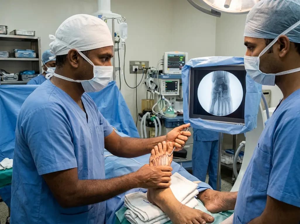
A recent clinical study identifies proximal intercuneiform joint disruption in 43.8% of low-energy Lisfranc injuries. Due to the low diagnostic sensitivity of standard radiographs and MRI, orthopedic surgeons must perform routine intraoperative stress testing to ensure complete anatomical reduction and fixation.
Today

A landmark study reveals that physiologic subtypes of gestational glucose intolerance, including insulin resistance and insulin deficiency, independently raise long-term risks of maternal prediabetes and diabetes, underscoring the urgent need for structured postpartum surveillance.
Today
