.webp&w=384&q=75)

Loading, please wait...
Loading, please wait...
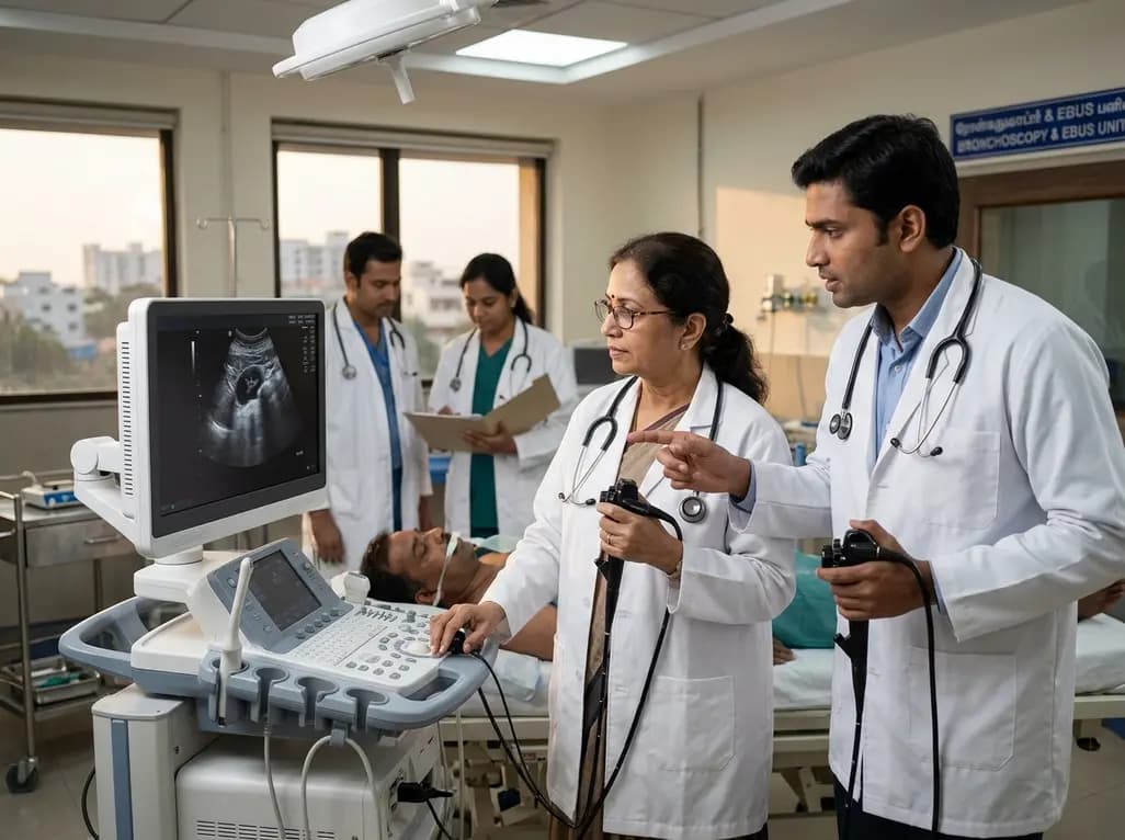
Clinicians frequently face intricate diagnostic challenges when evaluating thoracic adenopathy. Accurate tissue diagnosis dictates oncologic staging and steers therapeutic choices in granulomatous diseases. Historically, invasive surgical interventions carried notable risks and prolonged recovery periods. Fortunately, modern minimally invasive endosonography has radically reshaped this clinical domain. Today, thoracic specialists combine endobronchial ultrasound and transesophageal endoscopic ultrasound to evaluate mediastinal stations. However, optimal procedural selection requires meticulous preoperative planning. Consequently, recent clinical evidence demonstrates that a dedicated thoracic multidisciplinary team can systematically optimize mediastinal lymph node sampling. Furthermore, matching nodal accessibility with clinical indications ensures superior diagnostic accuracy and robust tissue acquisition.
Historically, surgical mediastinoscopy served as the benchmark for intrathoracic nodal staging. Although mediastinoscopy provided reliable tissue, it required general anesthesia and posed risks of serious vascular injury. Furthermore, surgical access could not easily reach posterior or inferior mediastinal nodal stations. The emergence of endobronchial ultrasound with transbronchial needle aspiration fundamentally transformed clinical practice. This bronchoscopic approach allows real-time ultrasound imaging and safe needle aspiration of paratracheal and hilar nodes. Additionally, clinicians recognized the complementary value of transesophageal endoscopic ultrasound. Using endoscopic ultrasound, operators easily sample station 4L, station 7, and lower mediastinal stations 8 and 9. Moreover, proceduralists can perform transesophageal aspiration using the ultrasound bronchoscope itself, a technique known as EUS-B. Consequently, combining transbronchial and transesophageal endosonography provides comprehensive access to nearly all mediastinal stations during a single sedation session. Major thoracic guidelines now strongly recommend endosonography as the primary staging modality over surgery. In addition, combining modalities significantly decreases procedural time and hospital costs compared to open surgical explorations. Therefore, structured decision pathways remain essential to prevent non-diagnostic procedures and patient complications.
A dedicated thoracic multidisciplinary team represents the cornerstone of contemporary mediastinal disease management. Uncoordinated referrals often produce fragmented clinical care and inconclusive diagnostic procedures. For example, an unguided bronchoscopy may fail to reach posterior subcarinal lesions that transesophageal access easily biopsies. Conversely, an integrated multidisciplinary team unites pulmonologists, thoracic surgeons, medical oncologists, diagnostic radiologists, and cytopathologists before tissue acquisition occurs. During these clinical conferences, the team examines computed tomography and positron emission tomography imaging in detail. In addition, specialists review patient cardiopulmonary status, vascular proximity, and specific tissue requirements. Suspected malignant lymphoma demands adequate tissue core volume for flow cytometry and immunohistochemistry. Similarly, infectious granulomatous diseases necessitate separate specimens for dedicated microbiological cultures. Therefore, predetermining whether to deploy EBUS, EUS-B, or conventional EUS prevents procedural redundancy. Furthermore, this collaborative planning optimizes needle selection and specimen triage. In fact, advance coordination allows pathologists to prepare appropriate preservation media beforehand. As a result, multidisciplinary coordination enhances initial diagnostic yields and minimizes the need for repeat invasive sampling.
A compelling study by Abdelsamad and colleagues provides strong real-world validation for multidisciplinary-guided endosonography. The authors evaluated 213 consecutive adult patients presenting with unexplained mediastinal or hilar lymphadenopathy. Within this mixed cohort, indications included 134 suspected carcinomas and 79 suspected benign or lymphoproliferative conditions. Notably, the overall diagnostic yield reached an impressive 92% across all enrolled participants. Furthermore, high diagnostic accuracy remained remarkably consistent regardless of underlying disease suspicion. The team achieved a 91.8% diagnostic yield in suspected carcinomas and a 92.4% yield in suspected benign or lymphoproliferative disorders. Consequently, this structured approach demonstrated equal reliability across diverse thoracic pathologies. In addition, the study established diagnostic concordance for benign conditions through three months of rigorous clinical and radiographic follow-up. This robust follow-up protocol prevented misinterpreting unrepresentative sampling as true negative findings. Moreover, the high procedural success rate translated into immediate clinical decision-making without surgical delays. Thus, pre-procedural multidisciplinary planning effectively resolves diagnostic uncertainty across broad clinical presentations.
Differentiating benign granulomatous disorders remains a frequent clinical challenge, particularly in regions with high tuberculosis endemicity. Clinicians routinely evaluate patients presenting with mediastinal lymphadenopathy where tuberculosis, sarcoidosis, and fungal infections share overlapping radiologic features. In these complex cases, cytological aspirates alone may fail to identify diagnostic non-caseating or caseating granulomas. Fortunately, modern endosonographic fine-needle biopsy needles can obtain intact histologic architecture. In the investigation by Abdelsamad and colleagues, predetermined modality selection secured definitive benign diagnoses in nearly ninety-three percent of suspected benign cases. Moreover, structured team planning ensured proper sample division at the bedside. Operators allocated dedicated needle passes for mycobacterial smear, cartridge-based nucleic acid amplification, and mycobacterial culture. As a result, patients avoided unguided empiric anti-tubercular therapy or inappropriate corticosteroid administration. Furthermore, confirming benign etiologies non-surgically spared vulnerable patients the morbidity of invasive mediastinoscopy. In addition, establishing rapid microbiological confirmation prevents disease progression and public transmission. Therefore, structured endosonography provides profound diagnostic utility for infectious and inflammatory thoracic diseases.
In modern thoracic oncology, establishing a histological cancer diagnosis represents only the initial step of workup. Pathologists must deliver extensive predictive biomarker profiling for advanced non-small cell lung carcinoma. These essential tests include EGFR, ALK, ROS1, BRAF, RET, MET, KRAS, and PD-L1 immunohistochemistry. Historically, clinicians questioned whether fine-needle endosonography could yield adequate cellular material for comprehensive next-generation sequencing. The findings by Abdelsamad and colleagues strongly refute this skepticism. Among 57 malignant cases where molecular testing was processed, the laboratory successfully completed analysis in 89.5% of specimens. This high completion rate reflects deliberate multidisciplinary planning before tissue acquisition. When the multidisciplinary team anticipates molecular profiling, operators obtain additional needle passes and optimize tissue handling protocols. In addition, rapid on-site cytologic evaluation confirms sample viability during the procedure. Similarly, immediate processing into cell blocks preserves high molecular integrity for targeted gene panels. Consequently, patients receive rapid genomic profiling without enduring repeat invasive biopsies. Therefore, multidisciplinary endosonography successfully fulfills the rigorous demands of personalized cancer therapy.
Combining endobronchial and transesophageal endoscopic ultrasound allows complete anatomical access to all intrathoracic lymph node stations. While EBUS effectively accesses anterior and paratracheal nodes along the tracheobronchial tree, EUS or EUS-B reaches posterior subcarinal and inferior paraesophageal stations. Consequently, this combined minimally invasive approach maximizes diagnostic reach, minimizes the risk of false negatives, and prevents unnecessary surgical mediastinoscopy in patients undergoing comprehensive thoracic staging.
Yes, modern endosonographic sampling reliably provides sufficient cellularity for comprehensive next-generation sequencing and biomarker analysis. By employing dedicated needle passes, rapid on-site evaluation, and newer fine-needle biopsy devices, proceduralists collect intact tissue cores rich in viable tumor cells. Real-world studies show that pre-procedural multidisciplinary planning achieves successful molecular profiling in approximately ninety percent of malignant cases, ensuring timely initiation of targeted oncologic therapies without requiring open surgical biopsies.
Benign granulomatous diseases like tuberculosis and sarcoidosis present with overlapping radiographic and clinical features, creating major diagnostic challenges. A multidisciplinary discussion aligns clinical data, imaging, and laboratory requirements before the biopsy occurs. This collaboration ensures operators collect adequate tissue for histology, mycobacterial cultures, and molecular tests. Consequently, clinicians achieve accurate definitive diagnoses, prevent misclassification, and avoid delays in initiating appropriate antimicrobial or anti-inflammatory treatments for affected patients.
Disclaimer: This content is for informational and educational purposes only and should not be considered medical advice. Always consult a qualified healthcare professional regarding any medical condition or treatment decisions. Refer to the latest local and national guidelines for clinical practice.
References

Read summarized clinical updates, watch expert medical content, and earn CME certifications right from your smartphone.
Open your smartphone camera to scan and install.





A retrospective study reveals that a multidisciplinary team-guided strategy combining EBUS, EUS-B, and EUS achieves a 92% diagnostic yield and 89.5% molecular adequacy in mediastinal and hilar lymphadenopathy across both benign and malignant indications.
4 weeks back

Recent research defines ferro-aging as a conserved iron-lipid axis driving organ decline across primates. Age-related iron dyshomeostasis stimulates ACSL4-mediated lipid peroxidation, promoting cellular senescence. Notably, vitamin C directly inhibits ACSL4, suppressing phospholipid oxidation and multi-organ decay.
Today
A retrospective cohort study evaluated 1,000 low-risk singleton births to assess maternal and neonatal outcomes during office-hours versus off-hours. Off-hours deliveries were significantly associated with higher odds of intrapartum cesarean sections and episiotomies, highlighting critical obstetric staffing considerations.
Today
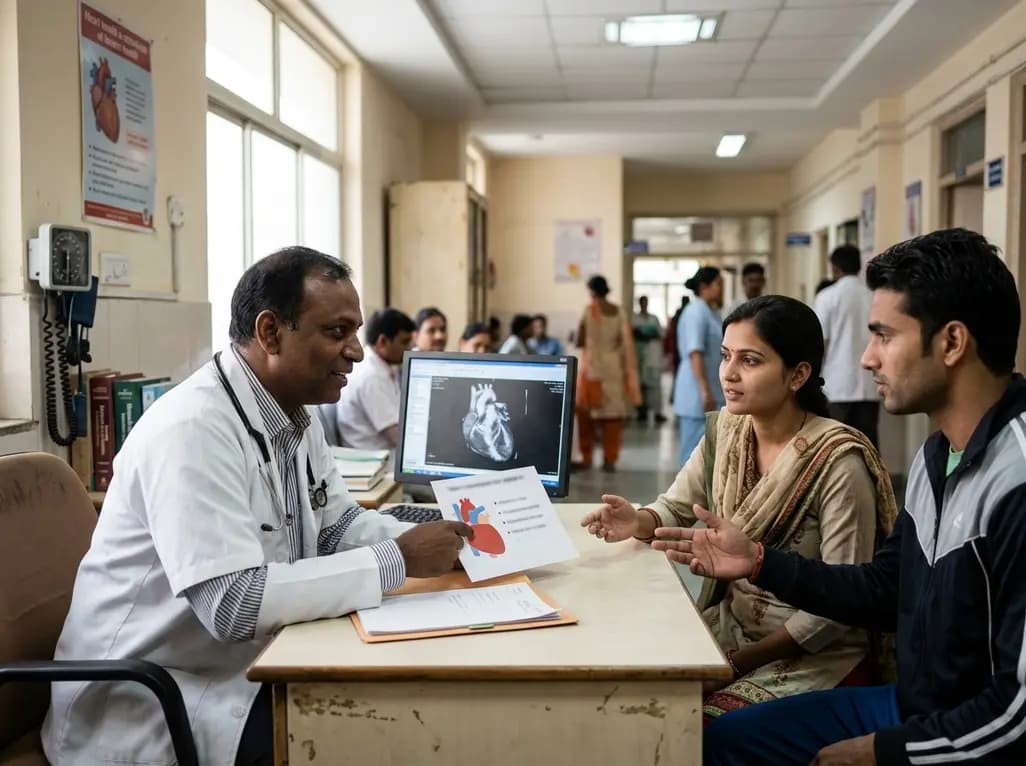
New clinical evidence demonstrates a shift away from strict exercise restrictions in genetic heart disease. While patients with channelopathies engage in more vigorous activity than those with cardiomyopathies, shared decision-making and individualized risk assessments ensure safe, beneficial exercise participation.
Today
Sarcopenia significantly impairs functional independence and amplifies fracture risks in patients with osteoarthritis and osteoporosis. Learn about modern diagnostic strategies, GLP-1 therapy considerations, and evidence-based non-pharmacological management across multidisciplinary clinical settings.
Today

Discover how to identify superimposed entrapment neuropathy in diabetic patients. This review details clinical evaluation, surgical decompression evidence, and a structured algorithm for primary care referrals to prevent ulcers and preserve limb function.
Today
