.webp&w=384&q=75)

Loading, please wait...
Loading, please wait...
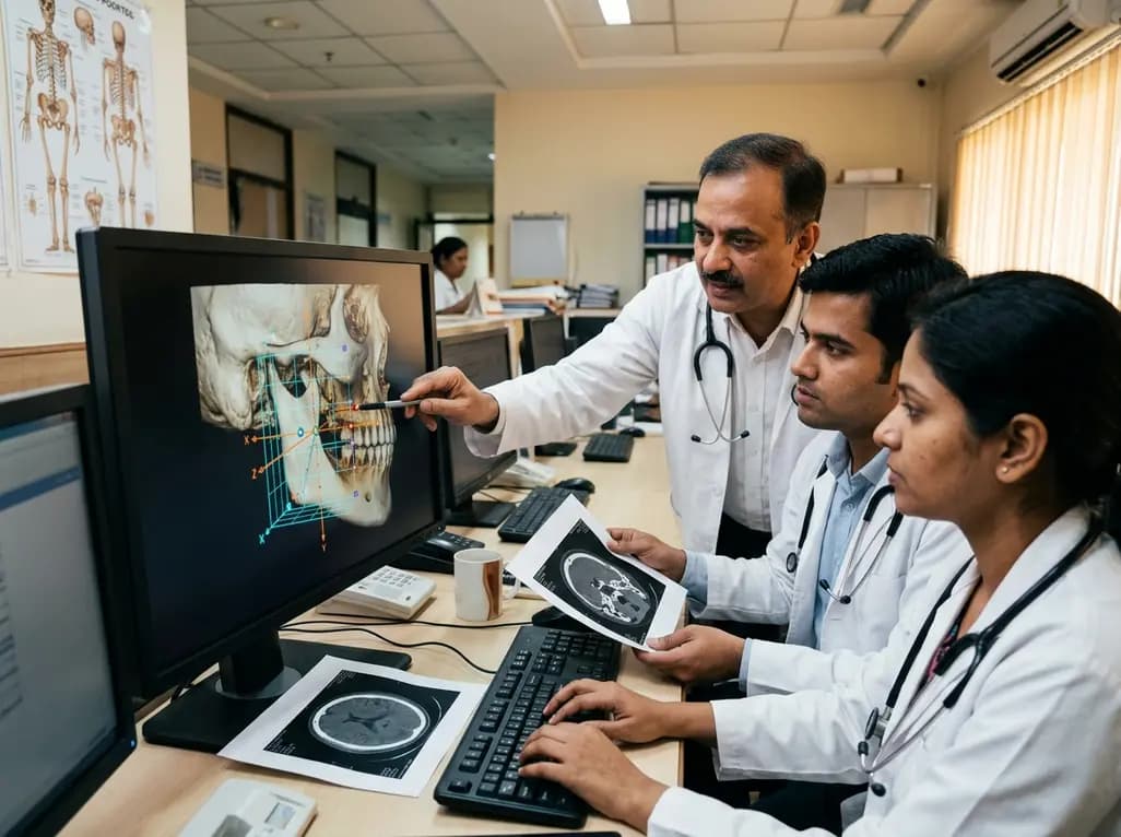
Precise assessment of mandibular ramus morphology remains essential for successful maxillofacial reconstructive surgery and targeted implant engineering. Surgeons frequently encounter complex anatomical deformities that require reproducible, landmark-anchored three-dimensional digital workflows. Consequently, standardizing spatial orientation across individual mandibular variations offers critical diagnostic clarity. A newly described technical report demonstrates an anatomical Cartesian coordinate system that resolves historic alignment challenges and enhances prosthetic manufacturing.
The mandibular ramus forms the primary structural bridge connecting the movable dental arch to the skull base. Therefore, any trauma, pathological lesion, or congenital asymmetry substantially alters temporomandibular joint biomechanics and occlusal harmony. When reconstructive surgeons perform orthognathic osteotomies or treat complex subcondylar fractures, they require exact dimensional measurements. Accurate evaluation of mandibular ramus morphology ensures proper restoration of vertical facial height, ramal inclination, and physiological joint loading. Historically, clinicians relied upon planar two-dimensional cephalometric radiographs to evaluate ramal dimensions. However, these traditional imaging modalities introduce projection distortion, magnification errors, and bilateral skeletal overlap. As three-dimensional computed tomography and surface tessellation language datasets became widespread, volumetric quantification improved dramatically. Nevertheless, spatial orientation in three-dimensional virtual space still requires standardized, objective reference axes. Without reproducible coordinate baselines, measurements across diverse patient cohorts remain unreliable. Maxillofacial specialists therefore need robust spatial frameworks that align digital models with true surgical anatomy. Standardizing these coordinates allows practitioners to anticipate bone remodelling vectors and avoid postoperative complications.
Modern computational workflows frequently use global iterative closest point or best-fit registration algorithms to compare skeletal geometries. Although these mathematical tools effectively overlay symmetrical structures, they demonstrate notable vulnerabilities during asymmetric mandibular analysis. Specifically, best-fit algorithms distribute geometric discrepancies evenly across the entire surface mesh rather than isolating true biological variations. If a patient exhibits severe post-traumatic condylar displacement or pathological bone resorption, the algorithm skews the entire alignment. Consequently, localized anatomical deformities distort the orientation of structurally intact ramal landmarks. Furthermore, postoperative physiological bone remodelling alters cortical contours, which misleads automated surface-matching software during longitudinal follow-up. These mathematical distortions prevent clinicians from distinguishing true functional adaptation from algorithmic registration artifacts. Maxillofacial surgeons must interpret skeletal morphology relative to biologically stable surgical landmarks rather than arbitrary geometric centroids. Therefore, relying exclusively on automated best-fit alignment compromises pre-surgical planning and diagnostic accuracy in complex trauma cases. Addressing these shortcomings demands a reliable coordinate system anchored strictly to verified anatomical landmarks.
To overcome the limitations of automated surface registration, investigators introduced a customized software environment named CTinA. This specialized software platform directly imports standard surface tessellation language files derived from volumetric computed tomography scans. Operators subsequently identify and mark validated anatomical landmarks by hand to establish a patient-specific spatial framework. In particular, the software references the posterior border of the mandibular ramus alongside the established Loukota line. The Loukota line extends horizontally through the deepest notch of the sigmoid groove, separating the condylar neck from the ramal base. By integrating these stable landmarks, the software constructs a reproducible, three-dimensional Cartesian coordinate system. This coordinate setup assigns mutually orthogonal X, Y, and Z axes to the ramus and condylar process. Because the system references concrete biological landmarks, it eliminates the bias inherent in best-fit geometric averaging. As a result, clinicians obtain an objective, clinically interpretable coordinate foundation that withstands pronounced anatomical variations. Furthermore, the coordinate system remains stable even when substantial condylar remodelling or pathological erosion occurs.
Standardized Cartesian coordinates provide transformative advantages for custom medical device engineering and patient-specific implant manufacturing. Currently, craniomaxillofacial fixations predominantly utilize permanent titanium plates and screws. Although titanium provides exceptional mechanical rigidity, it carries recognized drawbacks, including long-term stress shielding and secondary hardware removal surgeries. Consequently, biomedical engineers are actively developing biodegradable osteosynthesis plates fabricated from bioabsorbable magnesium alloys. Magnesium implants gradually dissolve in vivo while supporting native osteogenesis, eliminating the need for implant retrieval. However, designing effective magnesium plates requires precise knowledge of regional cortical morphology and dynamic biomechanical strain distribution. Because magnesium exhibits lower initial stiffness than titanium, plate geometry must accurately match the underlying mandibular surface. The landmark-based Cartesian coordinate system provides exact, repeatable morphological maps of the ramal surface contours. Engineers can therefore model standardized plate profiles that distribute masticatory loads evenly across the bone-implant interface. In addition, reproducible spatial measurements allow researchers to study progressive bone healing without interference from registration errors.
Integrating an anatomical Cartesian coordinate system into routine diagnostic pathways benefits diverse dental and maxillofacial specialties. Oral and maxillofacial surgeons manage complex trauma involving high subcondylar fractures and comminuted ramal injuries. By referencing reproducible anatomical landmarks, surgeons can simulate fracture reduction virtually with unmatched spatial precision. Similarly, orthognathic surgeons and orthodontists gain deeper insights into bilateral ramal asymmetries and rotational occlusal discrepancies. Standardized coordinate analyses clarify whether facial asymmetry stems from vertical ramal deficiency or horizontal condylar displacement. Furthermore, dental implantologists and prosthodontists evaluate spatial relationships between the mandibular canal, ascending ramus, and functional dentition more effectively. In post-oncologic mandibular reconstruction, this coordinate methodology guides vascularized fibular graft shaping to restore native mandibular contour. Ultimately, landmark-anchored coordinates bridge digital computer-aided design tools with practical surgical reality. Clinicians can confidently communicate findings across multidisciplinary teams, standardizing documentation and improving therapeutic predictability. As digital dentistry advances rapidly, standardized anatomical frameworks will undoubtedly form the cornerstone of personalized maxillofacial patient care.
The Loukota line runs from the deepest point of the mandibular notch perpendicular to the posterior border of the ascending ramus. Consequently, this reliable anatomical landmark cleanly delineates the condylar process from the mandibular ramus. By anchoring orthogonal axes to this distinct line, the software establishes an objective biological baseline. This reproducible alignment prevents coordinate skewing and ensures standardized morphometric analysis across diverse patient populations.
Magnesium alloys offer exceptional biocompatibility and mechanical properties that closely resemble human cortical bone. Unlike permanent titanium hardware, magnesium devices safely degrade over time through physiological resorption, releasing beneficial trace ions that stimulate bone healing. Therefore, patients avoid secondary operations for plate removal and experience reduced implant-related stress shielding. Precise morphological coordinate data optimize magnesium plate geometry, ensuring adequate structural support throughout the entire osseous healing period.
Iterative best-fit surface registration algorithms mathematically minimize total surface discrepancies across entire three-dimensional meshes. Consequently, pronounced local abnormalities, such as severe condylar fractures or marked osteolytic resorption, distort the overall alignment of healthy bone segments. In contrast, landmark-based Cartesian coordinate systems anchor spatial axes directly to verified, stable anatomical structures. This biological orientation isolates regional pathology without compromising whole-jaw alignment, ensuring clinically interpretable and anatomically consistent assessments.
Disclaimer: This content is for informational and educational purposes only and is not intended to serve as medical advice, diagnosis, or treatment. Healthcare professionals should rely on their clinical judgment and consult relevant medical literature before making patient care decisions. Neither the author nor the publisher assumes any liability for any injury and/or damage to persons or property arising from this publication. Refer to the latest local and national guidelines for clinical practice.
References

Read summarized clinical updates, watch expert medical content, and earn CME certifications right from your smartphone.
Open your smartphone camera to scan and install.





A new technical report introduces an anatomical landmark-based Cartesian coordinate system to evaluate mandibular ramus morphology. This reproducible approach refines 3D surgical planning and optimizes patient-specific osteosynthesis plate design.
last month

Discover the crucial role of glymphatic dysfunction in stroke, where perivascular clearance failure and neuroinflammation exacerbate cerebral edema and post-stroke brain injury.
Today

A prenatal case highlights severe glutaric acidemia type II presenting as bilateral hyperechoic cystic kidneys at 18 weeks. Whole-exome sequencing revealed a novel homozygous ETFDH frameshift variant, demonstrating the power of rapid genetic testing in fetal renal anomalies.
Today

Review how tailored exercise programs benefit frail older patients with heart failure. Explore pathophysiological mechanisms, individualized training prescriptions, and strategies to overcome adherence barriers in clinical practice.
Yesterday

This comparative study evaluates posterior labral tears with and without glenohumeral instability, highlighting distinct demographic patterns, MRI underestimation in stable shoulders, and successful arthroscopic repair outcomes.
Yesterday

A clinical study demonstrates that early intervention with tocilizumab, either as monotherapy or bridged with corticosteroids, achieves significant proptosis reduction and resolves active inflammation in treatment-naive thyroid eye disease.
Yesterday
