.webp&w=384&q=75)

Loading, please wait...
Loading, please wait...
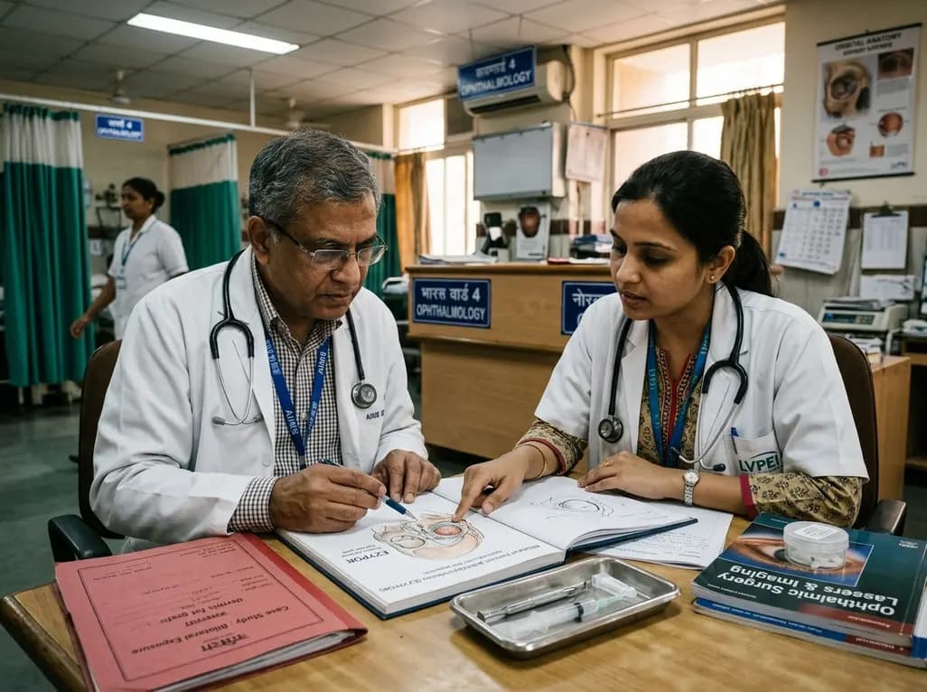
Orbital volume replacement remains a fundamental objective of anophthalmic socket surgery following enucleation or evisceration. Modern oculoplastic surgery relies heavily on integrated biomaterials designed to promote fibrovascular ingrowth and improve prosthetic motility. However, post-surgical complications can still jeopardize surgical success. Among these adverse events, orbital implant exposure represents a significant challenge that can compromise structural integrity and aesthetic outcomes. A recent clinical case highlighted a 72-year-old female who developed bilateral exposure of EZYPOR porous polyethylene enucleation implants eighteen months after staged surgeries. This complex presentation ultimately required bilateral socket reconstruction using autogenous dermis fat grafts. Clinicians must understand how implant architecture, tissue handling, and systemic patient factors interact to provoke late structural breakdown. Consequently, ophthalmic surgeons must recognize early warning signs and evaluate mechanical biomaterial properties. This clinical review explores the pathophysiology of porous implant breakdown, discusses potential contributing variables, and details salvage strategies. By analyzing these critical elements, surgeons can optimize patient selection and refine operative techniques to minimize anophthalmic socket complications.
Porous polyethylene implants have gained widespread popularity in oculoplastic surgery over several decades. Specifically, high-density porous polyethylene allows host fibrovascular tissue ingrowth through interconnected micro-channels, thereby anchoring the device and reducing catastrophic extrusion rates. The EZYPOR implant features an integrated suturing platform designed to simplify extraocular muscle reattachment directly onto the implant body. Therefore, surgeons can achieve stable muscle fixation without requiring an additional wrapping material such as donor sclera or pericardium. While this streamlined design reduces operative time and avoids foreign wrap morbidity, it introduces unique mechanical considerations. The raised suturing ridges and surface characteristics can exert localized mechanical pressure on overlying tissue layers. Furthermore, rigid edges may generate excessive focal friction against the Tenon capsule during routine ocular movements. If fibrovascular ingrowth progresses slowly, the anterior surface remains vulnerable to micro-ischemia and progressive tissue thinning. In addition, an unwrapped porous structure might abrade delicate anterior socket layers if conjunctival closure lacks sufficient laxity. Thus, understanding the physical dynamics of customized suturing platforms is essential when selecting orbital biomaterials.
The development of orbital implant exposure usually stems from a multifactorial interplay between patient biology, operative execution, and implant mechanics. Systemic host factors significantly influence socket healing and long-term tissue viability. For instance, advanced age often leads to thinned conjunctival tissue, decreased microvascular perfusion, and reduced orbital fat resilience. Consequently, elderly patients experience slower vascularization of porous matrices, leaving the implant surface relatively avascular for prolonged intervals. Similarly, systemic microvascular comorbidities, such as diabetes mellitus or peripheral vascular disease, further compromise wound remodeling. Beyond intrinsic patient factors, surgical technique plays a crucial role in maintaining anterior barrier integrity. Inadequate mobilization of Tenon capsule or excessive tension during anterior layered closure predisposes the socket to wound dehiscence. Moreover, choosing an oversized spherical implant creates persistent anterior pressure that gradually leads to conjunctival erosion. In bilateral cases, subtle symmetry discrepancies and cumulative prosthetic friction can aggravate underlying mechanical tension. Therefore, surgeons must meticulously evaluate tissue elasticity, perform tension-free closures, and carefully choose implant dimensions to prevent delayed socket breakdown.
Patients suffering from exposed anophthalmic devices typically present with persistent mucopurulent discharge, socket discomfort, chronic conjunctival inflammation, and prosthetic instability. Clinical slit-lamp examination reveals a visible defect in the conjunctiva and Tenon capsule, unmasking the rough anterior surface of the porous polyethylene sphere. In the early stages, the exposure defect may measure only a few millimeters; however, chronic exposure facilitates microbial colonization. Bacterial biofilm formation on porous biomaterials creates an intractable nidus of infection that rarely responds to conservative topical or systemic antimicrobial therapy alone. Furthermore, chronic exposure causes peripheral conjunctival cicatrization, fornix foreshortening, and socket contracture, which ultimately prevents the patient from retaining an ocular prosthesis. If clinicians neglect early exposure, the surrounding vascularized tissues retract further, accelerating implant extrusion and socket architecture collapse. In bilateral presentations, functional disruption and psychological distress multiply considerably. Therefore, prompt clinical diagnosis and early staging of the exposure defect are essential to determine whether conservative tissue preservation remains viable or whether definitive surgical reconstruction is mandatory.
When porous orbital implant exposure becomes extensive or fails primary closure, autogenous dermis fat grafting represents the gold standard for definitive socket salvage. In severe cases involving large defects or unvascularized porous matrices, surgeons must remove the exposed implant entirely to eliminate the nidus of chronic inflammation. Autogenous dermis fat provides both volume replenishment and healthy, vascularized surface epithelialization simultaneously. During the reconstructive procedure, the surgeon harvests an appropriately sized dermal-fat graft, typically from the lower abdominal wall, buttock, or lateral thigh. Subsequently, the deep fatty portion of the graft fills the intraconal volume defect, while the deepithelialized dermal surface bridges the conjunctival gap. The recipient conjunctival margins are meticulously sutured to the dermal perimeter, establishing a robust blood supply from surrounding socket vasculature. Dermis fat grafts demonstrate remarkable biocompatibility, completely eliminating foreign-body exposure risks and allowing excellent conjunctival re-epithelialization over time. Consequently, this versatile autologous technique restores deep conjunctival fornices, supports comfortable ocular prosthetic wear, and reliably restores anatomical socket symmetry without synthetic implant recurrence.
Preventing implant breakdown demands strict adherence to rigorous surgical principles and sustained postoperative vigilance. Surgeons should ensure meticulous multilayered anatomical closure during primary enucleation, securing posterior Tenon capsule, anterior Tenon capsule, and conjunctiva independently without excessive suture tension. Furthermore, selecting appropriate implant sizes—typically ranging between 18 mm and 20 mm in adults—prevents anterior socket overcrowding and reduces tissue ischemia. When using porous polyethylene devices with integrated suturing platforms, surgeons must orient the platforms deeply and carefully smoothen any irregular edges. Postoperatively, clinicians must schedule regular long-term follow-up examinations to detect subtle conjunctival thinning before full-thickness breakdown develops. Educating patients to report localized irritation, sudden discharge changes, or prosthesis dislodgement facilitates rapid clinical evaluation. In addition, oculists and ocularists must coordinate closely to ensure that the prosthetic shell does not exert localized vaulting pressure on the anterior apex of the socket. Through proactive socket surveillance and evidence-based surgical refinements, surgical teams can sustain excellent reconstructive outcomes and prevent late implant complications.
Orbital implant exposure typically arises from multifactorial causes, including poor fibrovascular ingrowth, excessive wound tension during layered closure, and mechanical friction from ocular prostheses. Furthermore, patient comorbidities such as microvascular disease, advanced age, or prior socket radiation can severely impair tissue healing. In some instances, rough implant edges or prominent suturing platforms generate focal pressure that gradually erodes the overlying conjunctiva and Tenon capsule.
An autogenous dermis fat graft provides both structural volume and a biological soft-tissue surface to reconstruct contracted or damaged anophthalmic sockets. When clinicians remove an exposed synthetic implant, the dermal component integrates directly with surrounding conjunctiva, while the underlying adipose tissue replaces lost orbital volume. Consequently, this autologous graft eliminates foreign-body extrusion risks, restores deep fornices, and provides stable support for a functional ocular prosthesis.
Small, early superficial exposures without active infection occasionally respond to conservative medical therapies, such as topical antibiotics, temporary prosthesis removal, or lubricants. However, porous polyethylene implants have interconnected porous structures that readily harbor bacterial biofilms once exposed to the external environment. Therefore, conservative management often proves insufficient for large or persistent defects, and definitive surgical intervention with patch grafts or dermis fat grafting is usually required.
Disclaimer: This content is for informational and educational purposes only and does not constitute medical advice, diagnosis, or treatment recommendations. Always consult a qualified healthcare professional regarding any medical condition or clinical decision. Refer to the latest local and national guidelines for clinical practice.
References

Read summarized clinical updates, watch expert medical content, and earn CME certifications right from your smartphone.
Open your smartphone camera to scan and install.





A recent clinical report details bilateral porous polyethylene (EZYPOR) enucleation implant exposure requiring dermis fat grafting. This article examines the biomechanical risks, surgical factors, and reconstruction strategies for managing exposed orbital implants.
last month
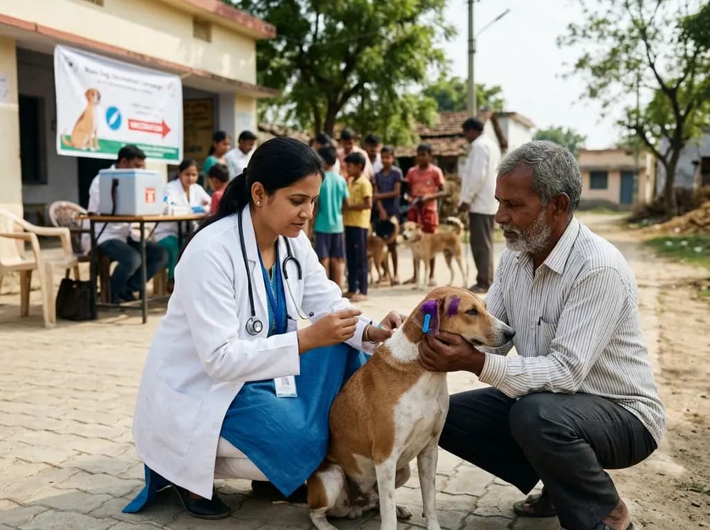
A landmark cluster randomized trial demonstrates that community-based mass dog vaccination achieves superior coverage and sustains protective herd immunity against rabies far better than conventional campaign models, offering vital lessons for global and Indian elimination targets.
Today
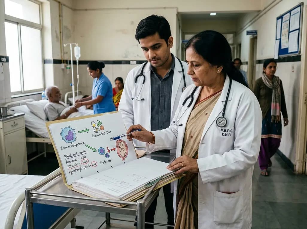
Explore the crucial link between dietary protein and immunocompetence. This review examines how low-protein nutrition shifts metabolic resources away from pathogen defense toward early reproduction, compromising host survival and immunity against severe bacterial infections.
Today

Differential oxygenation during peripheral TA-NRP can cause undetected coronary hypoxemia due to arch vessel clamping. A novel circuit with continuous aortic root monitoring detects desaturation early, facilitating left ventricular unloading and preventing allograft dysfunction and unnecessary heart discard.
Today

A longitudinal study reveals that patients undergoing staged bilateral hip arthroscopy experience progressive contralateral labral deterioration during surgical intervals. However, structural degradation does not compromise 2-year postoperative clinical outcomes or patient-reported satisfaction scores.
Today

A post hoc analysis of the PARM-T2D study shows pemafibrate exerts baseline-dependent effects on LDL-C and particle size in type 2 diabetes and hypertriglyceridemia. It lowers triglycerides and remnants while favorably remodeling atherogenic LDL particles based on initial lipid control status.
Today
