.webp&w=384&q=75)

Loading, please wait...
Loading, please wait...
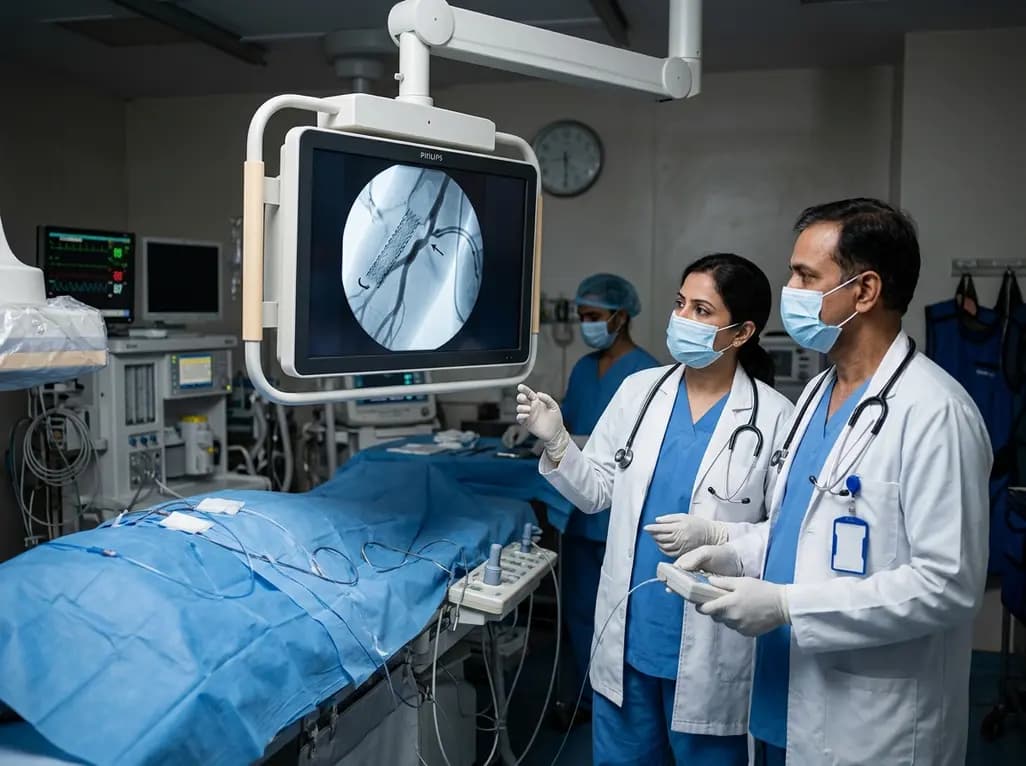
Maintaining functional vascular access remains the lifeline for end-stage renal disease patients undergoing regular dialysis. Although covered endovascular prostheses offer superior patency over standard balloon angioplasty, secondary failures still challenge clinicians worldwide. Recent clinical evidence demonstrates that longitudinal geographic miss significantly accelerates circuit failure and reduces overall access survival. Vascular specialists frequently encounter restenosis at the margins of covered devices, which underscores the urgent need for precise deployment. Consequently, identifying procedural pitfalls enables interventionalists to tailor their techniques and improve long-term dialysis outcomes. Understanding lesion dynamics and deploying appropriate stent sizes directly influence patient safety and longevity.
Geographic miss occurs when an endovascular device fails to adequately cover the entire target lesion or when clinicians select an inappropriate diameter. Specifically, longitudinal geographic miss represents residual stenosis exceeding thirty percent within five millimeters of either device margin. When operators deploy a stent graft without covering adjacent diseased segments, turbulent flow and compliance mismatch provoke rapid intimal hyperplasia. Consequently, vigorous tissue proliferation develops at the uncovered vascular boundary, precipitating early re-stenosis. In hemodialysis circuits, where shear stress remains exceptionally high, unaddressed marginal disease acts as a severe hemodynamic trigger. Moreover, vessel trauma from balloon pre-dilation outside the covered zone creates a pro-inflammatory microenvironment. Smooth muscle cells migrate aggressively across the uncovered margin, which inevitably leads to luminal loss. Therefore, vascular specialists must meticulously evaluate the entire target segment using high-resolution angiography or intravascular ultrasound prior to deployment. Recognizing subtle marginal pathology allows interventionalists to achieve complete anatomical coverage and minimize turbulent shear stress.
Recent prospective clinical registry data involving 203 patients illuminate the prognostic impact of edge-related procedural errors. Among these patients, eighty-one percent had arteriovenous grafts and nineteen percent had arteriovenous fistulas. The investigation revealed a one-year stent graft patency rate of 63.1% and an overall access primary patency rate of 22.7%. Importantly, multivariable Cox regression analysis showed that longitudinal geographic miss independently increased the hazard of stent graft patency loss by 2.62 times. Furthermore, the risk of losing primary access patency rose dramatically with a hazard ratio of 2.76. These statistically significant associations persisted even after extensive adjustment for baseline clinical risk factors. Consequently, the presence of edge stenosis emerged as a dominant modifiable determinant of vascular circuit failure. When operators achieve complete lesion coverage from healthy vessel to healthy vessel, access survival improves substantially. Thus, these robust findings highlight that procedural precision directly governs long-term clinical durability in hemodialysis maintenance.
Beyond edge-related coverage errors, the clinical registry identified several other critical variables that govern long-term circuit patency. For instance, pre-existing diabetes mellitus notably aggravated vascular endothelial dysfunction, leading to accelerated neo-intimal proliferation. Similarly, patients presenting with acute thrombosis experienced markedly worse patency compared to those undergoing elective maintenance for non-occlusive stenosis. Intraprocedural complications, particularly vessel rupture or extensive dissection, also correlated with premature circuit failure. Furthermore, anatomical characteristics played a major role; smaller stent diameters and excessive graft lengths increased the likelihood of turbulent flow and thrombosis. When clinicians confront acute thrombosis in diabetic patients, the margin for procedural error narrows considerably. Therefore, interventional teams must adopt proactive risk-stratification strategies during every intervention. Selecting larger-caliber devices when anatomically feasible and avoiding aggressive over-dilation prevent catastrophic vessel wall injury. Integrating clinical risk assessment with refined procedural execution optimizes long-term salvage rates.
Preventing procedural miss requires comprehensive imaging protocols and rigorous device selection before endovascular deployment. First, operators should utilize orthogonal angiographic projections to fully characterize the proximal and distal extent of the stenotic plaque. Relying on a single view frequently leads to foreshortening and underestimation of lesion margins. Additionally, intravascular ultrasound provides real-time luminal assessment, allowing precise localization of healthy landing zones. Selecting a covered prosthesis that extends at least five to ten millimeters beyond each diseased edge ensures adequate structural bridging. Furthermore, clinicians must account for device foreshortening during deployment, which commonly occurs with braided self-expanding nitinol platforms. Carefully monitoring the leading edge under continuous fluoroscopy prevents unintentional migration during release. After deployment, completion angiography should confirm unrestricted run-off without residual marginal narrowing. If significant edge disease persists, operators should immediately perform post-dilation or deploy an overlapping extension to secure a clean anatomical transition.
Translating these valuable clinical insights into routine practice requires systematic surveillance protocols and multidisciplinary collaboration. Nephrologists, vascular surgeons, and interventional radiologists must coordinate closely to monitor dialysis parameters during routine hemodialysis sessions. Dynamic venous pressures, blood flow rates, and physical examination findings offer early clues regarding developing edge restenosis. When surveillance suggests progressive circuit dysfunction, timely duplex ultrasound evaluation can pinpoint marginal disease before total thrombotic occlusion occurs. Moreover, quality improvement initiatives within vascular access centers should track procedural metrics, specifically monitoring rates of longitudinal geographic miss. Standardizing endovascular technique and adopting advanced imaging guidance reduce inter-operator variability across diverse clinical centers. By addressing modifiable procedural targets, clinical teams can significantly decrease the frequency of repeated endovascular re-interventions. Ultimately, optimizing initial procedural execution preserves precious vascular real estate and enhances overall quality of life for chronic hemodialysis patients.
Longitudinal geographic miss occurs when an endovascular stent graft fails to cover the entire diseased segment, leaving residual stenosis greater than 30% within five millimeters of either device edge. This procedural error frequently results from vessel foreshortening, inadequate pre-procedural imaging, or inaccurate device sizing. Consequently, persistent turbulence at uncovered margins provokes rapid intimal hyperplasia, which significantly shortens vascular access circuit survival.
Clinical studies demonstrate that longitudinal geographic miss more than doubles the hazard of both stent graft patency loss and primary access patency loss. Specifically, multivariable analyses reveal hazard ratios exceeding 2.6 for graft failure and 2.7 for access failure. Uncovered diseased edges trigger aggressive smooth muscle cell migration and thrombus formation, accelerating restenosis and requiring frequent salvage re-interventions.
Interventionalists can prevent geographic miss by utilizing multi-angle angiography and intravascular ultrasound to identify true lesion boundaries. Furthermore, operators should choose stent grafts that extend at least five millimeters into healthy adjacent tissue at both ends. Accounting for device foreshortening and performing immediate completion angiography allow operators to deploy extension stents if any marginal residual narrowing remains visible.
Disclaimer: This content is for informational and educational purposes only and does not constitute medical advice, diagnosis, or treatment recommendations. Clinical decisions should always be made by qualified healthcare professionals based on individual patient assessment and prevailing clinical standards. Refer to the latest local and national guidelines for clinical practice.
References

Read summarized clinical updates, watch expert medical content, and earn CME certifications right from your smartphone.
Open your smartphone camera to scan and install.





A clinical study highlights that longitudinal geographic miss significantly increases the risk of stent graft restenosis and circuit failure in hemodialysis access, emphasizing the critical importance of achieving complete lesion coverage during endovascular interventions.
last month

With post-monsoon dengue surges threatening South Asian health systems, physicians must refine triage and management. This guide covers vital warning signs, plasma leakage detection, crystalloid titration, and vector containment strategies to prevent mortality during seasonal arboviral epidemics.
Today

Findings from Project Viva indicate that sustained postpartum weight retention, rather than excessive gestational weight gain alone, directly worsens women's mid-life cardiovascular health, degrading blood pressure and glycemic parameters decades after delivery.
Today

A large UK Biobank study reveals that plasma proteomic signatures capture preclinical organ damage and improve multiorgan risk prediction across early CKM syndrome stages 0 to 2 beyond traditional PREVENT clinical models.
Today

A retrospective CBCT study reveals that superior TMJ space asymmetry correlates moderately with occipital condyle-atlas-axis asymmetry in unilateral TMJ osteoarthritis, highlighting an imaging correlate between jaw biomechanics and upper cervical alignment rather than a standalone diagnostic marker.
Today

A Bayesian multilevel meta-analysis reveals that aerobic training combined with moderate carbohydrate restriction modestly lowers HbA1c in type 2 diabetes. However, sparse data and very low certainty leave incremental benefits over exercise or diet alone unproven, highlighting the need for individualized care.
Today
