.webp&w=384&q=75)

Loading, please wait...
Loading, please wait...
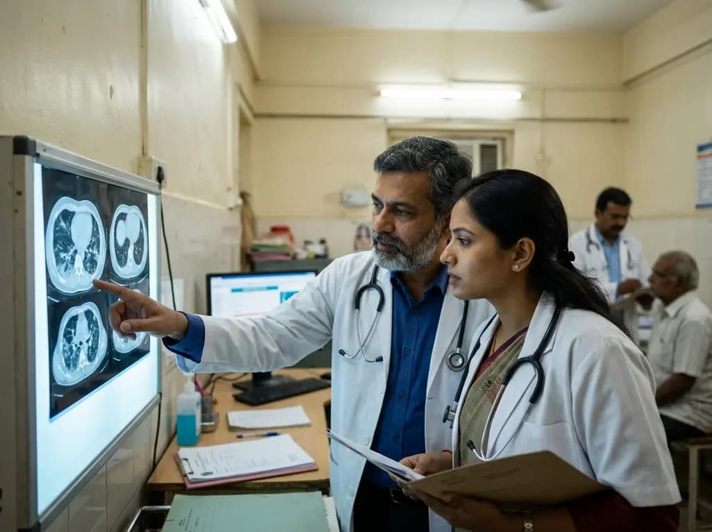
Following the initial recovery from acute SARS-CoV-2 infection, a significant proportion of patients continue to experience persistent respiratory symptoms. This phenomenon, widely known as Long-COVID or post-acute sequelae of COVID-19 (PASC), often manifests through lingering shortness of breath, chronic cough, and reduced exercise tolerance. Specifically, Long-COVID lung abnormalities represent a critical subset of these post-viral complications, necessitating a nuanced clinical approach to diagnosis and long-term management. These abnormalities encompass a spectrum of parenchymal changes, ranging from transient inflammatory ground-glass opacities to more permanent fibrotic remodeling. Understanding the nuances of these respiratory manifestations is vital for clinicians, as they directly impact a patient’s health-related quality of life and long-term functional status. Current clinical evidence suggests that while many of these changes are self-limiting, a minority of patients develop progressive pulmonary fibrosis. Therefore, early identification and tailored intervention remain the cornerstones of modern respiratory care. Furthermore, as the healthcare system continues to adapt to the post-pandemic landscape, multidisciplinary collaboration between pulmonologists, radiologists, and primary care physicians is essential to navigate the diagnostic and therapeutic challenges posed by this condition.
The development of Long-COVID lung abnormalities is driven by complex immune-mediated pathways that extend far beyond the acute viral clearance phase. During the initial infection, the virus triggers a robust inflammatory response characterized by a cytokine storm, endothelial damage, and microvascular thrombosis. Consequently, even after the virus is no longer detectable, persistent immune activation can lead to ongoing parenchymal injury. In addition, research into respiratory post-acute sequelae has identified specific molecular signatures and immune cell profiles, such as altered monocyte and T-cell responses, that contribute to sustained lung inflammation. These biological processes are similar to those seen in other forms of interstitial lung disease, where aberrant wound healing leads to excessive collagen deposition. Notably, the severity of the acute phase often correlates with the extent of subsequent lung damage; however, even patients with mild initial infections can present with significant radiographic abnormalities. Moreover, the role of the lung microbiome and its interaction with the systemic immune system is an area of growing interest, as dysbiosis may further exacerbate inflammatory pathways. Ultimately, these mechanisms underline the heterogeneous nature of the condition, where some individuals achieve complete resolution while others face permanent architectural distortion of the lung tissue.
Establishing an accurate diagnosis for Long-COVID lung abnormalities requires a comprehensive and multifaceted approach. High-resolution computed tomography (HRCT) remains the gold standard for visualizing parenchymal changes and distinguishing between inflammatory and fibrotic phenotypes. Clinicians typically observe patterns such as ground-glass opacities, reticular patterns, and traction bronchiectasis, which help in phenotyping the disease. Furthermore, pulmonary function tests (PFTs) are indispensable for quantifying physiological impairment, with many patients demonstrating a reduced diffusing capacity for carbon monoxide (DLCO) despite having normal spirometry results. In addition to imaging and lung function testing, functional assessments like the 6-minute walk test (6MWT) provide valuable insights into the patient’s exercise capacity and oxygen desaturation during exertion. Specifically, international consensus guidelines now recommend performing a chest CT scan in patients with persistent or progressive symptoms at least three months after the initial infection. Significant emphasis is placed on using low-dose protocols to minimize radiation exposure during serial follow-up examinations. By combining these objective measures with patient-reported symptom burden scales, healthcare providers can develop a holistic view of the patient’s respiratory health and monitor the temporal evolution of any detected abnormalities over time.
Post-COVID pulmonary fibrosis has emerged as one of the most concerning long-term complications, particularly in survivors of severe pneumonia or acute respiratory distress syndrome (ARDS). This condition involves the thickening and scarring of lung tissue, which can lead to progressive respiratory decline if not managed appropriately. Significantly, unlike idiopathic pulmonary fibrosis (IPF), which is typically progressive and irreversible, post-COVID fibrotic changes often show a tendency to stabilize or even partially resolve over several months. Nevertheless, approximately 20% to 30% of hospitalized survivors may exhibit some degree of radiographic fibrotic patterns during follow-up. Key risk factors for developing these permanent changes include advanced age, male sex, prolonged mechanical ventilation, and pre-existing comorbidities. Radiologically, these patients often present with persistent reticulation and architectural distortion that may mimic early-stage interstitial lung disease. Therefore, distinguishing post-COVID residual abnormalities from progressive fibrotic conditions is crucial to avoid unnecessary long-term treatments. Ongoing research aims to identify biomarkers that can predict which patients are at higher risk for non-resolving fibrosis, allowing for more targeted monitoring. Clinical management currently focuses on early identification through HRCT and PFTs, ensuring that patients receive timely pulmonary rehabilitation and symptomatic support to maximize their functional independence and lung health.
Currently, the therapeutic landscape for Long-COVID lung abnormalities is largely supportive and organ-specific, as definitive FDA-approved treatments for this syndrome are still under investigation. For patients with persistent inflammatory changes, systemic corticosteroids like prednisolone have shown promise in improving symptoms and gas exchange by dampening the immune response. Additionally, antifibrotic agents such as nintedanib and pirfenidone, which are traditionally used for IPF, are being evaluated in clinical trials for their potential to halt the progression of post-viral scarring. Significantly, the use of antivirals during the acute phase of infection, such as remdesivir or nirmatrelvir/ritonavir, has been suggested to reduce the likelihood of developing late-stage complications by limiting early viral-mediated damage. Beyond pharmacological interventions, multidisciplinary rehabilitation programs that include tailored exercise, nutritional counseling, and psychological support are vital for improving quality of life. Furthermore, prevention remains the most effective strategy against long-term sequelae. COVID-19 vaccination has been shown to reduce the risk of developing Long-COVID by approximately 30% to 40% in various population studies. By preventing severe acute illness and promoting faster viral clearance, vaccines significantly lower the probability of sustained parenchymal injury. Consequently, maintaining up-to-date vaccination status and early diagnosis through surveillance remain the best defenses against the long-term respiratory impacts of the virus.
Looking ahead, the management of Long-COVID lung abnormalities will continue to evolve as more long-term longitudinal data becomes available. Future research is likely to focus on the genetic and molecular drivers of fibrosis resolution, which could unlock new therapeutic pathways for other interstitial lung diseases. In addition, the integration of artificial intelligence and quantitative CT analysis may enhance the accuracy of detecting subtle parenchymal changes and predicting disease trajectory. For instance, advanced imaging algorithms can already identify microvascular changes that are often missed on standard radiologic reviews. Furthermore, the development of specialized post-COVID clinics in India and globally will facilitate more standardized care and better data collection for clinical trials. As we move deeper into the post-pandemic era, the emphasis will shift toward personalized medicine, where treatments are tailored based on the specific immune phenotype of the patient. Ultimately, while the acute threat of COVID-19 has subsided, the commitment to understanding and treating its long-term effects remains a priority for the global medical community. Continued surveillance and dedicated research will ensure that patients suffering from lingering respiratory effects receive the most effective, evidence-based care available.
The most frequent imaging findings in patients with Long-COVID include ground-glass opacities, which represent ongoing inflammation or resolving pneumonia, and reticular patterns that indicate scarring. Some patients also show traction bronchiectasis or subpleural bands. While many ground-glass opacities resolve within six months, fibrotic-like changes such as architectural distortion may persist. Standardizing terminology using the Fleischner Society glossary is recommended to help clinicians accurately track these temporal changes through serial low-dose CT scans.
The primary distinction lies in the temporal progression and clinical history. Post-COVID pulmonary fibrosis typically follows a severe acute viral insult and has a significant tendency to stabilize or partially regress over time, which is rare in idiopathic pulmonary fibrosis (IPF). Clinically, IPF is a chronic, progressive disease of unknown etiology, whereas post-COVID changes are reactive. Repeated high-resolution CT scans and pulmonary function tests over 6 to 12 months are essential to confirm the non-progressive nature of post-viral abnormalities.
Vaccination is a powerful tool for reducing the incidence and severity of Long-COVID. Studies indicate that vaccinated individuals are significantly less likely to develop persistent symptoms and lung-specific abnormalities compared to those who are unvaccinated. By reducing the severity of the acute infection and the risk of hospitalization or ARDS, vaccines prevent the extensive parenchymal damage that serves as a precursor to fibrosis. Maintaining up-to-date booster doses remains the most effective way to mitigate the risk of post-acute sequelae.
Disclaimer: This content is for informational and educational purposes only and does not constitute medical advice, diagnosis, or treatment. Always seek the advice of a qualified healthcare provider with any questions you may have regarding a medical condition. Refer to the latest local and national guidelines for clinical practice.
References
Fésü D et al. [Long-COVID syndrome and lung-specific abnormalities following COVID-19]. Orv Hetil. 2026 Jul 05. doi: 10.1556/650.2026.33584. PMID: 42402140.
Yoon SH et al. Best Practice: International Multisociety Consensus Statement for Post–COVID-19 Residual Abnormalities on Chest CT Scans. Radiology. 2025 Jul 22. doi: 10.1148/radiol.250037.
Sala M et al. Long COVID patients with abnormal lung CT scans and molecular drivers of pulmonary fibrosis. Nature Immunology. 2024 Oct 07. doi: 10.1038/s41590-024-01978-y.
Shyam T et al. Scars from the pandemic: understanding post-COVID-19 interstitial lung disease. Breathe (Sheff). 2025 Aug 28; 21: 250037. doi: 10.1183/20734735.0037-2025.

Read summarized clinical updates, watch expert medical content, and earn CME certifications right from your smartphone.
Open your smartphone camera to scan and install.





A comprehensive clinical review of Long-COVID syndrome focusing on lung-specific abnormalities, pulmonary fibrosis management, and the crucial role of diagnostic imaging and vaccination in improving patient outcomes.
3 months ago

Review how tailored exercise programs benefit frail older patients with heart failure. Explore pathophysiological mechanisms, individualized training prescriptions, and strategies to overcome adherence barriers in clinical practice.
Today

Leucine Rich Bio has introduced South Asia's first comprehensive vaginal microbiome test, BugSpeaks. By profiling microbial dysbiosis, pathogens, and antimicrobial resistance, this next-generation diagnostic platform provides clinicians with deeper insights into recurrent infections, fertility, and reproductive health.
Today

In high-risk pulmonary embolism complicated by severe anemia and active uterine bleeding, systemic thrombolysis is contraindicated. Large-bore aspiration thrombectomy provides rapid hemodynamic rescue without fibrinolysis, supported by inferior vena cava filter placement and multidisciplinary critical care.
Today

This comparative study evaluates posterior labral tears with and without glenohumeral instability, highlighting distinct demographic patterns, MRI underestimation in stable shoulders, and successful arthroscopic repair outcomes.
Today

A clinical study demonstrates that early intervention with tocilizumab, either as monotherapy or bridged with corticosteroids, achieves significant proptosis reduction and resolves active inflammation in treatment-naive thyroid eye disease.
Today
