
Loading, please wait...
Loading, please wait...
Beta Thalassemia Major is a significant genetic health challenge, particularly in the Indian subcontinent. This hereditary disorder is characterized by defective beta-globin chain synthesis, which necessitates lifelong blood transfusions. Consequently, these repeated treatments lead to progressive liver iron accumulation. If clinicians do not manage this overload effectively, it eventually results in severe organ damage and fibrosis. Therefore, identifying accurate and non-invasive monitoring tools remains a top priority for pediatric hematologists.
A recent prospective study analyzed 77 children diagnosed with Beta Thalassemia Major to determine the efficacy of shear wave elastography (SWE) and magnetic resonance imaging (MRI) apparent diffusion coefficient (ADC) measurements. Researchers sought to correlate these modalities with Liver Iron Concentration (LIC) and T2* values. Specifically, the study found that median liver elasticity values differed significantly across various iron overload severity groups. The analysis revealed that elasticity was notably higher in patients with severe iron loading compared to those in mild or normal categories. Furthermore, the results suggest that these imaging parameters offer a promising window into the degree of hepatic involvement.
In the clinical landscape of India, serum ferritin often serves as the primary marker for iron stores due to its cost-effectiveness. However, ferritin levels can fluctuate due to inflammation or infection, potentially masking the true extent of liver iron accumulation. Because shear wave elastography is becoming more widely available in urban diagnostic centers, it may serve as a vital adjunct to standard MRI T2* protocols. Additionally, using ADC values alongside elastography provides a more comprehensive view of the liver microenvironment in these young patients. Nevertheless, practitioners must integrate these findings with existing clinical guidelines to optimize chelation therapy.
Since the body cannot actively excrete excess iron from transfusions, the metal deposits in vital organs. In the liver, this causes oxidative stress, which eventually leads to fibrosis, cirrhosis, and potentially liver failure if not managed with chelation.
While MRI T2* is currently the gold standard for quantifying iron, elastography is highly effective at measuring liver stiffness. Therefore, it acts as a complementary tool that helps detect early structural damage caused by iron overload.
Yes, both shear wave elastography and MRI ADC are non-invasive and do not involve ionizing radiation. Consequently, they are safe for frequent monitoring in children who require regular assessment of their iron status.
Disclaimer: This content is for informational and educational purposes only and does not constitute medical advice. Always seek the advice of a qualified healthcare provider with any questions regarding a medical condition. Refer to the latest local and national guidelines for clinical practice.
References

Read summarized clinical updates, watch expert medical content, and earn CME certifications right from your smartphone.
Open your smartphone camera to scan and install.


New research evaluates liver shear wave elastography and MRI ADC as non-invasive tools for monitoring liver iron accumulation in pediatric thalassemia patie...
4 months ago
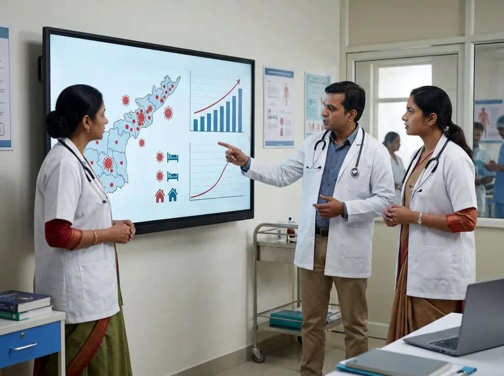
Andhra Pradesh reported 10 new Covid-19 cases, taking the state tally to 49 while deaths remain at four. With 24 patients hospitalized and 16 under home isolation, the Health Department has intensified monitoring. Medical professionals should review regional distribution, diagnostic protocols, and management plans.
Today

An 11-year Swedish registry study of 618 uterine sarcoma patients found that minimally invasive surgery yielded survival comparable to open surgery in early stages. However, adjuvant chemotherapy conferred no survival benefit in localized or advanced disease, highlighting stage and histology as key outcomes.
3 days back
A cross-sectional study evaluates post-intensive care syndrome in cardiac patients 2-4 weeks post-ICU discharge, highlighting cognitive, psychological, and functional impairments and the need for structured multidisciplinary rehabilitation.
3 days back
Anterior cruciate ligament reconstruction failure lacks uniform definition. A narrative review proposes an integrative framework incorporating objective and subjective instability, persistent pain, restricted motion, graft rupture, and secondary meniscal injury to standardize clinical reporting.
3 days back

With World Obesity Atlas data warning that over 41 million Indian children are overweight or obese, ICMR and NIN have unveiled a 10-point policy roadmap. The initiative calls for mandatory front-of-pack labeling, HFSS taxes, strict marketing bans, and healthier school environments to curb non-communicable diseases.
Today
