.webp&w=384&q=75)

Loading, please wait...
Loading, please wait...

Cataract formation represents one of the most frequent complications of chronic intraocular inflammation. Furthermore, surgical rehabilitation in these eyes presents substantial challenges due to compromised ocular structures. Recent clinical data highlight that lens subluxation in uveitis occurs at a notably higher frequency following cataract surgery when compared to non-uveitic controls. Surgeons operating on these delicate eyes encounter altered anterior segment anatomy, progressive zonular fragility, and intense postoperative inflammatory surges. Consequently, achieving stable intraocular lens positioning demands thorough preoperative risk stratification and rigorous surgical planning. Clinicians must understand how specific uveitic subtypes, demographic factors, and chronic structural alterations influence surgical outcomes. This article reviews recent multi-center registry data, details underlying pathophysiologic mechanisms of zonular compromise, and outlines practical perioperative strategies to optimize visual outcomes and minimize postoperative lens instability.
Recent evidence from the United States TriNetX database provides critical insight into postoperative stability in uveitic eyes. Specifically, researchers queried electronic health records between January 2013 and December 2023 to evaluate 18,973 patients with antecedent uveitis undergoing cataract extraction. The mean patient age was 68 years, with a predominant female representation of 58.57%. Furthermore, Caucasian patients constituted 57.23% of the total cohort, and non-Hispanic individuals comprised 72.65%. Across all evaluated subgroups, uveitic eyes showed a significantly elevated incidence of postoperative lens subluxation and dislocation compared to matched non-uveitic controls.
In addition, investigators analyzed pediatric cohorts within the multi-center database. However, limited sample sizes and strict data suppression thresholds precluded robust statistical conclusions in children. Therefore, clinicians should interpret pediatric sub-analyses cautiously while acknowledging observed baseline trends. Demographic variations also emerged across different racial and ethnic groups within the adult population. Ultimately, these real-world epidemiological findings decisively confirm that intraocular inflammation markedly impairs long-term capsular stability across diverse clinical settings.
The TriNetX analysis demonstrated substantial variation in lens instability rates across different uveitic etiologies and anatomical classifications. In particular, eyes with infectious posterior uveitis exhibited the highest incidence of lens subluxation at 0.416%, representing 416 cases per 100,000 surgeries. Similarly, non-infectious panuveitis demonstrated a pronounced risk, showing an incidence of 0.286% or 286 per 100,000 cases. These heightened rates highlight the destructive impact of posterior segment inflammation on the ciliary body and zonular apparatus.
In contrast, isolated anterior uveitis displayed lower overall subluxation rates, although the risk still exceeded general population baselines. Moreover, chronic recurrent inflammation causes persistent breakdown of the blood-aqueous barrier, accelerating local tissue degradation. Consequently, clinicians must recognize that posterior and diffuse intraocular inflammation confers an intrinsically higher risk of zonular dehiscence. Meticulous anatomical staging before surgical intervention remains essential for identifying susceptible patients and preparing tailored surgical plans.
Understanding why uveitic eyes develop capsular instability requires an examination of underlying biochemical and cellular mechanisms. Chronic intraocular inflammation promotes continuous proteolytic enzyme release, including matrix metalloproteinases that degrade fibrillin-rich zonular fibers. Furthermore, recurrent ciliary body inflammation leads to ciliary atrophy, directly impairing zonular anchorage and structural support. In addition, extensive posterior synechiae and pupil seclusion create mechanical shear forces during surgical pupil dilation and capsulotomy.
Chronic topical or systemic corticosteroid therapy may also alter tissue elasticity and accelerate anterior segment collagen breakdown. In severe cases, the formation of cyclitic membranes exerts progressive traction on the ciliary processes and lens periphery. When these inflammatory membranes contract postoperatively, they cause progressive zonular dehiscence and late intraocular lens tilt. Thus, both inflammatory biochemical degradation and chronic mechanical forces combine to compromise zonular integrity over time.
Preventing postoperative lens subluxation in uveitis requires meticulous planning before entering the operating room. First, surgeons must ensure complete ocular quiescence for at least three consecutive months prior to elective surgery. Prophylactic systemic or periocular corticosteroids should be initiated several days preoperatively to suppress subclinical inflammatory pathways. Intraoperatively, surgeons should employ gentle surgical manipulation to avoid stressing fragile zonules.
The use of flexible iris retractor hooks or pupillary expansion rings reduces direct traction on the anterior capsule during pupil enlargement. Furthermore, surgeons should carefully evaluate zonular integrity immediately following capsulorrhexis. If zonular laxity or focal dehiscence appears, early placement of a capsular tension ring redistributes mechanical forces evenly across the remaining zonules. Moreover, selecting a hydrophobic acrylic intraocular lens with broad haptic contact enhances capsular stability and prevents optical decentration.
Following surgery, uveitic patients require persistent, structured monitoring to detect early signs of optical decentration or subluxation. Even after uneventful surgery, late-onset in-the-bag dislocation can develop years later due to progressive zonulopathy and capsular contraction. Therefore, clinicians must routinely assess intraocular lens centration, anterior chamber depth symmetry, and subtle phacodonesis during follow-up visits.
If symptomatic subluxation occurs, management depends on the degree of optical axis compromise and ongoing ocular inflammation. Minor decentration without visual distortion can often be managed conservatively with refractive correction and continued inflammation control. Conversely, substantial dislocation into the vitreous or anterior chamber necessitates surgical re-intervention, such as pars plana vitrectomy with lens repositioning or exchange. Scleral-sutured or sutureless intrascleral haptic fixation techniques offer durable alternatives when capsular support fails completely.
The findings from this large cohort carry direct implications for everyday ophthalmic surgical practice. Because patients with uveitis face significantly higher odds of zonular failure, preoperative counseling must explicitly address the lifelong risk of lens dislocation. Furthermore, ophthalmologists should document baseline zonular status, pupil dynamics, and anterior segment morphology prior to surgery. This documentation establishes crucial benchmarks for postoperative comparison.
Additionally, coordinating care with medical uveitis specialists ensures optimal immunomodulatory control before and after cataract extraction. Surgeons should also maintain a low threshold for utilizing capsular stabilization devices during the primary procedure. By anticipating zonular weakness, employing tissue-preserving surgical steps, and sustaining vigilant long-term surveillance, clinicians can mitigate risks and safeguard visual outcomes for patients with uveitis.
Uveitis generates persistent intraocular inflammation that activates matrix metalloproteinases and proteolytic enzymes, degrading fibrillin within the zonular apparatus. Additionally, recurrent cyclitis induces ciliary body atrophy, weakening zonular anchor points. When combined with mechanical traction from contracting cyclitic membranes and anterior segment manipulation during complex cataract removal, these biological changes dramatically increase the long-term risk of capsular bag instability, optical decentration, and complete intraocular lens dislocation.
Large registry data indicate that infectious posterior uveitis and non-infectious panuveitis carry the highest risk of postoperative lens subluxation, reaching incidence rates of 0.416% and 0.286% respectively. Because these subtypes involve extensive posterior segment inflammation, they cause more widespread ciliary body damage and cyclitic membrane formation than isolated anterior uveitis. Consequently, surgeons must maintain heightened vigilance and consider capsular tension devices when operating on eyes with posterior or panuveitis.
When surgeons identify intraoperative zonular laxity, they should promptly minimize mechanical stress using low fluidic parameters and dispersive viscoelastics. Placing a capsular tension ring redistributes forces along the capsular equator, stabilizing the bag for safe lens implantation. If severe focal dehiscence exceeds three clock hours, sutured capsular tension segments or alternative fixation strategies, such as intrascleral haptic fixation, become necessary to secure long-term intraocular lens stability.
Disclaimer: This content is for informational and educational purposes only... Refer to the latest local and national guidelines for clinical practice.
References

Read summarized clinical updates, watch expert medical content, and earn CME certifications right from your smartphone.
Open your smartphone camera to scan and install.





A large-scale TriNetX analysis reveals that patients with uveitis experience higher rates of lens subluxation or dislocation after cataract surgery than controls. Patients with panuveitis and posterior uveitis carry the greatest risk, underscoring the necessity of meticulous perioperative inflammation control.
Today
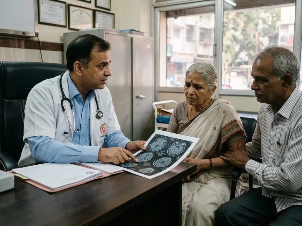
With dementia cases in India projected to double, clinicians must recognize neurodegenerative decline before irreversible neuron loss occurs. This review explores differential diagnostic strategies for memory loss, the growing disease burden, and the targeted application of newly approved anti-amyloid therapies.
Today
A school-based intervention using the Health Action Process Approach (HAPA) significantly enhanced adolescents' perceived benefits of consulting healthcare providers and promoted proactive menstrual health behaviors, offering key clinical insights for managing adolescent dysmenorrhea and endometriosis.
Today

A multicenter propensity-matched study evaluated DOAC plus single versus dual antiplatelet therapy after carotid artery stenting. Findings revealed comparable intracranial hemorrhage and ischemic stroke rates, supporting DOAC plus SAPT as a viable antithrombotic strategy in high-risk patients.
Today

A cross-sectional study reveals that sarcopenic obesity independently increases functional impairment and severe disc degeneration in patients with degenerative lumbar spinal stenosis, highlighting the need for body composition assessment.
Today

At Homa CME 2026 at Hyderabad's T-Hub, healthcare leaders emphasized moving beyond routine metrics through digital calculators and precision stratification. Discover why characterizing individual insulin secretion, resistance patterns, and vascular risks transforms chronic metabolic care in India.
Yesterday
