.webp&w=384&q=75)

Loading, please wait...
Loading, please wait...
Acute myocardial infarction typically results from atherosclerotic plaque rupture and subsequent thrombus formation. However, non-atherosclerotic mechanisms account for a significant proportion of atypical presentations, especially among younger adults and patients lacking standard cardiovascular risk factors. Among these uncommon etiologies, left atrial myxoma embolization remains a rare yet life-threatening cause of coronary occlusion. Clinicians must recognize these atypical presentations quickly because standard treatment protocols for atherosclerotic coronary disease often require modification when managing embolic non-atherosclerotic events. Consequently, understanding the unique pathophysiology, diagnostic nuances, and invasive procedural strategies for tumor-related emboli is paramount for emergency physicians, cardiologists, and cardiothoracic surgeons alike.
Primary cardiac neoplasms are exceptionally uncommon, and cardiac myxomas represent the vast majority of benign intracardiac tumors. Most myxomas arise in the left atrium, frequently attached by a pedicle to the fossa ovalis border of the interatrial septum. Although histologically benign, these gelatinous masses possess a high propensity for fragmentation and systemic embolization. The friable villous surface of the tumor, combined with continuous intracardiac shear stress, facilitates the detachment of tumor fragments or overlying surface thrombi into the arterial circulation. Consequently, systemic emboli frequently travel to the cerebral, visceral, or peripheral vascular beds. However, coronary artery embolization remains distinctly unusual, occurring in less than two percent of recognized cases. When embolization into coronary circulation occurs, tumor fragments most frequently lodge in the left anterior descending artery due to preferential diastolic hemodynamics. Therefore, mechanical occlusion induces rapid transmural myocardial ischemia, precipitating acute ST-segment elevation myocardial infarction despite entirely healthy underlying coronary vessels.
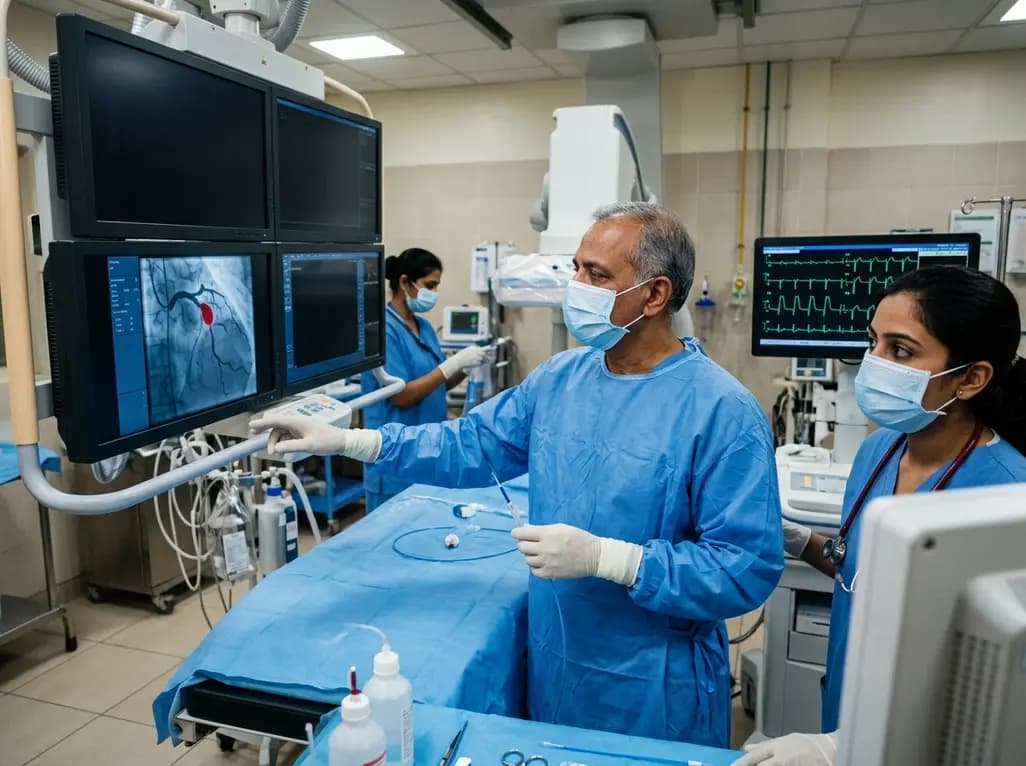
When patients present with acute anterior STEMI without conventional cardiovascular risk factors, emergency coronary angiography represents the initial diagnostic step. Angiographic evaluation typically reveals an abrupt cutoff or filling defect within a major epicardial vessel, often without background coronary calcification or luminal irregularities. In this clinical scenario, interventional cardiologists must maintain a high index of suspicion for non-atherosclerotic coronary embolisms. Routine percutaneous coronary intervention paradigms usually prioritize immediate balloon angioplasty followed by stent implantation. However, placing a permanent metallic stent over a detached myxomatous fragment can induce localized inflammatory aneurysms, stent malapposition, or inadequate wall apposition. Therefore, clinicians should consider alternative etiology when angiograms show smooth, pristine coronary trees alongside an isolated occlusion. Subsequently, operators must carefully evaluate the vessel architecture before committing to irreversible stent deployment, ensuring that invasive reperfusion matches the underlying mechanical etiology.
Intravascular imaging modalities, particularly intravascular ultrasound (IVUS) and optical coherence tomography (OCT), serve as indispensable diagnostic tools during emergency catheterization. When invasive operators restore antegrade vessel patency, intravascular imaging can definitively evaluate the arterial wall architecture. Specifically, IVUS clearly differentiates between disrupted atherosclerotic plaque, spontaneous coronary artery dissection, and purely intraluminal embolic material. In cases of embolic coronary occlusion, intravascular ultrasound demonstrates intact intimal and medial layers without plaque rupture, ulceration, or intramural hematoma. Furthermore, histopathologic evaluation of retrieved aspirate provides definitive confirmation of the diagnosis. Pathologists readily identify characteristic stellate or globular myxoma cells embedded within an abundant, loose acid mucopolysaccharide-rich myxoid stroma. Thus, combining real-time intravascular imaging with careful microscopic examination prevents misdiagnosis and directs subsequent clinical investigations toward identifying an intracardiac embolic source.
Re-establishing coronary perfusion represents the primary therapeutic objective during acute STEMI management. In the setting of tumor-induced coronary occlusion, mechanical or manual aspiration thrombectomy offers significant therapeutic advantages over immediate stenting. Manual aspiration effectively extracts the occluding myxomatous fragment, rapidly restoring TIMI grade 3 antegrade epicardial flow and microvascular perfusion. Furthermore, complete extraction of the embolic mass allows operators to avoid unnecessary stent implantation in disease-free vessels. Eliminating the need for permanent stents also reduces subsequent risks of stent thrombosis, restenosis, and prolonged dual antiplatelet therapy. Nevertheless, operators must exercise caution during aspiration to prevent distal fragmentation or microvascular embolization of gelatinous debris. If aspiration alone fails to achieve adequate luminal diameter or hemodynamic stability, gentle low-pressure balloon angioplasty or bailout stenting may serve as secondary options. Ultimately, achieving complete vessel patency while sparing healthy coronary architecture represents the optimal catheterization goal.
Following successful coronary reperfusion, multimodality cardiovascular imaging is essential to locate the embolic source. Transthoracic echocardiography (TTE) and transesophageal echocardiography (TEE) readily identify the size, mobility, and attachment site of intracardiac masses. Cardiac magnetic resonance imaging (CMR) further characterizes tumor vascularity and tissue composition, distinguishing myxomas from thrombi or malignant neoplasms. Once imaging confirms a left atrial mass, urgent surgical resection becomes imperative. Surgical excision under cardiopulmonary bypass prevents catastrophic recurrent embolization, acute mitral valve obstruction, and sudden cardiac death. Cardiac surgeons typically perform complete full-thickness resection of the tumor stalk alongside a margin of adjacent interatrial septum, followed by primary or patch reconstruction. Postoperative prognosis remains outstanding, with long-term survival mirroring that of the general population. Nonetheless, periodic echocardiographic surveillance remains mandatory to monitor for rare late tumor recurrences.
Cardiac myxomas have a friable, gelatinous consistency that easily fragments under intracardiac hemodynamic stress. These detached tumor pieces, or fresh thrombi forming on the tumor surface, embolize into the systemic arterial circulation. When fragments enter the coronary arteries, they lodge mechanically in distal branching vessels, obstructing blood flow and causing transmural myocardial infarction.
Aspiration thrombectomy directly retrieves the occlusive myxoma fragment, restoring normal coronary blood flow without requiring permanent foreign material. Because the underlying coronary artery has no atherosclerotic disease, avoiding stent implantation eliminates long-term risks like stent thrombosis or in-stent restenosis while sparing the patient from prolonged dual antiplatelet therapy.
Urgent surgical excision is necessary because residual mobile tumor tissue in the left atrium carries an ongoing, elevated risk of recurrent systemic embolization to the brain, visceral organs, or other coronary vessels. Furthermore, a large mobile myxoma can prolapse into the mitral valve orifice, causing acute left ventricular inflow obstruction and sudden death.
Disclaimer: This content is for informational and educational purposes only and is not intended as medical advice. Refer to the latest local and national guidelines for clinical practice.
References

Read summarized clinical updates, watch expert medical content, and earn CME certifications right from your smartphone.
Open your smartphone camera to scan and install.





Left atrial myxoma embolization represents a rare, non-atherosclerotic etiology of acute STEMI. This review highlights coronary angiography findings, aspiration thrombectomy, intravascular imaging, histopathologic confirmation, and definitive surgical resection strategies.
4 weeks back

With post-monsoon dengue surges threatening South Asian health systems, physicians must refine triage and management. This guide covers vital warning signs, plasma leakage detection, crystalloid titration, and vector containment strategies to prevent mortality during seasonal arboviral epidemics.
Today

Findings from Project Viva indicate that sustained postpartum weight retention, rather than excessive gestational weight gain alone, directly worsens women's mid-life cardiovascular health, degrading blood pressure and glycemic parameters decades after delivery.
Today

A large UK Biobank study reveals that plasma proteomic signatures capture preclinical organ damage and improve multiorgan risk prediction across early CKM syndrome stages 0 to 2 beyond traditional PREVENT clinical models.
Today

A retrospective CBCT study reveals that superior TMJ space asymmetry correlates moderately with occipital condyle-atlas-axis asymmetry in unilateral TMJ osteoarthritis, highlighting an imaging correlate between jaw biomechanics and upper cervical alignment rather than a standalone diagnostic marker.
Today

A Bayesian multilevel meta-analysis reveals that aerobic training combined with moderate carbohydrate restriction modestly lowers HbA1c in type 2 diabetes. However, sparse data and very low certainty leave incremental benefits over exercise or diet alone unproven, highlighting the need for individualized care.
Today
