.webp&w=384&q=75)

Loading, please wait...
Loading, please wait...

Auricular reconstruction represents one of the most intricate challenges in facial plastic and reconstructive surgery. Historically, clinicians have focused intense operative effort on carving intricate cartilage convolutions. However, achieving absolute structural perfection remains difficult. A key clinical goal centers on creating an auricle that seamlessly integrates into the facial profile without attracting unwanted gaze. In a landmark study, researchers evaluated how the general public assesses ear reconstruction asymmetry. Their findings reveal that casual observers notice vertical positioning discrepancies far more readily than minute contour variations. Consequently, reconstructive surgeons must adapt their operative priorities to ensure optimal macroscopic balance.
Reconstructing the human ear requires surgical precision and deep anatomical insight. Whether addressing congenital microtia or traumatic tissue loss, surgeons rebuild intricate cartilaginous landmarks. Autologous costal cartilage frameworks remain the gold standard worldwide, particularly across reconstructive units in India. During these extensive procedures, clinicians meticulously carve the antihelix, scapha, and tragus. However, postoperative wound healing, skin contraction, and edema inevitably soften subtle contours over time.
Surgeons frequently examine their operative outcomes from inches away under intense theatrical illumination. Conversely, patients interact with society across conversational distances where casual observers perceive the face as a unified whole. Therefore, clinicians must re-examine what constitutes true aesthetic success. When evaluating ear reconstruction asymmetry, does the public actually scrutinize subtle contour definition, or do they notice broader spatial positioning? Until recently, surgeons lacked objective data identifying which auricular differences draw visual attention. Establishing these perceptual priorities allows surgical teams to allocate operative time wisely, reduce patient morbidity, and achieve superior real-world aesthetic integration.
To define how casual observers perceive auricular deformities, researchers designed a robust crowdsourced study. The investigators digitally altered six standardized facial portraits across four distinct morphological features. These parameters included contour definition, auriculocephalic projection, overall ear size, and vertical placement. By manipulating baseline symmetrical models across graded magnitude steps, the authors produced 138 unique experimental visual stimuli.
Subsequently, a cohort of 550 adult laypersons reviewed 50 randomized images via an online platform. Participants freely identified any facial feature that drew immediate visual attention without knowing the study's specific focus on ears. In addition, the researchers employed generalized linear mixed-effects models to evaluate predictors of observer gaze fixation.
Remarkably, the study revealed that baseline ears rarely attracted visual attention. Across all experimental presentations, participants selected ears in only 3.8% of total responses. This important baseline demonstrates that the general public does not routinely scrutinize ears during ordinary social encounters. However, specific structural modifications quickly disrupted this visual neutrality, directing clinicians toward the exact aesthetic dimensions that demand intraoperative priority.
The study revealed stark differences in how distinct auricular asymmetries influence visual gaze. Among all tested variations, vertical position displacement served as the primary trigger for observer scrutiny. Specifically, an upward vertical displacement of merely 10% caused 32.1% of lay observers to identify the ear as distinctly noticeable. Similarly, a downward displacement of 10% produced a 21.5% identification rate. Statistical modeling confirmed that increasing vertical displacement magnitude significantly raised the odds of visual gaze distraction.
In sharp contrast, isolated variations in contour definition and lateral projection rarely attracted attention. Lay observers routinely overlooked blunted helical rims or mild projection discrepancies when the ear occupied its proper vertical coordinates. Furthermore, overall ear size demonstrated moderate visual impact, as a 15% size enlargement drew observer notice in 22.9% of evaluations.
These findings carry profound implications for reconstructive planning. While surgeons dedicate significant operative hours to carving subtle internal reliefs, casual observers focus primarily on global alignment. Therefore, maintaining precise vertical symmetry must take precedence over microscopic cartilage detail during auricular framework placement.
Translating these objective insights into clinical practice requires a deliberate refinement of operative strategy. When repairing unilateral microtia or major traumatic avulsions, the contralateral normal ear provides the essential anatomical reference. Surgeons routinely create an acrylic or x-ray film template to map exact proportions and landmark coordinates. To prevent vertical displacement, clinicians must align the superior helical margin with the ipsilateral brow line and lateral canthus.
Furthermore, surgeons must carefully position the framework relative to underlying skeletal landmarks. Patients with hemifacial microsomia often present with severely displaced low-set vestigial remnants. In these complex cases, the surgeon must not rely on abnormal local tissue landmarks. Instead, they should elevate the cutaneous pocket superiorly to harmonize with the unaffected contralateral auricle.
Additionally, intraoperative positioning must resist long-term soft-tissue contracture. Autologous cartilage frameworks placed beneath tight skin envelopes encounter persistent contraction forces during postoperative healing. By anchoring the framework securely to the temporal periosteum at the intended vertical height, surgeons prevent secondary migration. Prioritizing accurate vertical positioning ensures optimal long-term social inconspicuousness.
Congenital microtia occurs frequently across India, representing a significant reconstructive workload in pediatric plastic surgery centers. Costal cartilage grafting remains the preferred technique nationwide due to the cost and extrusion risks of synthetic implants. However, harvesting costal cartilage involves genuine donor-site morbidity, including postoperative pain, chest wall asymmetry, and potential thoracic complications.
Understanding lay perception provides valuable guidance for Indian surgical teams seeking operative efficiency. Surgeons frequently spend hours meticulously carving secondary reliefs like the fossa triangularis and cymba conchae. While detailed craftsmanship remains admirable, empirical data indicate that minor contour simplifications do not affect social acceptance. Consequently, surgeons can streamline carving protocols without sacrificing social aesthetic outcomes.
Moreover, Indian patients frequently present late during adolescence or adulthood, often bearing scarred tissue from trauma or folk piercings. In secondary reconstructions, creating crisp subcutaneous relief becomes technically challenging. Clinicians can confidently assure these patients that restoring accurate vertical alignment and global size delivers a completely natural social appearance, even if internal definition remains subtle.
Lay observers perceive human faces holistically rather than analyzing isolated anatomical subunits. An ear positioned 10% too high or too low disrupts horizontal facial alignment across the eyes and brow. In contrast, minor contour variations, such as softened antihelical folds, blend easily into the facial periphery. Consequently, observers quickly overlook minor shape imperfections, whereas vertical misalignment immediately triggers conscious visual notice.
Research indicates that an ear size increase of 15% significantly increases visual scrutiny, drawing attention in nearly 23% of observations. While observers tolerate minor volumetric variances, significant enlargement breaches normal facial balance. Therefore, reconstructive surgeons must carefully match contralateral auricular height and width during framework carving. This meticulous planning prevents noticeable size discrepancies that otherwise compromise an inconspicuous aesthetic result.
Surgeons must utilize precise radiographic templates derived from the contralateral normal ear to verify vertical coordinates. Aligning the superior helical rim with the lateral canthus and brow line ensures accurate placement. Additionally, securing the carved cartilage framework firmly to the deep temporal fascia and periosteum prevents migration. This rigid fixation resists persistent soft-tissue contraction and myofibroblastic forces during tissue maturation.
Disclaimer: This content is for informational and educational purposes only and should not be considered medical advice. Always consult a qualified healthcare professional regarding any medical conditions or treatments. Refer to the latest local and national guidelines for clinical practice.
References

Read summarized clinical updates, watch expert medical content, and earn CME certifications right from your smartphone.
Open your smartphone camera to scan and install.





A landmark study evaluates layperson perception of ear asymmetry, revealing that vertical ear position and overall size attract far more visual scrutiny than minor contour details. These findings provide vital clinical guidance for reconstructive surgeons planning microtia and post-traumatic repairs.
Today
A randomized trial shows oral DL-3-n-butylphthalide (200 mg thrice daily) significantly slows functional decline in spinocerebellar ataxia type 3 over 12 months, improving SARA and SCAFI scores with favorable safety and zero serious adverse events.
Today

A comprehensive 26-year cancer registry analysis reveals significant declines in cervical cancer incidence alongside stable rates of non-cervical anogenital malignancies. These epidemiologic findings provide critical insights for expanding HPV vaccination, diagnostic vigilance, and preventative oncology care.
Today

A Bayesian network meta-analysis evaluates exercise training in HFpEF, demonstrating that combined aerobic and resistance training, HIIT, and moderate continuous training significantly enhance peak VO2, while functional electrical stimulation uniquely improves quality of life without altering cardiac remodeling.
Today
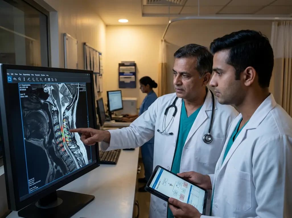
Cervi-AI achieves expert-level agreement in evaluating cervical spinal stenosis and spinal cord compression on MRI, while its automated imaging metrics reliably predict anterior versus posterior surgical approaches in multilevel degenerative cervical myelopathy.
Today

Hormonal shifts throughout puberty, menstruation, fertility treatments, pregnancy, and menopause cause profound glucose excursions in women with diabetes. Understanding these endocrine transitions and utilizing advanced continuous glucose monitoring enables clinicians to optimize personalized diabetes care.
Today
