.webp&w=384&q=75)

Loading, please wait...
Loading, please wait...
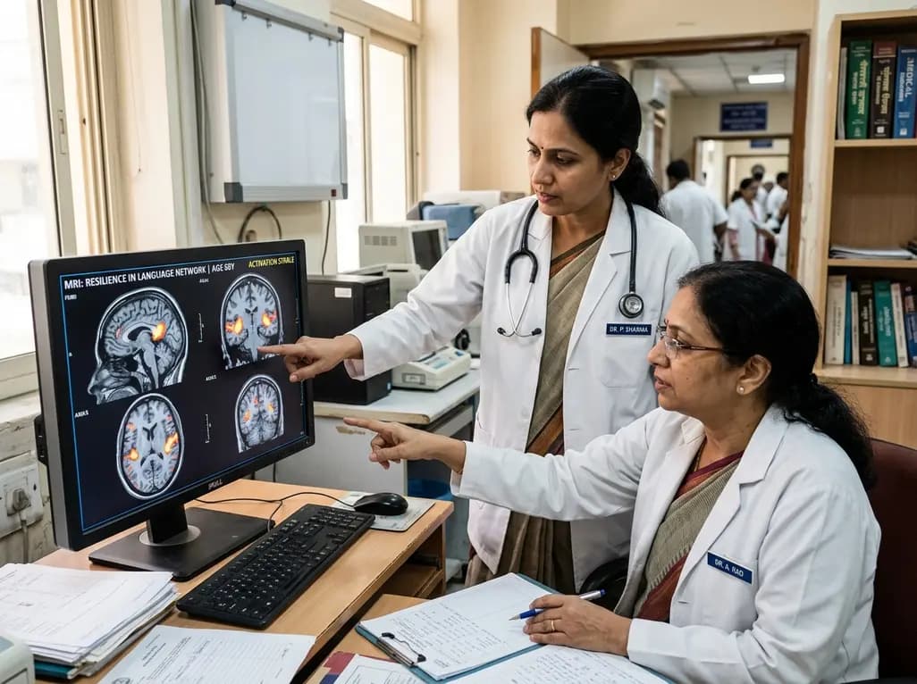
Understanding language network aging provides vital insights into the neurobiology of healthy cognitive senescence. As individuals grow older, many cognitive domains experience a gradual and well-documented decline. For instance, processing speed, working memory, attention, and executive problem-solving frequently diminish over time. However, linguistic capabilities demonstrate a strikingly different trajectory throughout human adulthood. Clinicians and neuroscientists have long observed that vocabulary, syntactic understanding, and verbal comprehension remain exceptionally robust across the lifespan. Recent neuroimaging research from the Massachusetts Institute of Technology and Boston University now clarifies the physiological basis behind this remarkable resilience. Consequently, these findings reshape how clinicians evaluate cognitive aging and distinct neural pathways.
During healthy aging, neural structures undergo subtle anatomical and physiological transformations. Nonetheless, distinct cortical circuits respond to the passage of time with varying vulnerability. A study published in Nature Communications evaluated neural activity across two cohorts, comparing adults aged 17 to 39 with those aged 41 to 80. By employing functional magnetic resonance imaging, researchers mapped how these distinct cohorts processed challenging cognitive tasks. Remarkably, during language-focused activities, the neural activation patterns within the language network of older adults appeared nearly identical to younger counterparts. Furthermore, functional synchronization across the left-hemisphere frontotemporal language nodes showed no significant age-related degradation. In contrast, non-specialized cortical regions displayed noticeable alterations in recruitment and synchronization. Therefore, the specialized language system maintains structural and functional integrity even when surrounding neural tissue undergoes physiological senescence. These results challenge historical assumptions that aging impairs all higher-order cognitive domains uniformly. Instead, specialized computational hubs in the human cortex demonstrate evolutionary and experience-dependent preservation.
To contextualize language preservation, researchers contrasted the language system with the multiple demand network. The multiple demand network comprises bilateral frontal and parietal cortical regions that support flexible executive control. Specifically, this distributed system orchestrates fluid intelligence, working memory, goal-directed behavior, and rapid task switching. When participants performed demanding spatial working memory exercises, older adults exhibited marked functional changes within this domain-general system. Specifically, the multiple demand network in older adults was measurably smaller in functional volume. Moreover, its internal functional connectivity was significantly less synchronized compared to younger participants. Additionally, the overall magnitude of signal activation diminished across the older cohort. Consequently, these executive networks reflect the typical vulnerability seen in general cognitive aging. However, domain-specific networks, such as the language processing system, operate under distinct organizational principles. Because language processing relies on highly specialized, modular neural architecture, it avoids the functional dispersion commonly observed in multipurpose executive networks.
Standard group-averaging neuroimaging methods often blur crucial individual anatomical variations. To overcome this limitation, the investigators utilized individual-subject functional localization techniques. This precision mapping allowed researchers to pinpoint the exact boundaries of functional networks in each participant. Consequently, the team could rigorously evaluate whether the topography, lateralization, and selectivity of the language cortex changed across age cohorts. The data revealed that language-selective voxels maintained identical lateralization toward the left hemisphere across both cohorts. Furthermore, the magnitude of blood-oxygen-level-dependent responses remained steady when participants read sentences or listened to narrative stories. In addition, the internal temporal synchrony between frontal and temporal language hubs remained tightly coupled in older individuals. Thus, precision neuroimaging confirms that functional specialization does not erode under normal physiological aging. These empirical findings provide an objective neural explanation for why older adults maintain crystallized knowledge and lexical depth.
The study also evaluated how older brains manage challenging and unfamiliar linguistic elements. For example, participants encountered complex syntactic structures, rare lexical items, and low-frequency vocabulary. In younger cohorts, these syntactic and semantic challenges reliably trigger elevated hemodynamic responses within the core language network. Crucially, the older cohort demonstrated the exact same elevated activation patterns when navigating linguistic complexity. Therefore, the brain does not lose its responsiveness to novel grammatical or lexical challenges as decades pass. Furthermore, older adults effectively process linguistic nuances without needing compensatory hyperactivation from adjacent executive networks. Many researchers previously hypothesized that older adults rely heavily on bilateral executive recruitment to compensate for language decline. However, these recent findings indicate that the primary language network handles complex linguistic computations autonomously. As an individual accumulates decades of verbal experience, the specialized linguistic circuit continually reinforces its internal connectivity.
These neuroimaging findings provide profound practical implications for practicing physicians, geriatricians, and neurologists. When evaluating elderly patients presenting with cognitive complaints, clinicians must distinguish between general executive slowing and linguistic impairment. Because healthy language network aging preserves syntax, semantic retrieval, and lexical comprehension, any marked linguistic breakdown warrants immediate clinical suspicion. For example, prominent word-finding difficulties, progressive anomia, or comprehension deficits should not be dismissed as benign normal aging. Instead, these symptoms may indicate neurodegenerative processes such as Primary Progressive Aphasia, frontotemporal lobar degeneration, or early Alzheimer's disease. Furthermore, recognizing linguistic resilience allows rehabilitation specialists to leverage intact language pathways during cognitive therapy. Clinicians can design therapeutic strategies that utilize robust verbal skills to compensate for deficits in spatial reasoning or fluid working memory. Ultimately, understanding which brain networks resist physiological decay empowers healthcare professionals to deliver targeted diagnostic assessments and personalized cognitive interventions.
A primary diagnostic challenge in geriatric medicine is differentiating normal age-related cognitive slowing from early neurodegenerative pathology. Because executive processing and processing speed naturally decline with advancing age, patients often report frustration with multitasking or spatial memory. Clinicians can reassure patients that modest changes in executive speed reflect common age-related shifts in the multiple demand network. In contrast, preserved verbal storytelling, rich conversational vocabulary, and intact grammar represent normal brain function in healthy older individuals. However, when an elderly patient exhibits severe semantic loss or syntactic disintegration, physicians must pursue comprehensive diagnostic workups. Such investigations should include formal neuropsychological testing, structural neuroimaging, and metabolic assessments. Additionally, primary care physicians should routinely screen for reversible metabolic, vascular, or pharmacological contributors to cognitive impairment. By establishing clear baselines of preserved linguistic function, medical teams can promptly identify pathological deviations and initiate timely therapeutic support.
Q1: Why does language processing remain resilient while memory and decision-making decline with age?
Language processing relies on a dedicated, domain-specific neural network refined across decades of continuous verbal experience. In contrast, executive functions depend on the domain-general multiple demand network, which manages fluid problem-solving and rapid task-switching. While multipurpose executive circuits exhibit structural and functional decline during normal senescence, specialized linguistic hubs maintain robust connectivity and functional activation throughout healthy aging.
Q2: How do precision fMRI techniques help scientists understand the aging brain?
Traditional brain imaging often averages data across diverse subjects, obscuring subtle anatomical variations. Precision fMRI maps individual cortical networks within each participant, isolating specific functional regions with high accuracy. Consequently, researchers can precisely measure network synchronization, spatial extent, and activation magnitude. This individual-level mapping demonstrated that the functional architecture of the language network remains remarkably preserved across older populations.
Q3: When should a physician suspect pathological impairment rather than normal cognitive aging?
Mild slowing in processing speed and multitasking typically reflects normal changes in the multiple demand network. However, significant language deterioration, such as severe anomia, impaired comprehension, or grammatical breakdown, is abnormal. Clinicians should investigate progressive linguistic deficits promptly through formal neuropsychological testing and neuroimaging to evaluate for conditions like Primary Progressive Aphasia or Alzheimer's disease.
Disclaimer: This content is for informational and educational purposes only. It does not constitute medical advice or replace professional judgment. Refer to the latest local and national guidelines for clinical practice.
References

Read summarized clinical updates, watch expert medical content, and earn CME certifications right from your smartphone.
Open your smartphone camera to scan and install.





Groundbreaking neuroimaging research demonstrates that the brain's specialized language network remains highly resilient against age-related decline, unlike general executive networks. Discover why older adults preserve linguistic comprehension, syntax processing, and vocabulary despite natural cognitive changes.
last month

With post-monsoon dengue surges threatening South Asian health systems, physicians must refine triage and management. This guide covers vital warning signs, plasma leakage detection, crystalloid titration, and vector containment strategies to prevent mortality during seasonal arboviral epidemics.
Today

Findings from Project Viva indicate that sustained postpartum weight retention, rather than excessive gestational weight gain alone, directly worsens women's mid-life cardiovascular health, degrading blood pressure and glycemic parameters decades after delivery.
Today

A large UK Biobank study reveals that plasma proteomic signatures capture preclinical organ damage and improve multiorgan risk prediction across early CKM syndrome stages 0 to 2 beyond traditional PREVENT clinical models.
Today

A retrospective CBCT study reveals that superior TMJ space asymmetry correlates moderately with occipital condyle-atlas-axis asymmetry in unilateral TMJ osteoarthritis, highlighting an imaging correlate between jaw biomechanics and upper cervical alignment rather than a standalone diagnostic marker.
Today

A Bayesian multilevel meta-analysis reveals that aerobic training combined with moderate carbohydrate restriction modestly lowers HbA1c in type 2 diabetes. However, sparse data and very low certainty leave incremental benefits over exercise or diet alone unproven, highlighting the need for individualized care.
Today
