.webp&w=384&q=75)

Loading, please wait...
Loading, please wait...

Clinicians frequently encounter diagnostic dilemmas when evaluating painful anterior neck masses and thyrotoxic symptoms. Subacute thyroiditis (SAT), also termed de Quervain thyroiditis, represents a self-limiting yet debilitating inflammatory condition that typically follows viral upper respiratory infections. While conventional ultrasound displays characteristic hypoechoic, ill-defined areas with diminished vascularity, these sonographic patterns can mimic anaplastic carcinoma, aggressive lymphoma, or infiltrative malignancies. Consequently, clinicians require objective, noninvasive diagnostic modalities to resolve clinical uncertainty. Emerging research demonstrates that 2D shear wave elastography offers a reliable quantitative assessment of parenchymal stiffness, establishing a new paradigm for both initial diagnosis and longitudinal disease monitoring in modern clinical practice.
Standard B-mode ultrasonography provides anatomical detail, yet it cannot quantify the biomechanical alterations occurring during acute parenchymal inflammation. In contrast, 2D shear wave elastography utilizes acoustic radiation force impulses to generate transverse shear waves within the thyroid tissue. Specialized ultrafast imaging sequences track the propagation velocity of these shear waves in real time. Because shear waves travel markedly faster through rigid, noncompliant structures than through supple tissues, the system converts propagation velocity into tissue elasticity. Technicians express this elasticity quantitatively as the Young modulus in kilopascals (kPa).
Unlike conventional strain elastography, which depends heavily on manual, operator-dependent compression against underlying cervical structures, shear wave technology delivers reproducible, objective tissue quantification. The automated acoustic push eliminates manual compression artifacts, thereby minimizing interobserver variability. Clinicians receive a high-resolution, color-coded elastogram superimposed over standard grayscale anatomy alongside precise numerical values. Consequently, radiologists can systematically sample focal inflammatory patches and uninvolved parenchymal regions. This provides reproducible metrics that mirror underlying histopathological changes.
The remarkable rise in shear modulus observed during the active phase of subacute thyroiditis stems directly from microscopic cellular alterations. Histopathologically, the condition provokes extensive follicular destruction, acute interstitial edema, granulomatous infiltration, and foreign body giant cell reactions. As inflammatory cellular infiltrates densely pack the thyroid follicles, intraparenchymal pressure rises sharply. Furthermore, early microscopic fibroblastic proliferation and dense interstitial edema render the inflamed thyroid lobes markedly indurated and noncompliant.
On physical examination, clinicians readily appreciate this process as a firm, exquisitely tender thyroid gland. However, manual palpation cannot yield objective, standardized measurements. Recent clinical investigations indicate that baseline thyroid stiffness in active subacute thyroiditis frequently exceeds 120 kPa, contrasting sharply with healthy thyroid parenchyma that typically measures below 25 kPa. Interestingly, this acute mechanical rigidity often matches or exceeds values documented in malignant thyroid lesions. Clinicians must interpret elevated elasticity scores in conjunction with patient symptoms, high erythrocyte sedimentation rates, and suppressed thyrotropin levels to avoid misdiagnosing acute inflammatory stiffness as thyroid carcinoma.
Distinguishing subacute thyroiditis from other causes of thyrotoxicosis and focal neck pain represents a frequent challenge in outpatient endocrinology. Graves disease also causes thyrotoxic symptoms, yet it exhibits distinctly different elastographic properties. The diffuse hypervascularity and follicular hyperplasia characteristic of Graves disease create a softer, more compliant parenchymal architecture, typically registering values well below those recorded during acute granulomatous inflammation.
Similarly, Hashimoto thyroiditis presents with chronic autoimmune lymphocytic infiltration and variable fibrosis. Although chronic autoimmune thyroiditis yields stiffness measurements greater than normal tissue, the stiffness values rarely approach the extreme elevations seen in active granulomatous thyroiditis. Most importantly, shear wave elastography helps clinicians differentiate focal inflammatory patches from solid malignant thyroid nodules. Although both entities produce elevated stiffness readings, subacute thyroiditis demonstrates widespread, poorly demarcated, geographic stiffness maps that extend beyond discrete grayscale nodular borders. Furthermore, rapid normalization on serial follow-up scans effectively confirms a benign inflammatory process without requiring invasive fine-needle aspiration biopsies.
Beyond baseline diagnosis, shear wave imaging provides exceptional clinical utility for monitoring disease resolution and guiding therapeutic decisions. Standard management for subacute thyroiditis incorporates nonsteroidal anti-inflammatory drugs for mild discomfort or oral corticosteroid tapers for severe pain and systemic inflammation. However, serum inflammatory markers such as C-reactive protein occasionally normalize before parenchymal inflammation fully clears, leading to premature medication withdrawal and clinical relapse.
Prospective investigations demonstrate a progressive, predictable decline in parenchymal stiffness across serial evaluations. During active therapy, mean tissue stiffness drops significantly by four weeks, often halving its baseline value as interstitial edema and granulomatous infiltration recede. By eight to ten weeks, tissue elasticity typically returns to near-normal baseline ranges, reflecting architectural restoration and cellular healing. Therefore, serial stiffness measurements offer treating clinicians an objective radiological bio-marker. Incorporating follow-up elastography allows physicians to tailor steroid tapering schedules precisely, confirm complete resolution, and prevent recurrent inflammatory rebounds.
Integrating shear wave elastography into routine endocrine and radiological workflows requires strict adherence to standardized scanning protocols to ensure reproducible results. Sonographers must instruct patients to avoid swallowing, speaking, or deep breathing during image acquisition. Because external mechanical force distorts tissue elasticity calculations, examiners must apply minimal transducer pressure to the neck, using generous amounts of acoustic coupling gel.
Examiners should place regions of interest over representative hypoechoic inflammatory zones, taking care to avoid surrounding carotid vascular pulsations, the tracheal cartilage, and cystic necrotic regions. Averaging multiple consecutive acquisitions ensures statistical reliability. Clinicians should synthesize elastographic data alongside conventional B-mode features, color Doppler vascularity maps, and laboratory parameters, including free thyroxine and inflammatory indices. Adopting this multimodal framework allows physicians to maximize diagnostic certainty, minimize superfluous nodule biopsies, and deliver personalized, evidence-based care throughout the disease course.
Conventional ultrasound provides anatomical visualization, revealing nonspecific hypoechoic, ill-defined areas with low vascular flow that can resemble malignancy. Conversely, 2D shear wave elastography measures physical tissue stiffness quantitatively in kilopascals. This noninvasive technology offers objective biomechanical metrics reflecting underlying granulomatous inflammation. Consequently, it complements grayscale imaging by supplying functional diagnostic data and aiding clinical differentiation.
Because both subacute thyroiditis and thyroid malignancies display high tissue stiffness, absolute numerical values occasionally overlap. However, subacute thyroiditis exhibits diffuse, poorly circumscribed geographic stiffness patterns on elastograms, unlike the discrete, localized rigidity of carcinomas. Furthermore, subacute thyroiditis demonstrates dramatic stiffness reduction on serial scans after anti-inflammatory therapy, effectively confirming inflammatory etiology and ruling out malignancy.
Corticosteroid therapy rapidly suppresses clinical symptoms and serum inflammatory markers, but microscopic inflammation can persist within the thyroid tissue. Serial shear wave elastography tracks resolving parenchymal stiffness from initial diagnosis through four- and eight-week intervals. This objective decline confirms true tissue healing, assisting physicians in safely tapering steroids and minimizing the risk of symptom rebound.
Disclaimer: This content is for informational and educational purposes only and should not be considered medical advice or relied upon as a substitute for professional clinical judgement. Healthcare professionals should make diagnostic and therapeutic decisions based on their clinical experience and the specific circumstances of each patient. Guidelines and medical literature may change; please consult recent official recommendations. Refer to the latest local and national guidelines for clinical practice.
References
Golu I et al. Diagnostic and Monitoring Value of 2D Shear Wave Elastography in Subacute Thyroiditis: A Prospective Case-Control Study. Int J Endocrinol. 2026 undefined undefined. doi: 10.1155/ije/4035441. PMID: 42763572.
Ruchala M, Szczepanek-Parulska E, Zybek A, Moczko J, Czarnywojtek A, Kaminski G, Sowinski J. The role of sonoelastography in acute, subacute and chronic thyroiditis: a novel application of the method. Eur J Endocrinol. 2012;166(3):425-432. doi: 10.1530/EJE-11-0852.
Tekcan Sanli DE, Yildirim D. The value of shear wave elastography and velocity standard deviation value in the differentiation of subacute thyroiditis and chronic autoimmune thyroiditis. Cerrahpasa Med J. 2022;46(1):15-20. doi: 10.5152/cjm.2022.21048.
Cantisani V, Grazhdani H, Drakonaki E, et al. Strain and shear-wave elastography in thyroid pathology. Med Ultrason. 2014;16(3):233-240.

Read summarized clinical updates, watch expert medical content, and earn CME certifications right from your smartphone.
Open your smartphone camera to scan and install.





Subacute thyroiditis presents diagnostic dilemmas when differentiating it from malignancy or chronic thyroiditis. Discover how 2D shear wave elastography provides quantitative tissue stiffness metrics that enhance diagnostic accuracy and assist in tracking disease resolution during therapy.
Today
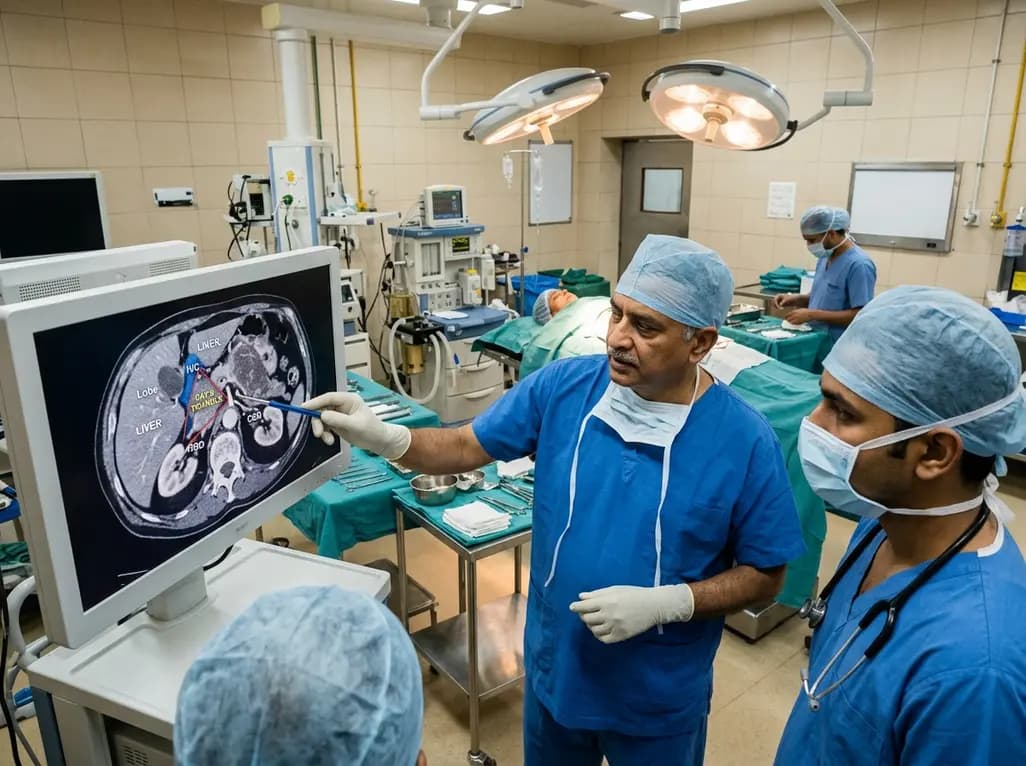
Researchers at AIIMS New Delhi have identified Ray's triangle, a high-risk surgical zone between the liver and inferior vena cava. Recognising this danger area allows surgical oncologists to achieve complete cytoreduction in advanced abdominal cancers while substantially lowering the risk of life-threatening hemorrhage.
Today

An updated systematic review and meta-analysis of 7,875 patients reveals that routine microaxial left ventricular support during nonemergent high-risk PCI does not reduce mortality or ischemic complications, while significantly increasing major bleeding events.
Yesterday
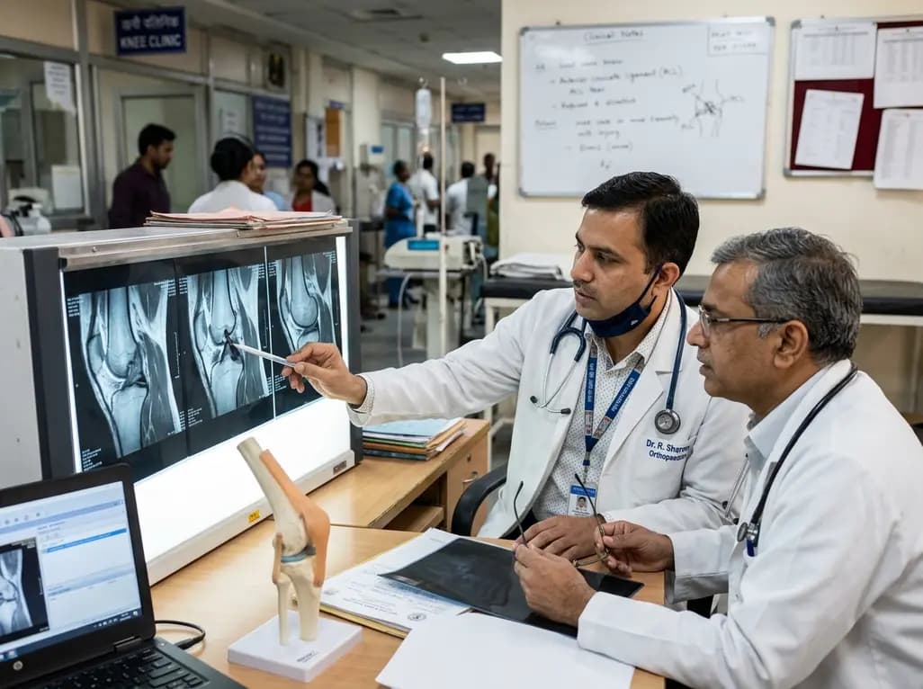
The FIRM-P framework redefines anterior cruciate ligament instability management by separating objective knee phenotypes from patient context and integrating arthrogenic muscle inhibition as a dynamic modifier to optimize individual surgical timing and outcomes.
Today
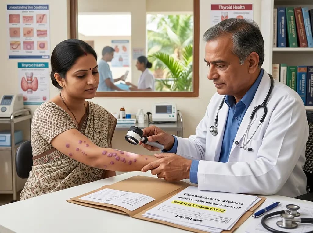
A case-control study demonstrates a significant association between lichen planus and thyroid disorders, with elevated TSH and anti-TPO antibody levels across cutaneous and follicular subtypes, emphasizing the critical value of routine thyroid screening.
Today
