.webp&w=384&q=75)

Loading, please wait...
Loading, please wait...

Periodontal assessment forms the cornerstone of effective oral healthcare, guiding early therapeutic interventions and preventing bone destruction. Historically, manual periodontal probing has remained the diagnostic standard, despite inherent limitations in tactile subjectivity and tissue trauma. Recent technological breakthroughs in high-resolution dental imaging present non-invasive alternatives. A pivotal clinical study demonstrates that intraoral ultrasonography provides outstanding agreement with conventional clinical probing depth, marking a significant evolution in periodontal diagnostics.
Traditional periodontal evaluation relies extensively on manual probes, such as the UNC-15 probe, to measure pocket depths around natural dentition. However, mechanical probing presents established biological limitations. Clinicians often encounter substantial variation caused by probing force, insertion angulation, and soft tissue inflammatory status. For instance, inflamed junctional epithelium permits excessive probe penetration, which creates false-positive measurements of tissue loss. Conversely, dense calculus deposits or tight subgingival anatomy can impede manual instruments and yield artificially shallow readings. Additionally, patients frequently report discomfort during aggressive probing of inflamed gingival tissues. Consequently, dental scientists have explored non-invasive modalities that offer objective, reproducible anatomic visualization. Intraoral ultrasonography utilizes high-frequency acoustic waves to map periodontal microarchitecture in real time without ionizing radiation. By reflecting sound waves off acoustic impedance boundaries between soft tissues, enamel, and bone, ultrasonographic transducers capture pristine sectional images of the periodontium. Therefore, this technological approach eliminates mechanical tissue displacement while delivering reliable structural measurements.
To assess clinical feasibility, investigators designed a rigorous proof-of-concept agreement trial involving sixty-nine human participants. The research team evaluated 963 standardized mid-buccal sites, encompassing 767 anterior teeth, 127 premolars, and 69 first molars. Examiners recorded clinical probing depths using standard manual UNC-15 periodontal instruments. Concurrently, clinicians acquired B-mode acoustic scans of identical anatomical sites using an 18-MHz intraoral ultrasound transducer. Two independent, blinded examiners analyzed the ultrasound images to measure ultrasound-derived gingival sulcus depth. Furthermore, investigators implemented generalized estimating equations and clustered intra-class correlation coefficients to properly account for within-patient data clustering. The trial also evaluated several critical confounding variables across comprehensive clinical subgroups. Specifically, the statistical protocol analyzed probing depth strata, local bleeding on probing, anatomical tooth morphology, subgingival calculus, restorations, orthodontic attachments, and tobacco smoking status. Through this standardized comparative setup, the researchers established an extensive benchmark to validate acoustic depth measurements against manual physical assessments across diverse oral environments.
The clinical study demonstrated exceptionally strong statistical agreement between ultrasound-derived measurements and manual probing depths. Overall, the intra-class correlation coefficient reached 0.942, indicating outstanding diagnostic consistency across all examined tooth types. Moreover, the mean absolute error remained remarkably low at 0.29 millimeters, accompanied by a negligible mean overall difference of only 0.023 millimeters. In clinical practice, precision margins dictate utility, and 85.0% of all measured sites differed by no more than 0.50 millimeters. In addition, 96.7% of evaluated sites differed by 1.00 millimeter or less, comfortably satisfying international clinical tolerance thresholds. Subgroup analyses confirmed robust performance regardless of gingival inflammation or smoking history, with subgroup correlations ranging from 0.786 to 0.952. Nevertheless, molar sites presented minor technical challenges, exhibiting the highest mean absolute error at 0.38 millimeters and a slight underestimation of 0.123 millimeters. Posterior anatomy and transducer positioning likely contributed to these subtle variations, yet the overall diagnostic reliability remained remarkably stable.
Integrating acoustic diagnostics into regular dental practice offers numerous distinct advantages for clinicians and patients alike. Because intraoral ultrasound requires no physical insertion into the gingival crevice, it completely eliminates patient pain and bleeding. Furthermore, the acoustic transducer generates permanent, objective digital cross-sections of the gingival margin, supracrestal tissues, and alveolar crest. Clinicians can archive these images directly into electronic health records and review them longitudinally across serial treatment visits. Thus, practitioners can track subtle changes in mucosal thickness, soft tissue architecture, and healing dynamics without disturbing fragile tissue attachments. This non-invasive capability proves particularly invaluable during the early postoperative healing phases following regenerative therapy or dental implant placement. During these critical recovery periods, traditional mechanical probing remains strictly contraindicated due to the risk of tearing delicate immature collagen fibers. Consequently, intraoral scanning provides clinicians with unparalleled diagnostic insight while preserving fragile surgical margins and improving patient comfort during routine monitoring.
Despite these promising findings, clinicians must recognize existing technical limitations before intraoral ultrasound enters routine dental practice. First, this proof-of-concept study focused exclusively on standardized mid-buccal sites, leaving complex interproximal pockets and lingual contours unexplored. Interproximal contact points present anatomical constraints that restrict bulky linear transducers from capturing pristine acoustic reflection angles. Therefore, bioengineers must develop ultra-miniaturized intraoral probes capable of negotiating tight interdental spaces and deep posterior crevices. Second, image acquisition and acoustic interpretation require substantial operator training to prevent probe angulation errors and acoustic shadowing artifacts. Handheld positioning and tissue compression can distort soft tissue thickness if operators fail to maintain uniform acoustic coupling. Furthermore, extensive multi-center studies featuring diverse clinical skill levels must validate these findings in advanced periodontitis cohorts. Nevertheless, as artificial intelligence algorithms and automated landmark recognition continue to mature, acoustic periodontal charting could soon transform conventional diagnostics into a rapid, automated, and painless chairside standard.
Intraoral ultrasonography utilizes high-frequency acoustic waves emitted from a specialized intraoral transducer placed against the gingival margin. These acoustic signals reflect back when they encounter boundaries between tissues with different acoustic impedances, such as gingival epithelium, connective tissue, enamel, and alveolar bone. Dedicated software interprets these returning echo patterns, calculating the physical distance between the gingival margin and sulcular base without requiring mechanical tissue penetration.
Currently, intraoral ultrasound serves as an effective non-invasive adjunct rather than an immediate complete replacement for manual probing. Although ultrasonography achieves excellent accuracy on mid-buccal sites, modern transducer dimensions limit easy access to narrow interdental spaces and deep lingual pockets. Consequently, clinicians must still use conventional probes for complete full-mouth six-point examinations until engineers design smaller transducers tailored specifically for interproximal navigation.
Posterior molar sites demonstrated slightly higher mean absolute error due to restricted intraoral access, thicker cortical plates, and complex anatomical root curvatures. Maneuvering rigid ultrasound transducers in the posterior oral cavity challenges operator ergonomics and makes perpendicular probe orientation difficult to maintain. Furthermore, dense calculus, cheek interference, and restoration margins can cause minor acoustic shadowing that slightly impairs landmark visualization around molar crowns.
Disclaimer: This content is for informational and educational purposes only... Refer to the latest local and national guidelines for clinical practice.
References

Read summarized clinical updates, watch expert medical content, and earn CME certifications right from your smartphone.
Open your smartphone camera to scan and install.





A clinical agreement study demonstrates that intraoral ultrasonography achieves high concordance with manual probing depth across anterior teeth, premolars, and molars. This non-invasive imaging method accurately maps the gingival sulcus, providing an objective, pain-free adjunct to conventional periodontal probing.
Today

A retrospective study shows that symptom-monitored loading with lorazepam, chlordiazepoxide, or gabapentin significantly reduces hospital length of stay compared to CIWA-Ar-only therapy in severe alcohol withdrawal, providing a safer, more proactive management strategy.
Today
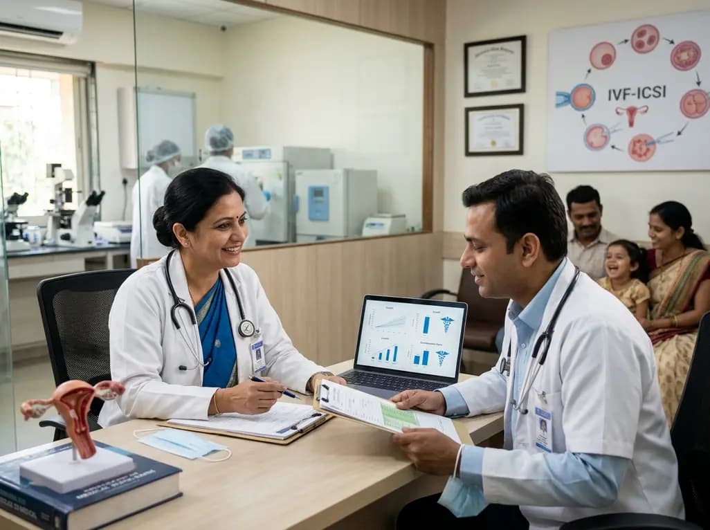
A retrospective cohort study of 5,481 children aged 3–6 years evaluated whether surgical sperm retrieval for ICSI affects offspring cognitive and physical growth compared with ejaculated sperm and IVF. Results demonstrate comparable IQ scores and growth parameters, providing reassuring safety evidence for clinical practice.
Today
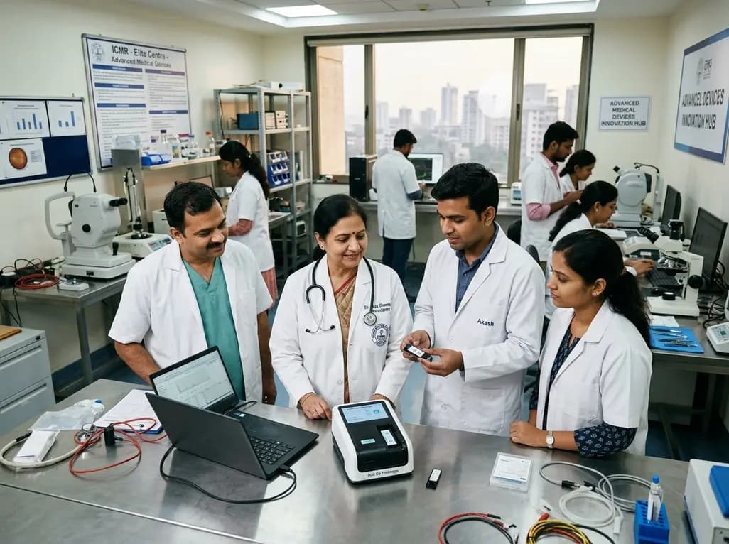
The Indian Council of Medical Research has funded seven top academic hubs to build indigenous healthcare technologies. Targeting point-of-care diagnostics, biomaterials, and ophthalmology, these multidisciplinary centres aim to tackle unmet clinical needs and make medical care affordable across India.
Today
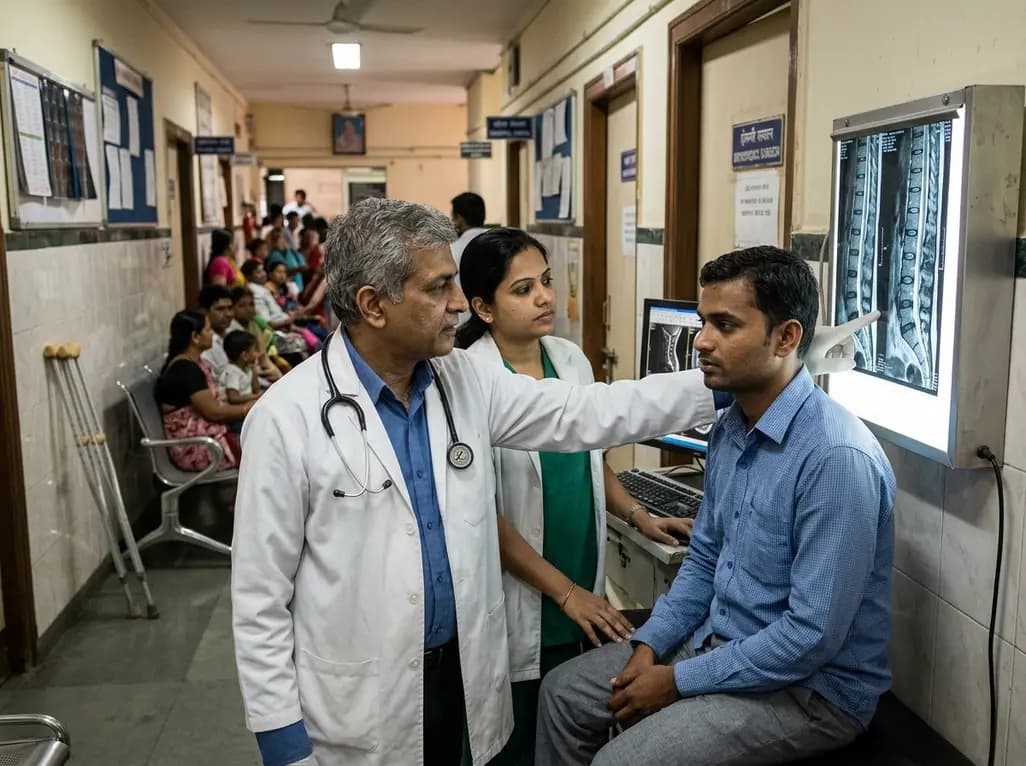
Diagnostic data shows that nearly one in four spine investigations now involve patients younger than 40 years. This comprehensive overview explores the drivers behind this demographic trend, evaluates ergonomic hazards, clarifies red-flag signs, and highlights clinical indications for judicious diagnostic imaging.
Today
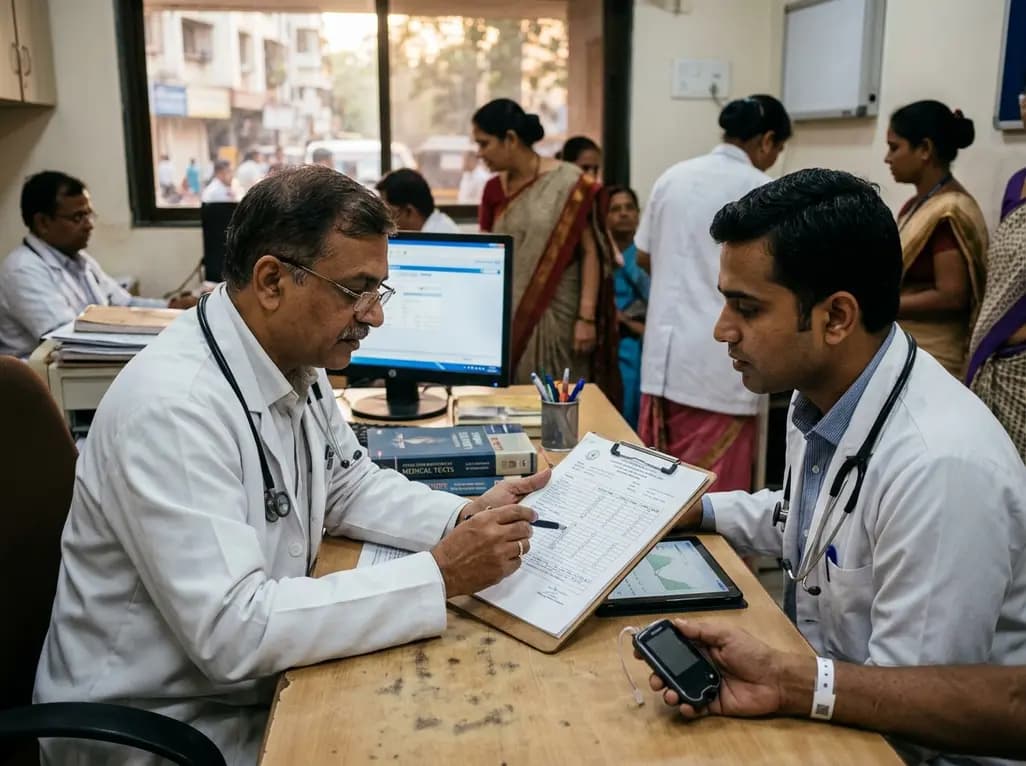
A multicenter cross-sectional study of 1,315 adults with type 1 diabetes reveals that the GMI/HbA1c ratio does not independently predict microvascular complications. Clinicians should prioritize sustained time in range and blood pressure management over glycation discordance metrics.
Today
