.webp&w=384&q=75)

Loading, please wait...
Loading, please wait...

Surgical intervention within the brainstem represents one of the most formidable challenges in modern neurosurgery due to high concentrations of vital neural pathways. Stereotactic biopsy provides essential histological and molecular diagnostics for brainstem neoplasms; however, the procedure still carries significant risks of post-procedural morbidity. Recent surgical advances demonstrate that intraoperative tract mapping performed directly through the needle cannula offers real-time physiological guidance. This novel technique allows neurosurgeons to identify critical tracts prior to tissue acquisition, thereby optimizing diagnostic yield while preserving vital neurological structures.
Intrinsic brainstem lesions encompass diverse pathological entities, including diffuse midline gliomas, focal astrocytomas, vascular malformations, and inflammatory or infectious tuberculomas. Consequently, accurate histopathological and molecular characterization is essential to establish targeted therapy regimens and determine long-term prognosis. Nevertheless, stereotactic biopsies in this region carry documented morbidity risks ranging between eight and twelve percent. Because dense motor, sensory, and cranial nerve fibers traverse extremely tight anatomic corridors, minor needle deflections can cause catastrophic neurological damage. Traditional frame-based and frameless stereotaxy rely almost exclusively on static pre-acquired magnetic resonance imaging. However, brain shift, mechanical needle bending, and subtle patient motion frequently introduce registration inaccuracies. Furthermore, structural imaging cannot confirm physiological pathway boundaries within tissue deformed by infiltrating tumors. Although open cranial procedures routinely utilize neurophysiological mapping, clinicians historically could not apply standard direct stimulation methods through closed stereotactic instrumentation. Therefore, neurosurgeons required a dedicated mechanism to confirm real-time functional boundaries at the needle tip before opening biopsy side windows.
To overcome traditional stereotactic limitations, surgical teams have evaluated an innovative technique that implements intraoperative tract mapping directly through the biopsy cannula. Specifically, the surgeon advances an outer cannula along the stereotactic trajectory to the precalculated target depth. Next, the operative team introduces a specialized, miniature dual epidural D-wave electrode down the inner lumen of the biopsy needle. This electrode acts as a stimulating probe positioned precisely at the designated sampling site. Neurophysiologists deliver short-train, low-intensity electrical pulses to map the surrounding microenvironment. Meanwhile, recording channels continuously monitor triggered electromyography responses from peripheral limbs and cranial nerve musculature, alongside somatosensory evoked potentials. Because electrical current thresholds correlate directly with anatomical distance, this setup provides high spatial resolution around the needle tip. In addition, the system requires no bulky cranial instrumentation, maintaining complete compatibility with standard stereotactic headframes. Consequently, surgical teams can integrate functional electrical interrogations into regular biopsy workflows without extending procedural duration excessively.
In a prospective feasibility study involving sixteen patients with intrinsic brainstem lesions, investigators assessed the clinical efficacy of this mapping method. Remarkably, electrophysiological responses emerged in fifty-six percent of the enrolled cohort, confirming the close proximity of the needle tip to functional white matter tracts. The medial lemniscus generated the most frequent signals, appearing in thirty-eight percent of cases. Additionally, corticospinal tract responses occurred in thirty-one percent of patients, whereas corticonuclear pathway responses appeared in twelve percent. These quantitative findings demonstrate that standard structural planning alone often positions the cannula adjacent to highly eloquent descending motor and ascending sensory tracts. When electrical thresholds triggered positive evoked potentials, clinicians promptly recognized that indiscriminate mechanical aspiration or cutting could disrupt intact axons. Conversely, the absence of evoked potentials confirmed a functionally silent zone, permitting confident tissue procurement. Thus, intraoperative electrophysiology transformed a static mechanical trajectory into an interactive, functionally monitored intervention.
The primary clinical value of this methodology lies in actionable real-time guidance. Whenever intraoperative stimulation generated pathway responses, surgeons refrained from immediate tissue sampling. Instead, the surgical team actively adjusted needle orientation to avoid functional injury. Operators first rotated the side-cutting aperture away from the elicited response vector. If electrical stimulation continued to indicate critical tract proximity, the team adjusted the cannula depth by several millimeters. Subsequently, biopsy cutting proceeded exclusively at coordinates devoid of tract activation. Overall, post-procedural neurological worsening occurred in twenty-five percent of patients, but severe permanent deficits arose solely when lesions directly infiltrated pathways. For instance, the most profound deficit occurred in a medullary lesion where persistent corticobulbar responses confirmed direct tract invasion. Most importantly, patients without tract responses and without computed tomography-verified hemorrhagic complications experienced zero permanent functional deterioration. Therefore, dynamic needle reorientation successfully prevented accidental tract transection in anatomically vulnerable corridors.
This technical evolution holds profound implications for tertiary neurosurgical and neuro-oncology centers across India. In Indian clinical practice, neurosurgeons regularly encounter complex brainstem lesions, including diffuse brainstem gliomas and atypical granulomatous infectious masses such as tuberculomas. Because infectious lesions, demyelinating processes, and aggressive malignancies can present with identical radiological signs, biopsy remains indispensable for avoiding erroneous empirical therapy. Moreover, Indian neurosurgical departments increasingly manage complex cranial cases using awake procedural protocols under local scalp blocks. Because the cannula-based mapping hardware uses miniature coaxial electrodes, clinicians can readily apply this setup during awake stereotactic biopsies. Patient cooperation further allows parallel continuous clinical testing during electrical stimulation. Although high-end intraoperative magnetic resonance suites remain restricted to select apex institutes, electrophysiological mapping hardware is relatively portable and affordable. Consequently, widespread adoption of cannula-based functional mapping can significantly reduce surgical morbidity and enhance diagnostic safety across diverse healthcare tiers.
As neurosurgical oncology moves toward individualized molecular therapeutics, obtaining adequate diagnostic tissue with minimal surgical morbidity becomes non-negotiable. Cannula-based mapping bridges the longstanding gap between stereotactic navigation and real-time intraoperative functional verification. Future technological innovations will likely integrate tractographic fiber tracking with algorithmic current-distance calibration, providing surgeons with automated proximity warnings. Furthermore, miniaturizing multiplex electrodes could permit simultaneous multi-directional stimulation, mapping a three-hundred-and-sixty-degree perimeter around the needle tip before sample excision. Additionally, combining electrophysiological verification with optical biopsy techniques, such as Raman spectroscopy or confocal laser endomicroscopy, could ensure maximal diagnostic yield from viable tumor regions while avoiding critical tracts entirely. As clinical trials expand to larger cohorts, standardized threshold algorithms will help establish unified safety margins. Ultimately, this approach redefines brainstem stereotactic surgery, transforming closed blind procedures into precise, physiology-guided diagnostic interventions.
When electrical stimulation through the cannula detects adjacent eloquent tracts, surgeons do not take tissue blindly. Instead, they carefully rotate the biopsy window away from the responsive neural tract. If tract responses persist, the surgeon modifies needle depth, sampling exclusively from functionally silent sites to prevent mechanical injury.
Yes, surgical teams can seamlessly implement this technique during awake frame-based procedures. Because the dual epidural electrode fits directly inside standard stereotactic cannulas, it introduces no extra discomfort. Awake testing enables clinicians to pair electrical monitoring with real-time patient neurological evaluations, significantly increasing overall surgical safety.
The primary clinical risks include focal tissue microtrauma, local hemorrhage, and transient neurological deterioration from mechanical pressure or microvascular injury. In rare cases where intrinsic pathology directly infiltrates critical tracts, even careful biopsy sampling may provoke permanent functional deficits despite meticulous electrophysiological monitoring.
Disclaimer: This content is for informational and educational purposes only. It should not be used as a substitute for professional medical advice, diagnosis, or treatment. Always consult your physician or qualified healthcare provider with any questions you may have regarding a medical condition. Clinical decisions must always be guided by clinical judgment, individual patient evaluation, and multidisciplinary institutional protocols. Refer to the latest local and national guidelines for clinical practice.
References

Read summarized clinical updates, watch expert medical content, and earn CME certifications right from your smartphone.
Open your smartphone camera to scan and install.





A prospective study demonstrates that intraoperative tract mapping through the biopsy cannula during stereotactic brainstem biopsy detects proximity to eloquent white matter tracts, enabling real-time needle repositioning and helping preserve critical neurological function.
Today

Bacteroides thetaiotaomicron utilizes an intricate multiprotein extracellular relay and outer membrane vesicles for cobamide scavenging in the human gut. This review examines its transport mechanics, riboswitch regulation, and potential clinical implications for host-microbe vitamin competition and nutrient absorption.
Today

Novel preclinical research demonstrates that antisense oligonucleotides targeting essential genes such as DARS1, DYNC1I2, and EIF2S3 selectively induce apoptosis in ovarian and lung cancer cells. Conditional biomarker activation provides a viable strategy to spare normal tissue while overcoming therapy resistance.
Today
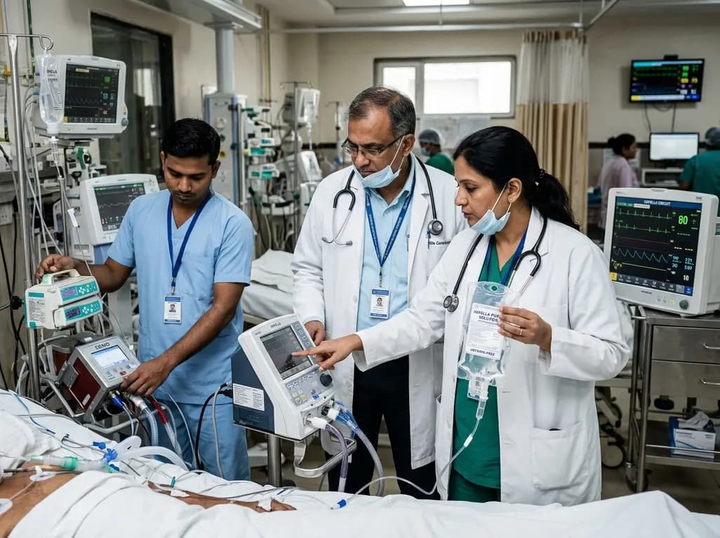
This clinical analysis examines heparin-free versus heparin-containing Impella purge solutions in patients receiving concomitant ECMO support (ECPELLA). Evidence suggests anticoagulant-free purges maintain device patency while reducing bleeding risks, offering a viable strategy alongside systemic anticoagulation.
Today

A 10-year Markov state-transition model shows minimally invasive tubular decompression dominates open laminectomy for pure lumbar spinal stenosis, yielding 5.95 vs 5.91 QALYs and saving $4,167 per patient through reduced complications and lower index costs.
Today

A preclinical study demonstrates that naringin-loaded phytosomes significantly enhance oral bioavailability and provide neuroprotective efficacy in diabetic neuropathy by dampening inflammation, reducing oxidative stress, and relieving neuropathic pain.
Today
