

Loading, please wait...
Loading, please wait...

During neurosurgical procedures near eloquent cerebral cortex, preserving functional pathways remains a fundamental priority for neurosurgeons. Consequently, intraoperative SSEP monitoring has emerged as an essential modality to safeguard sensory pathways and localize the central sulcus. Although clinicians routinely depend on evoked responses, the diagnostic accuracy of isolated signal changes for predicting postoperative neurological deficits requires thorough investigation. Recent clinical evidence offers valuable clarity regarding the prognostic boundaries of somatosensory signals during supratentorial craniotomies.
Neurosurgical oncology demands an intricate equilibrium between achieving maximal safe resection and preserving neurological function. Therefore, neurosurgical teams routinely utilize neurophysiological adjuncts to assess neural pathway integrity in real time. Intraoperative SSEP monitoring specifically tracks dorsal column-medial lemniscal pathways and cortical sensory processing areas. Surgeons frequently employ phase reversal techniques across the central sulcus to establish anatomical landmarks before initiating tumor resection. Nevertheless, questions remain regarding how accurately somatosensory fluctuations predict diverse postoperative functional deficits. Surgical manipulation, thermal injury, and regional ischemia can directly disrupt these neuroelectrical circuits. Moreover, brain shift and microvascular vasospasm frequently produce transient electrophysiological perturbations. Historically, practitioners assumed that signal loss corresponded strictly to focal sensory impairment. However, current neurosurgical evidence demonstrates that sensory pathway disturbances often signify broader regional tissue stress, making rigorous evaluation of this monitoring tool necessary.
A comprehensive retrospective investigation evaluating 511 patients undergoing craniotomy between 2019 and 2023 highlighted critical diagnostic characteristics of evoked potentials. Notably, intraoperative SSEP monitoring demonstrated an impressive specificity of 95% for predicting postoperative neurological deficits. This remarkable specificity underscores that intact, stable waveforms reliably indicate preserved baseline neurological status. Consequently, surgeons can proceed with confidence when signals remain robust throughout critical operative steps. Conversely, the analysis identified a sensitivity of only 16%, revealing a significant diagnostic limitation. Many patients who developed postoperative deficits never exhibited warning changes during recording. Because somatosensory pathways occupy specific anatomical corridors, adjacent motor or linguistic fiber tracts can sustain injury without triggering sensory alerts. Therefore, surgical teams cannot rely on normal waveforms as an absolute guarantee of surgical safety. Recognizing this diagnostic disparity prevents false reassurance during challenging cortical resections.
Waveform alterations during neurosurgery generally follow two distinct trajectories: transient fluctuations that recover or persistent signal loss. Importantly, the recent clinical study revealed that irreversible signal deterioration carries significantly higher prognostic value than reversible changes. Irreversible changes exhibited a positive predictive value of 72%, whereas reversible changes yielded a positive predictive value of 47%. This statistical difference underscores the clinical urgency required when somatosensory potentials degrade. Whenever alerts occur, surgical teams must immediately pause resection, irrigate the cavity with warm saline, and optimize systemic arterial pressure. Furthermore, surgeons should release aggressive brain retractors to alleviate focal cerebral ischemia. When signals recover promptly following these corrective maneuvers, the likelihood of permanent neurological injury decreases substantially. In contrast, persistent waveform loss strongly indicates structural parenchymal insult or irreversible microvascular occlusion, alerting the team to anticipate postoperative neurological deficits.
Intriguingly, intraoperative somatosensory signal degradation does not correlate solely with sensory impairment. In the analyzed patient cohort, SSEP alterations strongly correlated with postoperative motor deficits, yielding a diagnostic odds ratio of 6.1. Additionally, signal perturbations predicted postoperative language deficits, demonstrating a diagnostic odds ratio of 2.6. These notable associations occurred despite tumor size, anatomical location, and baseline eloquence remaining comparable across patient cohorts. Consequently, neurophysiologists conclude that evoked potential degradation serves as a surrogate marker for broader peri-Rolandic cortical dysfunction. Regional edema, venous congestion, and subcortical ischemia rarely respect discrete cytoarchitectonic boundaries in the brain. Therefore, vascular compromise within the middle cerebral artery territory simultaneously jeopardizes primary somatosensory cortex, motor pathways, and adjacent language networks. Recognizing this interconnected vulnerability allows clinicians to interpret sensory signal deterioration as a generalized warning of imminent peri-Rolandic compromise.
Because isolated sensory monitoring exhibits limited sensitivity, reliance on a single modality introduces clinical vulnerability. Therefore, modern neurosurgical practice strongly advocates for multimodal intraoperative neuromonitoring protocols. Combining somatosensory evoked potentials with transcranial motor evoked potentials significantly closes the diagnostic sensitivity gap. Motor evoked potentials provide continuous assessments of the corticospinal tract, which frequently suffers injury independently of sensory pathways. Furthermore, subcortical monopolar or bipolar stimulation enables millimeter-level proximity mapping during deep tumor resections. In patients with tumors situated near eloquent language areas, awake craniotomy with direct cortical mapping remains the definitive benchmark for preserving communication. Additionally, neuroanesthesiologists must maintain stable total intravenous anesthesia, avoiding volatile anesthetics that suppress evoked waveform amplitudes. When surgical teams integrate somatosensory recordings into a multimodal framework, they maximize patient safety and optimize functional oncological outcomes.
The definitive evidence regarding somatosensory evoked potentials provides practical insights for operative decision-making. First, clinicians must remember that high specificity makes any intraoperative alert an urgent, actionable event. Thus, surgical teams must establish standardized response checklists whenever amplitude drops exceed fifty percent or latency increases surpass ten percent. Second, because sensitivity remains low, surgeons must maintain meticulous microdissection technique even when evoked responses appear completely undisturbed. Third, operative teams must systematically record whether intraoperative alerts fully reverse before wound closure. Documenting waveform recovery assists in risk-stratifying patients who need prioritized intensive care monitoring and early postoperative neuroimaging. Ultimately, understanding the exact capabilities and limitations of somatosensory monitoring empowers surgical teams to protect delicate neural networks while pursuing aggressive tumor cytoreduction.
What is the primary role of SSEP monitoring during brain surgery?
Intraoperative SSEP monitoring continuously assesses functional conduction through dorsal column-medial lemniscal sensory pathways. Furthermore, surgeons utilize phase reversal techniques across the central sulcus to precisely locate primary sensory and motor cortices before resecting supratentorial brain tumors.
Why does SSEP monitoring show low sensitivity for neurological deficits?
SSEP monitoring specifically tracks ascending sensory tracts rather than descending motor or complex language networks. Consequently, focal damage to adjacent motor fibers or speech networks can occur without disrupting sensory pathways, leading to low sensitivity when evaluated as an isolated monitoring tool.
How do irreversible SSEP changes impact patient prognosis?
Irreversible SSEP alterations carry a significantly higher positive predictive value of 72%, compared to 47% for reversible changes. Persistent signal loss strongly signals severe microvascular ischemia or structural damage, alerting clinicians to anticipate substantial postoperative motor, sensory, or language deficits.
Disclaimer: This content is for informational and educational purposes only and should not be considered as medical advice or used for self-diagnosis and treatment. Please consult with a healthcare professional for proper medical assessment, advice, and treatment. Refer to the latest local and national guidelines for clinical practice.
References
Mehra A et al. Diagnostic Accuracy of Somatosensory-Evoked Potential Changes for Predicting Postoperative Neurological Deficits Following Brain Tumor Surgery. World Neurosurg. 2026 Jun. doi: 10.1016/j.wneu.2026.124982. PMID: 41967574.
Gupta S, Siddiqui SA, Sinha U, et al. Multimodal Intraoperative Neurophysiological Monitoring in Cranial and Spinal Tumour Surgeries: A Descriptive Observational Study. Cureus. 2023;15(11):e49411. doi:10.7759/cureus.49411.
Mishra MK, Pandey N, Sharma HB, et al. Diagnostic Accuracy of Somatosensory Evoked Potential and Transcranial Motor Evoked Potential in Detection of Neurological Injury in Intradural Extramedullary Spinal Cord Tumor Surgeries: A Short-Term Follow-Up Prospective Interventional Study Experience from Tertiary Care Center of India. Asian J Neurosurg. 2024;19(2):210-220. doi:10.1055/s-0044-1787052.

Read summarized clinical updates, watch expert medical content, and earn CME certifications right from your smartphone.
Open your smartphone camera to scan and install.





A study of 511 craniotomy cases reveals intraoperative SSEP monitoring provides 95% specificity but low sensitivity for neurological deficits, with irreversible changes carrying a 72% PPV.
Today

A randomized crossover trial evaluated fifth-year medical students interacting with an AI standardized patient versus a human standardized patient. The study revealed key differences in Mini-CEX scores, active engagement, and interview dynamics.
Today

A comparative evaluation of INNO-LiPA, Cobas 4800, and Amplicor highlights significant assay-dependent variation in high-risk HPV detection, emphasizing the clinical distinction between analytical sensitivity and screening specificity.
Today

A Canadian nutrition study reveals that higher diet costs predict lower dietary energy density and higher fruit intake, but also increase sodium and processed meat consumption.
Today
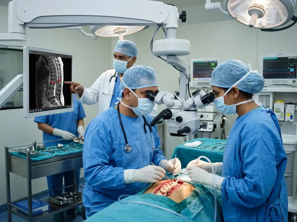
Idiopathic spinal cord herniation is a rare pathology presenting with progressive thoracic myelopathy. Early MRI identification and meticulous microsurgical reduction with dual bolstered dural defect repair prevent irreversible neurological deterioration.
Today

A novel smartphone-assisted biosensor utilizing molecularly imprinted polymers and catalytic nanozymes offers antibody-free, enzyme-free urinary microalbumin testing, enabling rapid, low-cost point-of-care screening for early chronic kidney disease.
Today
