.webp&w=384&q=75)

Loading, please wait...
Loading, please wait...

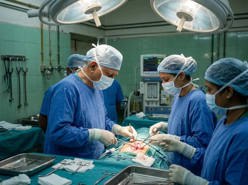
Spinal procedures present significant technical challenges because neural structures remain vulnerable to traction, compression, and vascular compromise during exposure and instrumentation. Consequently, intraoperative neuromonitoring spine surgery has evolved from an optional safeguard into an indispensable clinical modality for modern spine teams. Surgical teams frequently combine transcranial motor-evoked potentials (MEP) and somatosensory-evoked potentials (SSEP) to detect nascent neural distress before irreversible parenchymal damage occurs. While clinicians widely acknowledge the protective diagnostic utility of intraoperative monitoring, questions persist regarding its ability to predict postoperative functional recovery or persistent deficit. Furthermore, spine specialists require precise prognostic metrics to guide immediate operative choices. When electrophysiological waveforms deteriorate, surgical teams must rapidly decide whether to alter trajectory, remove implants, or elevate arterial pressure. A prospective study conducted at the All India Institute of Medical Sciences (AIIMS), Jodhpur, provides crucial real-world data regarding these electrophysiological changes. This research demonstrates how real-time electrophysiological fluctuations directly correlate with functional motor and sensory outcomes, empowering clinicians to interpret dynamic waveform modifications with heightened clinical confidence.
To clarify the prognostic precision of neurophysiological alerts, investigators designed a prospective observational study involving 68 consecutive surgical patients. The investigators included patients undergoing varied spinal procedures across diverse etiologies, such as degenerative deformities, trauma, and tumor resections. In each operative case, the neurophysiology team continuously recorded bilateral somatosensory-evoked potentials and intermittently triggered transcranial motor-evoked potentials. The multidisciplinary team categorized participants into distinct cohorts based on real-time signal dynamics throughout the intraoperative period. Specifically, the observers tracked signal stability, transient amplitude decrements, persistent wave loss, and spontaneous waveform recovery. Following surgical closure, independent clinicians evaluated all participants at two standardized intervals, namely 48 hours postoperatively and at six-month follow-up visits. Consequently, this longitudinal framework allowed investigators to distinguish temporary physiological conduction blocks from permanent structural cord injuries. Furthermore, this dual-timeline clinical assessment captured early postoperative recovery trajectories as well as durable long-term functional adaptations across diverse surgical scenarios.
The study data revealed marked differences in diagnostic sensitivity and clinical specificity between sensory and motor monitoring modalities. Somatosensory-evoked potentials monitor the dorsal columns and posterior cord perfusion, whereas motor-evoked potentials directly evaluate the anterior and lateral corticospinal tracts. Because anterior cord pathways are particularly susceptible to direct mechanical distortion and ischemic insult during instrumentation, MEP signals exhibited superior sensitivity. In the study cohort, nine patients demonstrated persistent intraoperative MEP reduction exceeding established warning thresholds. Among these nine individuals, eight patients developed overt postoperative motor deficits, yielding exceptional positive predictive value for persistent motor injury. In contrast, intraoperative SSEP amplitude decline occurred in only three patients throughout the surgical cohort, with merely one individual developing measurable sensory loss. Consequently, isolated sensory pathway monitoring missed critical ventral pathway stress that directly produced motor impairment. Therefore, the findings confirm that motor-evoked potentials provide superior diagnostic agility and predictive accuracy compared to somatosensory-evoked potentials alone.
A pivotal observation from the AIIMS Jodhpur investigation involves the distinct prognosis associated with transient signal alterations versus persistent losses. Notably, patients who experienced transient intraoperative MEP decline did not exhibit any persistent postoperative motor deficits upon physical examination. Moreover, patients exhibiting transient signal suppression ultimately achieved neurological improvements that matched patients whose signals remained stable or improved above baseline. This finding indicates that prompt intraoperative corrective action successfully reverses acute ischemic or mechanical conduction failure before permanent cellular necrosis ensues. In addition, 38 patients in the cohort demonstrated marked intraoperative signal improvements above their initial pre-incision baseline tracings. Importantly, these electrophysiological gains directly correlated with notable postoperative clinical motor and sensory improvements at six-month follow-up. Thus, intraoperative signal enhancement serves as a reliable dynamic surrogate marker for successful spinal cord decompression, giving surgeons immediate biological feedback regarding therapeutic success.
Reliable neurophysiological recordings demand meticulous collaboration between spine surgeons, neurophysiologists, and neuroanesthesiologists inside the operating suite. Inhalation anesthetics suppress cortical synaptic transmission and significantly attenuate transcranial motor-evoked potential amplitudes in a dose-dependent manner. Therefore, neuroanesthetic protocols typically require total intravenous anesthesia using balanced infusions of propofol and remifentanil without continuous neuromuscular blockade. When an unexpected signal decrement occurs, the multidisciplinary team must immediately execute a standardized troubleshooting protocol. First, the surgical team pauses instrumentation, relieves mechanical traction, and inspects newly placed hardware. Simultaneously, the anesthesia team optimizes physiological parameters by elevating mean arterial blood pressure, correcting hypothermia, and addressing arterial hypoxemia. Furthermore, the neurophysiologist verifies electrode integrity and excludes technical interference before surgical adjustments proceed. Because the study confirmed that reversible MEP changes prevent permanent neurological deficits, rapid collaborative intervention directly rescues vulnerable neural tissue.
The evidentiary insights from this trial offer substantial clinical guidance for neurosurgical and orthopedic spine units in India. Because complex degenerative deformities, high-velocity spine trauma, and spinal tumors present substantial complication risks, electrophysiological guidance provides vital surgical security. Clinicians should incorporate multimodal monitoring protocols incorporating both MEP and SSEP modalities into routine operative planning for high-risk procedures. Furthermore, surgical units must establish institutional checklists that define clear threshold criteria and immediate corrective maneuvers for waveform decay. When motor amplitudes drop by more than fifty percent, immediate operative release and blood pressure elevation must follow without delay. Conversely, when decompressive maneuvers yield spontaneous waveform amplitude increases, surgeons receive immediate confirmation of adequate neural decompression. Consequently, this objective feedback helps avoid unnecessary structural manipulations and shortens operative exploration times, promoting predictable long-term functional recovery.
Motor-evoked potentials directly assess the corticospinal tracts traversing the ventral spinal cord, which receive blood supply from the vulnerable anterior spinal artery. Conversely, somatosensory-evoked potentials monitor dorsal sensory columns. Because mechanical compression and operative ischemia typically impact anterior motor pathways first, motor monitoring detects acute neural compromise far more rapidly and reliably than sensory monitoring.
When motor signals deteriorate, the surgical team must immediately halt mechanical manipulation, remove traction, and verify implant placement. Simultaneously, the anesthesiologist elevates mean arterial pressure above 85 mmHg, ensures adequate oxygenation, and reverses inadvertent neuromuscular blockade. If technical artifacts are excluded, the surgeon considers removing recently placed hardware or reversing deformity correction to restore cord perfusion.
Spontaneous intraoperative improvement or amplitude recovery indicates successful relief of mechanical pressure and prompt restoration of microvascular spinal cord perfusion. Research confirms that patients displaying signal recovery or enhancement do not experience new postoperative deficits. Instead, these individuals frequently demonstrate significant motor and sensory functional gains above their pre-surgical baseline examinations at six months.
Disclaimer: This content is for informational and educational purposes only and does not constitute formal medical advice, diagnosis, or treatment. Healthcare professionals must evaluate individualized clinical scenarios independently. Refer to the latest local and national guidelines for clinical practice.
References

Read summarized clinical updates, watch expert medical content, and earn CME certifications right from your smartphone.
Open your smartphone camera to scan and install.





A prospective study evaluates the prognostic power of SSEP and MEP in spine surgery. Findings demonstrate that motor-evoked potentials offer superior sensitivity and specificity compared to somatosensory potentials, showing that transient alerts prevent deficits while permanent loss portends motor impairment.
Today

A Mayo Clinic study reveals that applying lower extremity elastic bandages in acute decompensated heart failure significantly improves diuretic efficiency by 340 mL per 40-mg furosemide equivalent. This mechanical therapy reduces required diuretic doses while safely maintaining necessary overall daily fluid clearance.
Today

A 34-year-old postpartum woman suffered a pheochromocytoma crisis resulting in cardiac arrest and respiratory failure. Intensivists successfully deployed veno-arteriovenous ECMO to stabilize refractory arrhythmias and differential hypoxemia, bridging her safely to complete tumor resection and full clinical recovery.
Today

A study shows that the aggregate index of systemic inflammation (AISI) significantly associates with elevated myocardial injury markers, including cardiac troponin T and CK-MB, in patients with coronary heart disease, highlighting its potential utility for cardiovascular risk stratification.
Today

A narrative review clarifies key nuances of the AO Spine Primary Infection Classification, including canal boundaries dorsal to the PLL, Type 1 versus Type 2 criteria, neural compression thresholds, and modifier application in primary spinal infections.
Today
Calcimimetic therapy for primary hyperparathyroidism can induce profound tumor fibrosis, necrosis, and reduced vascularity. These changes alter ultrasound and 4D-CT imaging and cause dense recurrent laryngeal nerve adhesions, requiring heightened surgical vigilance during definitive parathyroidectomy.
Today
