.webp&w=384&q=75)

Loading, please wait...
Loading, please wait...
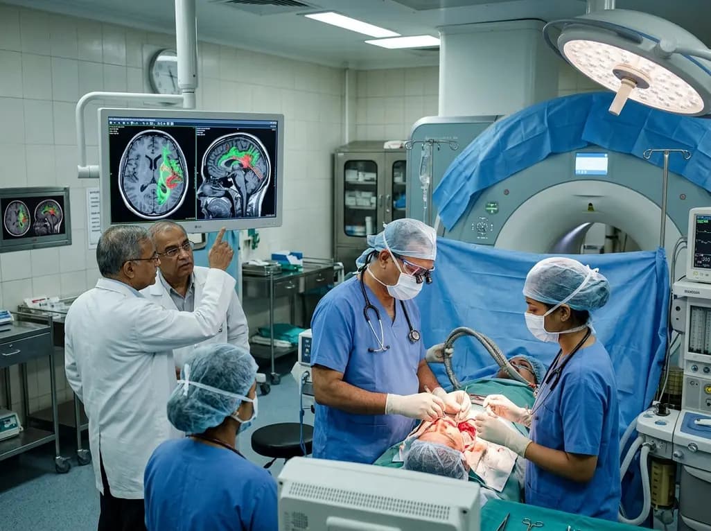
Surgical management of diffuse low-grade gliomas requires a delicate equilibrium between cytoreductive tumor clearance and functional neurological preservation. For decades, awake craniotomy utilizing direct electrical stimulation has set a remarkable benchmark. This approach consistently delivers overall survival exceeding twenty years alongside preserved neurocognition, low permanent neurological deficit rates below one percent, and return-to-work rates above ninety-three percent. However, the neurosurgical landscape has simultaneously seen widespread adoption of advanced technological adjuncts. Specifically, the integration of intraoperative MRI in LGG resections gained significant popularity worldwide over the past two decades.
Proponents argue that intraoperative imaging provides real-time anatomical feedback, effectively correcting for brain shift and maximizing the extent of resection. Nevertheless, critical questions remain regarding whether these radiological improvements truly translate into superior onco-functional longevity for patients. Because low-grade gliomas predominantly affect younger, working-age individuals, preserving neurocognitive networks is paramount. A comprehensive systematic review by Rosuel and Duffau critically re-evaluates the true clinical role of intraoperative MRI in LGG management. By scrutinizing published literature on long-term survival, functional deficits, and socioeconomic impact, this review challenges conventional assumptions surrounding technology-driven surgical strategies.
The systematic review analyzed nineteen dedicated studies encompassing 1,463 low-grade glioma patients undergoing resection with intraoperative MRI guidance. Across the analyzed cohorts, the median patient age was thirty-seven years, reflecting the characteristically young demographic afflicted by this disease. Notably, the mean surgical duration reached 6.2 hours, extending up to nine hours in several complex operative cases. This prolonged operating time underscores the logistical friction introduced by intraoperative scanning protocols, coil placement, and repeated imaging acquisitions.
Regarding cytoreduction, the reported mean extent of resection across the pooled series was 92.2 percent. While this volumetric figure appears robust on conventional post-resection imaging, the radiological gain must be weighed against biological and functional realities. High-field intraoperative scanners certainly assist neurosurgeons in identifying residual signal abnormalities before closing the craniotomy. However, non-enhancing low-grade glioma tissue diffusely infiltrates surrounding functional brain parenchyma well beyond visible radiological margins. Consequently, pursuing maximal radiological cytoreduction without direct functional guidance risks violating eloquent cortical and subcortical pathways. Therefore, relying primarily on image guidance rather than physiological testing creates an inherent trade-off between perceived volumetric clearance and functional neurological integrity.
Preserving functional independence and neurological integrity represents the primary determinant of post-surgical quality of life in low-grade glioma patients. Strikingly, the systematic review identified an average postoperative permanent neurological deficit rate of nine percent across eleven studies reporting functional morbidity. This complication rate is notably high when juxtaposed against the sub-one-percent deficit rates achieved with functional mapping and awake craniotomy. Thus, reliance on anatomical imaging alone may fail to protect delicate neural circuits during aggressive tumor debulking.
Furthermore, long-term functional recovery demonstrated substantial deficits across the reviewed literature. The mean return-to-work rate was only 64.8 percent, ranging widely between 45 and 84.6 percent. This reduced vocational reintegration highlights the profound downstream impact of subtle neurological impairments on daily life. In addition, formal neurocognitive assessments were exceptionally rare among the evaluated publications. Because standard neurological exams often miss subtle deficits in executive functioning, memory, and cognitive flexibility, the actual functional toll of surgery may remain vastly underreported. Consequently, measuring surgical success solely through extent of resection without rigorous neurocognitive follow-up provides an incomplete and potentially misleading clinical picture.
The ultimate benchmark of low-grade glioma management rests on extending progression-free and overall survival without accelerating malignant transformation. However, the systematic review uncovered substantial limitations in the existing evidence regarding long-term oncological outcomes. The mean duration of postoperative follow-up was only 3.8 years across nine reporting studies, which remains inadequate for evaluating an indolent disease with natural history spanning decades.
Moreover, only two studies provided progression-free survival data, documenting median intervals of 5.6 and 8.5 years. Similarly, overall survival was documented in just three publications, yielding a mean survival of 14.5 years. In comparison, contemporary awake functional mapping series routinely report overall survival figures surpassing twenty years. This marked discrepancy highlights that achieving high anatomical resection percentages through intraoperative imaging does not automatically translate into superior disease control. Indeed, overzealous resection guided strictly by anatomical boundaries cannot biologically eradicate diffuse glioma cells infiltrating critical functional tracts. Therefore, current surgical literature lacks robust evidence confirming that intraoperative MRI improves overall survival compared to standard functional neuro-oncological approaches.
Beyond clinical and onco-functional parameters, the practical deployment of intraoperative magnetic resonance suites imposes immense financial and logistical hurdles. The capital acquisition and infrastructure expenditure for a high-field intraoperative imaging suite averages an estimated additional cost of three million dollars. Furthermore, maintaining specialized non-ferromagnetic surgical instrumentation, specialized staff training, and ongoing service contracts adds substantial recurrent operational costs.
In addition to direct expenditures, the substantial increase in operative time impacts hospital throughput and increases anesthesia exposure. Prolonged general anesthesia carries independent risks for surgical complications, delayed extubation, and extended postoperative recovery. In healthcare landscapes across developing nations, including India, capital-intensive technologies present severe accessibility barriers. Allocating scarce resources toward multimillion-dollar imaging equipment that offers unproven survival superiority over lower-cost functional mapping raises major health economics questions. Clinicians and healthcare administrators must carefully evaluate whether such investments truly optimize patient-centered endpoints. Consequently, widespread routine utilization of intraoperative imaging suites remains difficult to justify when simpler, neurophysiological mapping techniques yield superior functional outcomes at a fraction of the cost.
The synthesized evidence necessitates a critical paradigm shift in contemporary neurosurgical oncology. For low-grade gliomas, maximizing resection cannot occur at the expense of permanent functional impairment. Instead of pursuing an image-guided resection strategy based purely on macroscopic boundaries, neurosurgeons should prioritize functional connectomics and individualized cortical-subcortical mapping.
Intraoperative functional mapping allows real-time assessment of language, motor, visuospatial, and executive functions during tumor removal. By identifying essential neural pathways, the surgical team can push the resection to individual functional boundaries rather than arbitrary radiological borders. While intraoperative imaging provides helpful anatomical orientation in select high-grade tumors or deeply situated lesions, its standalone utility in low-grade gliomas remains unproven. Ultimately, surgical teams should view neuroplasticity and functional preservation as the core pillars of long-term oncological success. Embracing an onco-functional philosophy supported by rigorous neuropsychological evaluations will ensure that patients achieve maximal longevity alongside optimal quality of life.
Systematic review data reveal an average permanent neurological deficit rate of 9% with intraoperative MRI guidance. In contrast, awake craniotomy with direct electrical stimulation maintains permanent deficits below 1%, demonstrating that image guidance alone does not adequately prevent functional neurological injury during aggressive tumor resection.
Current evidence does not demonstrate superior overall survival with intraoperative MRI in low-grade glioma surgery. Reporting series show a mean overall survival of 14.5 years with short follow-up, whereas functional mapping and awake resection cohorts consistently achieve median survival exceeding twenty years.
An intraoperative MRI suite requires approximately three million dollars in additional capital investment alongside expensive maintenance and non-ferromagnetic instruments. Furthermore, imaging acquisitions prolong operative duration by several hours, significantly increasing resource utilization, anesthesia risks, and healthcare expenditures without guaranteed functional benefits.
Disclaimer: This content is for informational and educational purposes only, and does not constitute medical advice, diagnosis, or treatment recommendations. Refer to the latest local and national guidelines for clinical practice.
References

Read summarized clinical updates, watch expert medical content, and earn CME certifications right from your smartphone.
Open your smartphone camera to scan and install.





A systematic review of 19 studies indicates that intraoperative MRI in low-grade glioma surgery increases surgical time and permanent neurological deficits (9%) without proven long-term survival benefits compared to awake functional mapping, while adding substantial economic and infrastructure burdens.
last month

Novel ex vivo evidence demonstrates that the next-generation triple combination vanzacaftor/tezacaftor/deutivacaftor partially rescues CFTR channel function in patients harboring the treatment-refractory L467F;F508del complex allele, overcoming non-responsiveness to elexacaftor/tezacaftor/ivacaftor.
Today

A pharmacometric and machine learning study reveals that specific vaginal bacteria alter cervical antiretroviral exposure, highlighting the need for precision HIV PrEP strategies.
Yesterday

Abdominal aortic aneurysm rupture remains a catastrophic vascular emergency. Groundbreaking research reveals that USP53 accelerates aneurysm progression by reprogramming smooth muscle cell metabolism toward aerobic glycolysis, highlighting a promising target beyond conventional diameter-based surveillance.
Today

A breakthrough study leverages natural language processing to accurately identify incident medication-related osteonecrosis of the jaw and track antiresorptive drug holidays from clinical narratives in electronic health records, advancing osteoporosis pharmacovigilance and dental safety.
Today

Recent research defines ferro-aging as a conserved iron-lipid axis driving organ decline across primates. Age-related iron dyshomeostasis stimulates ACSL4-mediated lipid peroxidation, promoting cellular senescence. Notably, vitamin C directly inhibits ACSL4, suppressing phospholipid oxidation and multi-organ decay.
Yesterday
