.webp&w=384&q=75)

Loading, please wait...
Loading, please wait...
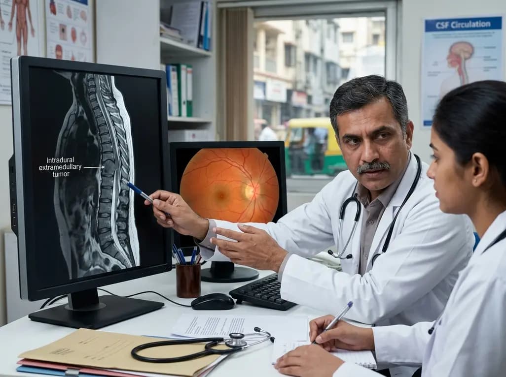
Physicians typically identify papilledema as a hallmark sign of intracranial pathology or idiopathic intracranial hypertension. However, recent clinical evidence demonstrates that intradural spinal tumors papilledema represents a rare but significantly critical association. The presence of bilateral optic disc swelling usually directs a clinician's attention toward brain lesions, venous sinus thrombosis, or hydrocephalus. In many instances, practitioners neglect the spinal column during the initial workup because the symptoms mimic intracranial hypertension. This narrow focus can lead to significant diagnostic delays and irreversible vision loss. A recent case series of three patients highlights the complexity of managing these vision-threatening complications. It also underscores the necessity of considering spinal pathology in patients with unexplained intracranial hypertension. When classic intracranial causes are absent on neuroimaging, the spine may hold the diagnostic answer. Consequently, maintaining a high index of suspicion for spinal lesions can transform the clinical trajectory. Early recognition is essential to prevent permanent damage to the optic nerve. Therefore, clinicians must look beyond the cranium when standard investigations for papilledema return negative results. This systematic approach ensures that rare but treatable causes of vision loss do not go unnoticed in the acute setting.
The link between spinal cord tumors and raised intracranial pressure is well-documented but remains poorly understood by many frontline physicians. Notably, these patients often present without ventriculomegaly or hydrocephalus, which further complicates the diagnostic process. Most clinicians associate raised pressure with a blockage in the ventricular system. However, spinal tumors can increase pressure through alternative pathways that do not immediately dilate the ventricles. Because the brain imaging appears normal, many patients receive a misdiagnosis of idiopathic intracranial hypertension. Furthermore, the typical demographic for spinal tumors may not match the young, female, and obese profile often seen in idiopathic cases. This discrepancy should serve as a major red flag for investigating secondary causes. Additionally, the rapid progression of visual symptoms in some cases requires immediate surgical intervention. If the clinician fails to identify the spinal source, they may perform unnecessary intracranial procedures. Such oversights highlight the importance of a comprehensive neurological and ophthalmological evaluation. By integrating spinal imaging into the workup of atypical intracranial hypertension, doctors can avoid the pitfalls of diagnostic tunnel vision. Ultimately, the goal is to stabilize the patient while identifying the root cause of the ophthalmic crisis.
The mechanism behind how spinal tumors cause intracranial hypertension involves complex cerebrospinal fluid (CSF) dynamics. Several theories exist to explain this phenomenon, with many focusing on the role of CSF protein. Specifically, intradural tumors often secrete high levels of protein or breakdown products into the subarachnoid space. This secretion significantly increases the viscosity of the cerebrospinal fluid. Consequently, the thickened fluid creates resistance at the arachnoid villi, which are the primary sites of CSF absorption into the venous system. When absorption slows down, the intracranial pressure rises, leading to optic disc swelling. Moreover, malignant cells from certain spinal tumors can directly infiltrate the subarachnoid pathways. This infiltration further impairs fluid circulation and drainage. Some researchers also suggest a \"water hammer effect,\" where the tumor reduces the compliance of the spinal subarachnoid space. This reduction in the spinal \"reservoir\" forces the intracranial compartment to absorb more pressure fluctuations. Additionally, subarachnoid hemorrhage from vascular spinal tumors can cause secondary arachnoiditis. These various mechanisms prove that the spine is not an isolated compartment. Instead, it is a continuous system where distal pathology can exert profound proximal effects on the brain and eyes.
One of the most deceptive features of these cases is the frequent absence of limb neurology. Usually, spinal tumors present with motor weakness, sensory changes, or bowel and bladder dysfunction. However, in the context of intradural spinal tumors papilledema, the patient may present with isolated visual symptoms. This lack of traditional spinal signs often lures physicians into focusing exclusively on the head. In the case series discussed, several patients had similar presentations where vision loss was the primary or only complaint. Without lower extremity symptoms, a physician might not think to order a spinal MRI. Consequently, the primary lesion remains hidden while the patient undergoes repeated lumbar punctures or brain scans. Therefore, it is imperative to \"think outside the box\" when evaluating disc swelling. If a patient presents with high opening pressures on a lumbar puncture but has a normal brain MRI, the spine should be the next area of focus. Furthermore, highly elevated CSF protein levels in the absence of infection should raise immediate suspicion for a spinal neoplasm. Recognizing these subtle clues can significantly shorten the time to diagnosis. This awareness is particularly vital in preventing the progression from papilledema to permanent optic atrophy.
In cases of vision-threatening papilledema, the preservation of sight must become the immediate priority. While surgical resection of the spinal tumor is the definitive treatment, it may not be enough to save vision if the pressure remains critically high. In some patients, the acuity of visual deterioration necessitates emergency CSF diversion. This can include the placement of an external ventricular drain or a lumboperitoneal shunt to bridge the gap until surgery. The case series highlights that different operative management strategies depend on the individual patient's risk profile. For example, some patients may require immediate tumor removal, while others need pressure management first. Additionally, the use of high-dose steroids can help reduce tumor-associated edema and potentially lower the protein load in the CSF. However, steroids are rarely a permanent solution. The ultimate goal is to remove the obstruction and restore normal CSF dynamics. Clinicians must closely monitor visual fields and visual acuity throughout the perioperative period. Success is not just measured by the successful removal of the tumor, but by the stabilization of the patient's vision. Early intervention remains the most reliable predictor of a positive ophthalmological outcome in these complex neuro-oncological cases.
For modern clinical practice, these findings suggest a shift in the diagnostic algorithm for unexplained intracranial hypertension. Physicians should consider spinal MRI as a standard part of the workup for papilledema when brain imaging is negative. This is especially true if the patient exhibits atypical features, such as advanced age or very high CSF protein. Furthermore, the presence of headache or back pain should further lower the threshold for spinal imaging. Notably, the delay in diagnosing these cases often spans months, during which the optic nerve undergoes progressive damage. Therefore, rapid communication between ophthalmologists, neurologists, and neurosurgeons is critical. Each specialist provides a piece of the puzzle, from identifying the disc swelling to locating the spinal lesion. Moreover, patient education regarding the importance of follow-up for visual changes is essential. Even after successful tumor resection, some patients may require ongoing monitoring for recurrence or persistent pressure issues. In conclusion, while intradural spinal tumors are a rare cause of papilledema, their impact on vision is devastating. By broadening the diagnostic horizon to include the entire neuroaxis, clinicians can ensure earlier detection. This proactive approach ultimately leads to better preservation of vision and improved overall quality of life for the patient.
Yes, intradural spinal tumors can occasionally present with isolated intracranial hypertension and papilledema. Many patients do not show motor or sensory deficits in their limbs, which often leads to diagnostic delays. Physicians must maintain a high index of suspicion and consider spinal imaging when intracranial causes for disc swelling are absent.
Spinal tumors can increase intracranial pressure by elevating CSF protein levels, which increases fluid viscosity. This thickened fluid then obstructs the arachnoid villi, impairing the body's ability to absorb cerebrospinal fluid. Additionally, mechanical blockages or tumor cell infiltration can disrupt normal CSF circulation throughout the entire subarachnoid space.
The primary treatment involves surgical resection of the spinal tumor to address the root cause. However, if vision is acutely threatened, emergency CSF diversion via shunting or drainage may be required first. Managing intracranial pressure immediately is vital to prevent permanent optic nerve damage and irreversible blindness in these patients.
Disclaimer: This content is for informational and educational purposes only and does not constitute medical advice, diagnosis, or treatment. Always seek the advice of your physician or other qualified health provider with any questions you may have regarding a medical condition. Refer to the latest local and national guidelines for clinical practice.
References
Sun R et al. Intra-cranial hypertension and vision-threatening papilloedema caused by intradural spinal tumours: a case series of three. Br J Neurosurg. 2025 Aug. doi: 10.1080/02688697.2023.2263087. PMID: 37807636.
Zisakis A, et al. Increased Intracranial Pressure Without Hydrocephalus Associated With Spinal Cord Tumor: Literature Review. J Neuroophthalmol. 2021 Mar;41(1):e44-e52. doi: 10.1097/WNO.0000000000001026.
Giannuzzi F, et al. Bilateral papilledema associated with retinal hemorrhagic appearance as earliest sign of spinal cord tumor. Eur J Ophthalmol. 2023 Sep;33(5):NP75-NP78. doi: 10.1177/11206721221132414.
"
Read summarized clinical updates, watch expert medical content, and earn CME certifications right from your smartphone.
Open your smartphone camera to scan and install.





Intradural spinal tumors rarely cause papilledema without hydrocephalus, posing a diagnostic challenge. This case series highlights the pathophysiology and the urgent need to prioritize vision preservation through spinal imaging and emergency CSF diversion when intracranial hypertension remains unexplained.
2 weeks back
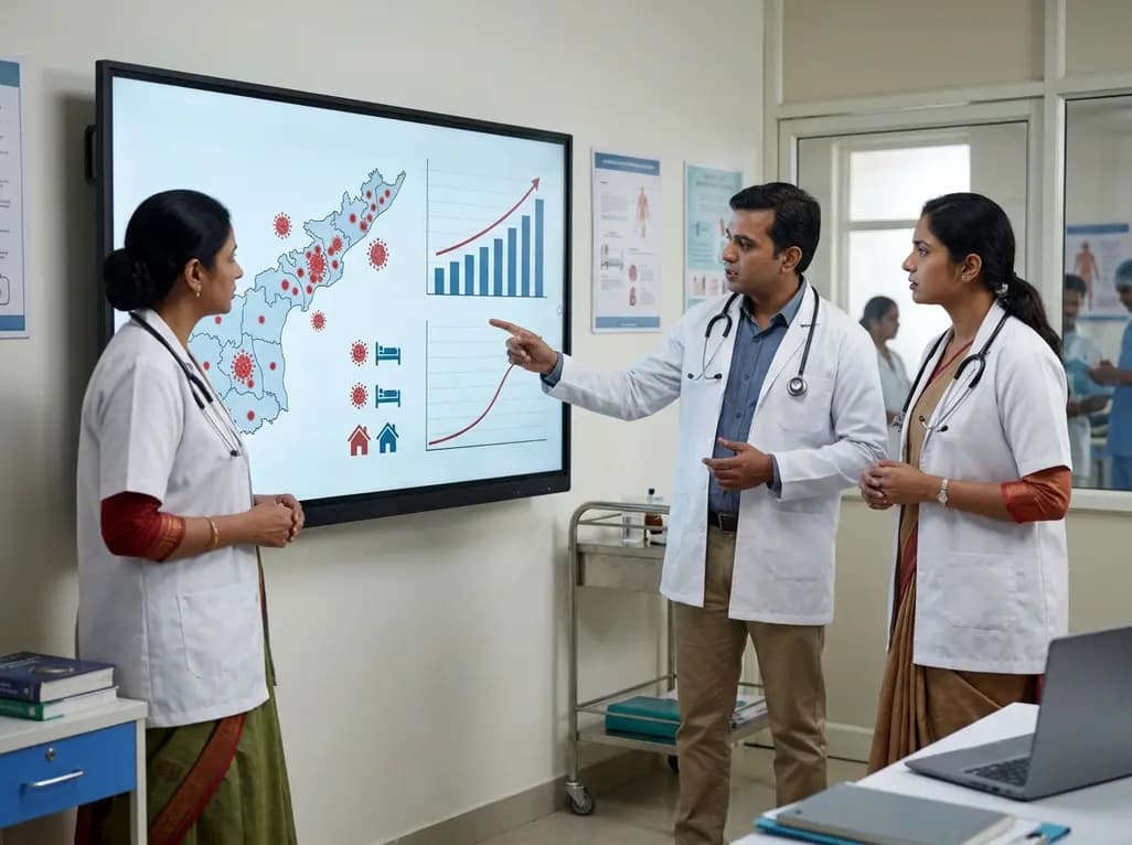
Andhra Pradesh reported 10 new Covid-19 cases, taking the state tally to 49 while deaths remain at four. With 24 patients hospitalized and 16 under home isolation, the Health Department has intensified monitoring. Medical professionals should review regional distribution, diagnostic protocols, and management plans.
Today

An 11-year Swedish registry study of 618 uterine sarcoma patients found that minimally invasive surgery yielded survival comparable to open surgery in early stages. However, adjuvant chemotherapy conferred no survival benefit in localized or advanced disease, highlighting stage and histology as key outcomes.
3 days back

A cross-sectional study evaluates post-intensive care syndrome in cardiac patients 2-4 weeks post-ICU discharge, highlighting cognitive, psychological, and functional impairments and the need for structured multidisciplinary rehabilitation.
3 days back

Anterior cruciate ligament reconstruction failure lacks uniform definition. A narrative review proposes an integrative framework incorporating objective and subjective instability, persistent pain, restricted motion, graft rupture, and secondary meniscal injury to standardize clinical reporting.
3 days back

With World Obesity Atlas data warning that over 41 million Indian children are overweight or obese, ICMR and NIN have unveiled a 10-point policy roadmap. The initiative calls for mandatory front-of-pack labeling, HFSS taxes, strict marketing bans, and healthier school environments to curb non-communicable diseases.
Today
