.webp&w=384&q=75)

Loading, please wait...
Loading, please wait...
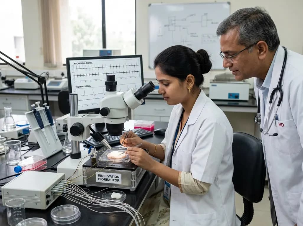
The study of somatosensory processing has achieved a major milestone with the development of a fully functional innervated human skin equivalent. For decades, researchers struggled to measure sensory encoding dynamics in artificial tissues using direct electrical readouts. This groundbreaking bioengineering breakthrough successfully bridges native tissue complexity with advanced electrophysiological recording systems. Consequently, clinicians and neuroscientists gain deeper insights into peripheral neural pathways, tactile perception, and cutaneous pain signaling mechanisms.
Traditional in vitro models of human integument have historically faced significant biological and technical bottlenecks. Most conventional engineered platforms lack functional neural integration, thereby preventing the study of authentic sensory transduction. When researchers previously added neuronal elements to skin cultures, they generally relied on indirect optical indicators or chemical stimulation. However, optical voltage dyes and calcium imaging cannot capture rapid millisecond-scale electrical spikes. Furthermore, chemical modulators fail to replicate physiological touch or temperature shifts. As a result, investigators could not observe real-time somatosensory encoding dynamics as they occur in living organisms.
To resolve these longstanding barriers, investigators combined regenerative tissue engineering with high-density microelectrode arrays. This bioengineered construct incorporates full-thickness dermal and epidermal compartments alongside functional neural circuits. Moreover, the tissue provides a robust extracellular matrix that nurtures cell differentiation and survival. Therefore, this platform effectively replaces animal testing while establishing a physiological human baseline for neurodermatology research.
The architecture of this innervated human skin equivalent closely mirrors native human tissue hierarchy. Bioengineers first created a living dermis populated with human fibroblasts to secrete endogenous extracellular matrix proteins. Subsequently, they cultivated stratified keratinocytes atop the dermal layer, guiding them to differentiate into basal, spinous, granulosum, and cornified strata. This stratified organization ensures authentic barrier properties and normal cellular signaling cascades.
Crucially, researchers introduced human sensory neurons and supporting Schwann cells directly into the construct. Axonal projections gradually elongated across the dermal matrix and penetrated the basal membrane of the epidermis. Consequently, these sensory fibers organized into specialized free nerve ending-like structures throughout the epithelial tissue. In addition, Schwann cells wrapped around the axons, facilitating proper nerve guidance and structural stabilization. By interfacing this complex 3D construct directly with high-density microelectrode arrays, researchers secured continuous electrical access to individual sensory terminals without disrupting tissue viability.
The hallmark feature of this engineered system is its ability to generate stimulus-specific action potentials. When researchers applied calibrated mechanical indentation to the tissue surface, the sensory endings instantly discharged rhythmic electrical signals. Furthermore, increased mechanical force produced higher spike frequencies, mirroring natural physiological adaptation. This precise frequency modulation demonstrates that engineered sensory afferents actively encode physical pressure.
Similarly, localized thermal stimulation evoked clear electrophysiological discharges across the microelectrode arrays. Applying distinct temperature shifts elicited characteristic burst firing patterns from the terminal arborizations. Importantly, detailed analysis of the recorded waveform morphology revealed distinct electrical signatures that differentiate mechanical pressure from thermal excitation. Temporal dynamics, including inter-spike intervals and peak amplitudes, varied predictably based on stimulus modality. Thus, the tissue demonstrates genuine sensory discrimination in vitro, proving that bioengineered free nerve endings can replicate complex peripheral receptor functions.
This biomimetic platform provides unprecedented utility for investigating peripheral neuropathy and neurodermatological disorders. Chronic pruritus, diabetic peripheral neuropathy, and post-herpetic neuralgia involve profound dysregulation of cutaneous sensory nerve fibers. However, clinicians often lack human-specific translational models to study how damaged nerve terminals initiate aberrant spontaneous firing. Because this tissue incorporates human cells, it avoids the interspecies variations commonly observed in rodent assays.
Additionally, pharmacologists can utilize this platform for high-throughput screening of novel analgesics and antipruritic agents. Researchers can test ion channel blockers, topical formulations, and neuroregenerative compounds under tightly controlled physical conditions. Furthermore, monitoring real-time electrical activity reveals immediate therapeutic responses and potential neurotoxicity. Consequently, the model will accelerate drug discovery pipelines for chronic cutaneous pain syndromes while reducing reliance on animal experimentation.
Beyond clinical therapeutics, this innovation holds transformative potential for neuroprosthetics and bioinspired robotics. Modern prosthetic limbs often lack bidirectional feedback, which leaves amputees without natural tactile sensation. By establishing an electrophysiological interface with living human tissue equivalents, bioengineers can design sophisticated tactile sensors that emulate biological sensory encoding.
Furthermore, biohybrid interfaces could one day bridge engineered cutaneous tissues directly with residual peripheral nerves in amputee patients. Such integration would restore authentic thermal and tactile sensations, significantly improving fine motor control and user embodiment. Moreover, robotics engineers can harness the underlying electrical encoding rules to develop neuromorphic electronic skins for intelligent automation. Thus, this breakthrough establishes a vital foundation for next-generation biomimetic sensory restoration systems.
Looking forward, researchers aim to expand the cellular diversity within the innervated skin equivalent. Integrating specialized mechanoreceptors, such as Meissner corpuscles and Merkel cell complexes, will enable even richer sensory discrimination. Additionally, incorporating vascular networks and immune cells will create fully immunocompetent cutaneous models. Such advanced systems will allow scientists to investigate neuroimmune interactions, wound repair mechanisms, and inflammatory hyperalgesia under realistic conditions. Therefore, continued convergence of biomaterials, neuroengineering, and microelectronics promises to revolutionize both experimental medicine and clinical care.
The construct integrates functional sensory neurons and Schwann cells within a stratified dermal-epidermal matrix. These neurons extend axonal projections that terminate as free nerve ending-like structures in the epidermis. When researchers apply mechanical pressure or localized temperature changes, specialized ion channels activate, generating action potentials. High-density microelectrode arrays record these electrical signals in real time, revealing distinct waveform morphologies and firing rates for each stimulus type.
Traditional in vitro skin models generally rely on indirect chemical assays or optical imaging techniques, such as calcium fluorescent dyes. While useful, these methods lack the millisecond temporal resolution necessary to capture rapid neuronal firing dynamics. In contrast, this bioengineered platform directly records electrophysiological activity using high-density microelectrode arrays. Furthermore, it incorporates human-derived cells, avoiding the physiological discrepancies and species differences inherent in animal models.
This platform offers immense potential for modeling peripheral neuropathies, chronic itch, and burn wound healing. Clinicians and pharmacologists can evaluate novel analgesics, topical medications, and neuroprotective agents by directly measuring changes in sensory firing patterns. Additionally, the technology informs the design of biohybrid neural interfaces, advanced prosthetics with sensory feedback, and neuromorphic tactile sensors for robotic and regenerative applications.
Disclaimer: This content is for informational and educational purposes only. It is not intended to provide medical advice or establish a standard of care. Clinical decisions should always be based on the individual clinical presentation, independent medical judgment, and established treatment guidelines. Refer to the latest local and national guidelines for clinical practice.
References

Read summarized clinical updates, watch expert medical content, and earn CME certifications right from your smartphone.
Open your smartphone camera to scan and install.





Researchers have engineered an innervated full-thickness human skin equivalent that electrically encodes mechanical and thermal stimuli, opening new avenues for sensory restoration and neurobiology.
last month

A secondary analysis of an HIV cure-related trial in South Africa highlights the acceptability and safety of serial excisional lymph node and gut biopsies among young women. Clear communication, empathetic care, and altruistic motives helped mitigate initial apprehension and sustain longitudinal trial engagement.
Today

A nationwide study covering 3.7 million Swedish births reveals that prenatal exposure to TORCH pathogens significantly elevates long-term risks of intellectual disability and autism. Healthcare providers must recognize clinical indicators, promote prenatal prevention, and track early pediatric neurodevelopment.
Today
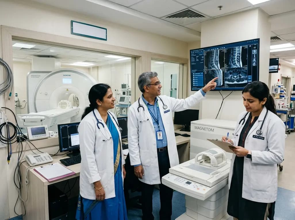
Under India's PLI initiative, domestic production of 57 critical medical devices has commenced, including MRI and CT scanners, cath labs, and heart valves. Global partnerships and local investments are expanding high-end technology, curbing import reliance, and strengthening clinical accessibility nationwide.
Today

A longitudinal study reveals that patients undergoing staged bilateral hip arthroscopy experience progressive contralateral labral deterioration during surgical intervals. However, structural degradation does not compromise 2-year postoperative clinical outcomes or patient-reported satisfaction scores.
Today
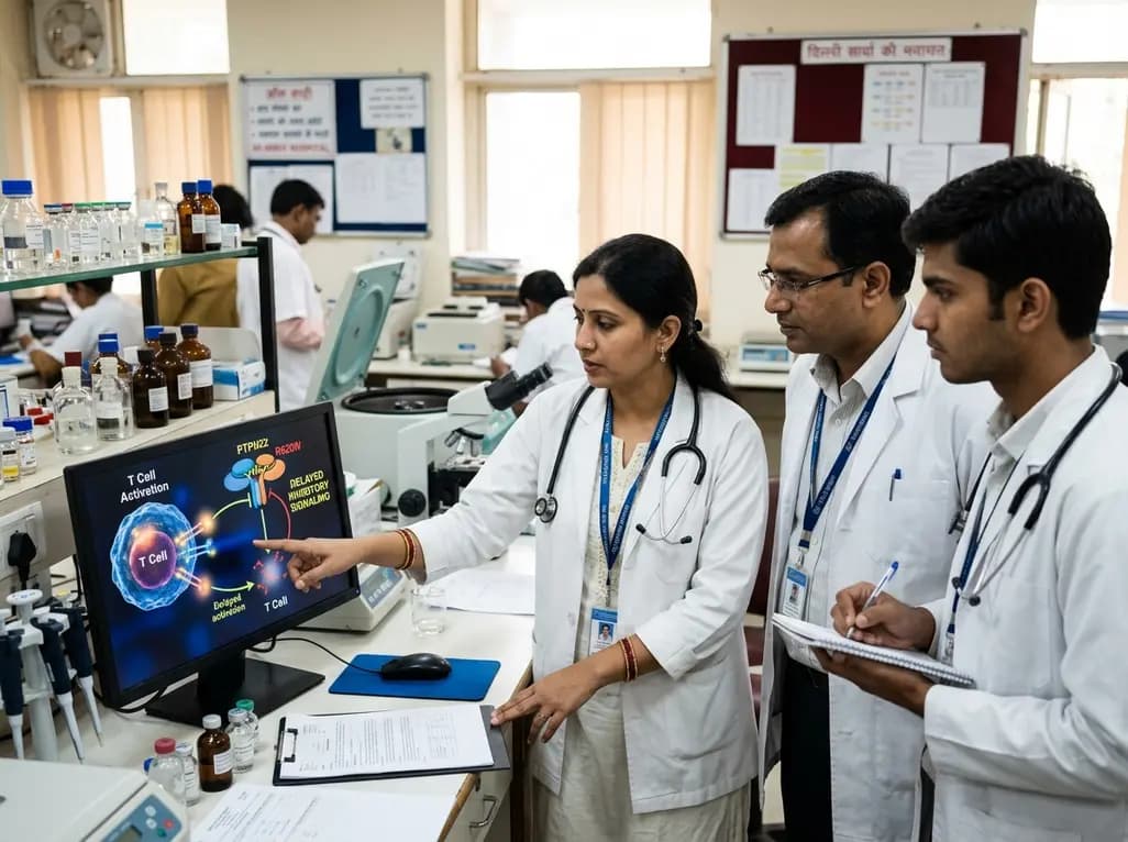
New immunological research reveals how PTPN22 forms a delayed inhibitory signaling complex to restrain T cell activation, explaining how the autoimmune-linked R620W variant disrupts feedback control and promotes persistent inflammation in conditions like rheumatoid arthritis and type 1 diabetes.
Today
