.webp&w=384&q=75)

Loading, please wait...
Loading, please wait...

Secondary lymphedema presents a challenging progressive condition that impairs quality of life. In modern reconstructive surgery, lymphaticovenular anastomosis offers an effective physiological solution for fluid drainage. Surgeons connect subdermal lymphatic channels directly into adjacent venules. Consequently, this supermicrosurgical procedure bypasses obstructed lymphatic pathways and alleviates progressive limb swelling. However, surgical success hinges on precise localization of functional lymphatic channels and viable recipient venules. Historically, microsurgeons relied heavily on exploratory incisions without direct visualization. Therefore, early interventions carried inconsistent success rates. Over the past decade, technological innovations have revolutionized preoperative mapping strategies. Today, clinicians utilize near-infrared optical fluorescence and high-frequency sonography to visualize subtle microvascular structures. Accurate lymphatic mapping directly influences incision placement, procedural time, and overall clinical outcomes. In countries like India, where cancer-related and post-infectious lymphedema impose substantial disease burdens, optimizing these microsurgical techniques remains crucial. Clinicians increasingly look for robust imaging algorithms to prevent premature surgical cancellations. Hence, understanding the comparative strengths of each imaging modality has become an essential priority for reconstructive surgeons worldwide. Specifically, combining dynamic functional assessments with anatomical tracing allows teams to design personalized operative roadmaps for every patient.
Contrast-enhanced ultrasound has recently emerged as an innovative modality for evaluating extremity lymphedema. Clinicians inject sulfur hexafluoride or perflubutane microbubbles intradermally to opacify lymphatic collectors. Subsequently, sonographers trace individual vessels in real time with high spatial clarity. Furthermore, ultrasound simultaneously displays the exact depth, caliber, and orientation of lymphatic channels beneath the epidermis. In addition, ultrasound evaluates neighboring venules, helping surgeons assess potential recipient matches before making skin cuts. Despite these distinct advantages, the technique presents notable practical limitations. For example, microbubble dispersion can remain unpredictable across fibrotic or chronically congested tissue layers. Consequently, deeper collectors or very narrow channels may fail to show sufficient contrast enhancement. In addition, operator experience substantially influences image acquisition and interpretation during examination. When ultrasound fails to delineate adequate vessels, surgeons often face difficult clinical choices. Specifically, some teams prematurely abandon or defer planned reconstructions due to perceived vessel absence. Thus, relying solely on negative ultrasound findings can inadvertently deny patients beneficial restorative interventions. Therefore, surgeons require an alternative diagnostic framework to corroborate negative sonographic results before concluding that viable vessels do not exist. Moreover, integrating secondary verification tools prevents unnecessary delays in vital surgical management.
Indocyanine green lymphography remains the gold standard imaging modality for evaluating superficial lymphatic architecture. During this procedure, clinicians inject small amounts of dye into the interdigital web spaces. Near-infrared camera systems then detect fluorescent emission signals as lymphatic channels transport the tracer proximally. As a result, the technique provides a panoramic, real-time map of regional superficial lymphatic drainage. Furthermore, indocyanine green lymphography accurately delineates characteristic pathological patterns, including linear channels, splash patterns, stardust distributions, and diffuse dermal backflow. These dynamic visual signatures highlight localized lymphatic hypertension and progressive valvular incompetence. In contrast to ultrasound, indocyanine green covers wide anatomical territories in a single comprehensive view. Therefore, surgeons can easily identify transition zones where residual linear channels meet dermal backflow. Even when ultrasound misses narrow or compressed channels, optical lymphography reveals subtle proximal tracer migration. Moreover, intraoperative fluorescence cameras allow continuous reassessment during dissection. Consequently, the technique serves as an invaluable functional safeguard. Clinicians can confidently direct targeted exploratory incisions toward areas showing faint functional flow, preserving surgical opportunities even after equivocal sonographic exams. Thus, optical fluorescence guidance reassures clinicians when deeper imaging modalities encounter technical difficulties.
Recent clinical evidence vividly illustrates the value of multimodal lymphatic imaging during complex reconstructive decision-making. In a highlighted case report, intraoperative contrast-enhanced ultrasonography failed to detect viable lymphatic collectors of adequate caliber. Under standard single-modality protocols, such negative sonographic findings often lead surgeons to cancel reconstructive procedures. However, the surgical team refused to abandon the intervention prematurely. Instead, they carefully revisited preoperative indocyanine green lymphography maps. The fluorescence images revealed localized dermal backflow alongside faint linear channels in the affected extremity. Consequently, the surgeons placed targeted empirical incisions directly over the fluorescent markers. During surgical exploration, the team identified functional lymphatic vessels alongside suitable recipient venules measuring between 0.3 and 0.4 millimeters. The microsurgeons then performed high-quality end-to-end anastomoses under supermicrosurgical visualization. Postoperatively, the patient experienced substantial clinical improvement. Specifically, the maximum interlimb circumference difference dropped from 2.5 centimeters to 1.7 centimeters, delivering meaningful symptomatic relief. This successful outcome demonstrates that sonographic failure does not confirm lymphatic absence. Furthermore, it highlights how individualized fluorescence data can salvage reconstructive procedures and restore physiological fluid transport. Therefore, practical empirical exploration guided by optical mapping remains an indispensable strategy in microsurgical care.
This paradigm shift underscores why reconstructive teams must integrate multiple imaging technologies into standard protocols. Relying on a single diagnostic modality creates dangerous blind spots during operative preparation. Ultrasound offers exceptional depth resolution and cross-sectional measurements, yet it can overlook subtle microvascular channels. Conversely, indocyanine green lymphography delivers unparalleled panoramic mapping of superficial functional flow, though it lacks deep penetration. Therefore, combining both diagnostic modalities creates complementary synergy. When clinicians encounter negative sonography, they should review dynamic fluorescence findings before canceling surgery. In addition, surgeons must recognize that dermal backflow zones frequently harbor usable lymphatic collectors along their borders. Targeted exploration guided by fluorescence can uncover vessels suitable for supermicrosurgery. Furthermore, adopting this multimodal diagnostic workflow holds significant relevance for high-volume surgical centers across India. In these settings, patients often present with late-stage secondary lymphedema after oncological resections or parasitic infections. By preventing inappropriate case cancellations, microsurgical teams maximize clinical throughput and optimize limb salvage. Ultimately, thoughtful integration of multimodal imaging empowers surgeons to convert initial diagnostic failures into successful vascular flow. As a result, patients achieve lasting volumetric reduction and improved long-term health.
Contrast-enhanced ultrasound can fail because microbubble diffusion depends on tissue interstitial pressure and vessel patency. Severe fibrosis, chronic adipose deposition, or collapsed lumina in advanced lymphedema often impede microbubble uptake. Furthermore, ultrasound offers a narrow focal field, making small or tortuous subdermal channels difficult to locate. Consequently, false-negative evaluations occur when patent vessels exist beyond the acoustic focus or contain minimal microbubble concentrations during examination.
Indocyanine green lymphography provides wide-field functional visualization of dermal lymphatics. Even when collectors fail to appear on ultrasound, fluorescence imaging reveals subtle linear pathways and dermal backflow zones. Experienced surgeons place targeted incisions along the perimeter of these backflow regions. Consequently, surgical exploration frequently uncovers patent subdermal collectors measuring 0.3 to 0.4 millimeters. Therefore, fluorescence mapping transforms equivocal diagnostic findings into actionable incision sites for successful anastomosis.
Following successful lymphaticovenular anastomosis, patients typically experience progressive reductions in limb circumference and tissue tension. Diverting lymph fluid into the venous system reduces interstitial fluid accumulation and dermal congestion. Furthermore, patients frequently report subjective lightness, enhanced mobility, and substantial reductions in recurrent episodes of cellulitis. Ultimately, restoring physiological lymphatic drainage preserves extremity function and significantly enhances overall health-related quality of life over prolonged recovery periods.
Disclaimer: This content is for informational and educational purposes only. It is not intended as medical advice, diagnosis, or treatment. Refer to the latest local and national guidelines for clinical practice.
References

Read summarized clinical updates, watch expert medical content, and earn CME certifications right from your smartphone.
Open your smartphone camera to scan and install.





Negative contrast-enhanced ultrasound does not rule out viable channels for lymphaticovenular anastomosis. Using indocyanine green lymphography, surgeons can identify functional lymphatic vessels and venules, converting failed imaging into successful surgical flow and limb circumference reduction.
Today
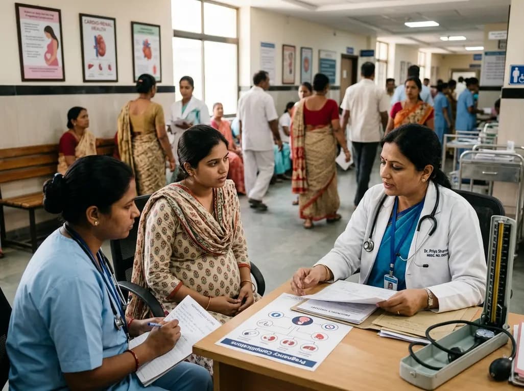
A large UK cohort study of 1.4 million women shows that pregnancy complications, including gestational diabetes, hypertension, and preterm birth, substantially raise long-term risks for hypertension, chronic kidney disease, type 2 diabetes, and cardiovascular events, highlighting early postpartum intervention needs.
Today
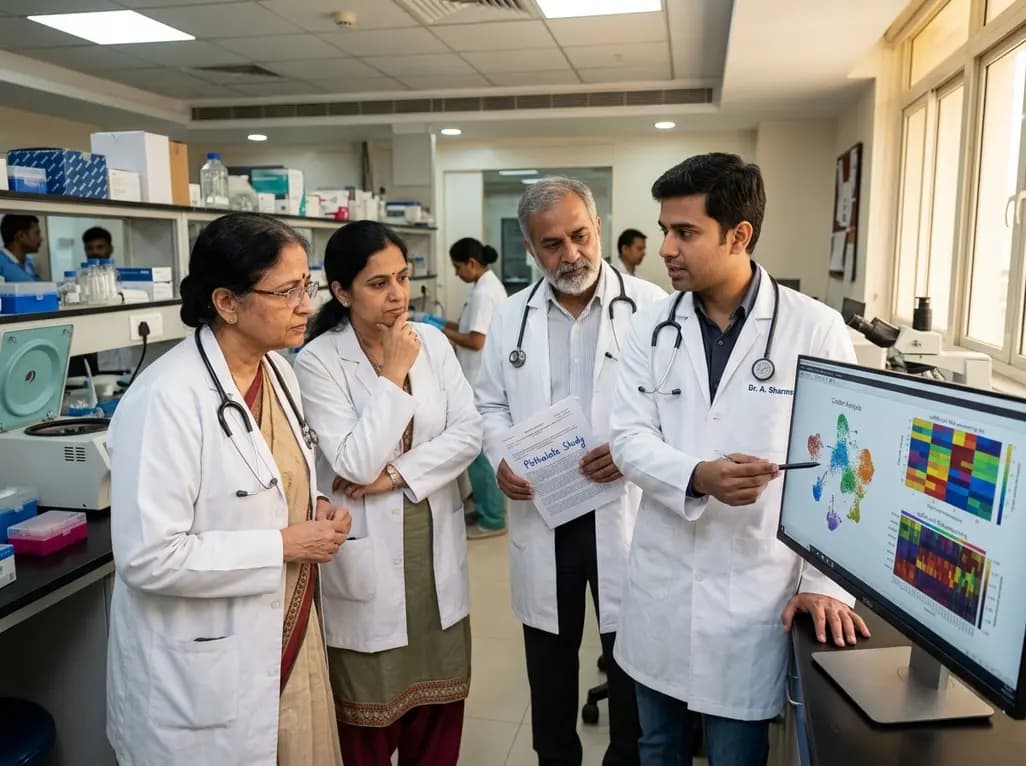
A landmark single-cell study reveals that MEHP, a major phthalate metabolite, damages human ovarian tissue beyond follicles. By disrupting glial-stromal communication, cellular adhesion, and mitochondrial respiration, environmental plasticizers impair ovarian integrity at realistic exposure levels.
Today
Giant cardiac hemangiomas are rare benign vascular tumors that can cause severe hemodynamic compromise and arrhythmias through mass effect. This comprehensive review covers clinical presentation, advanced multimodality imaging, histopathological diagnosis, surgical resection, and long-term surveillance protocols.
Today
This clinical overview examines recent advancements in lumbar disc herniation surgery, comparing microscopic and full-endoscopic discectomy while highlighting biportal techniques and emerging motion-preserving innovations for spine specialists.
Today

The World Health Organization has issued its first global guidelines for managing child and adolescent obesity. The recommendations strongly endorse family-based dietary interventions, regular physical activity, and behavioral support as frontline therapy, reserving GLP-1 therapies and surgery for select cases.
Today
