.webp&w=384&q=75)

Loading, please wait...
Loading, please wait...
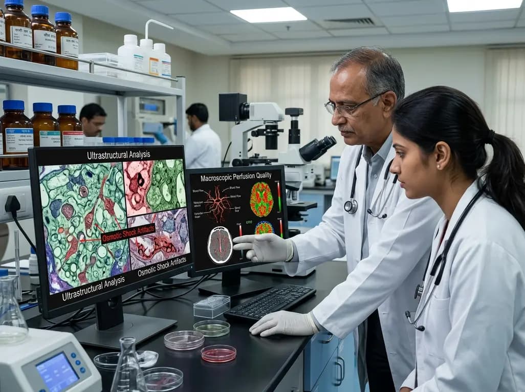
Perfusion fixation remains the gold standard method for preparing mammalian brain tissue for advanced microstructural examination. Recently, neuroscientists have investigated hyperosmotic brain perfusion protocols to facilitate large-scale connectomic reconstruction in whole brains. In typical laboratory and autopsy environments, researchers evaluate fixation success by assessing macroscopic indicators such as tissue firmness, vascular clearance, and cross-sectional neuroimaging. Consequently, clinicians and researchers frequently assume that uniform perfusate distribution guarantees pristine cellular architecture throughout the cerebrum.
However, recent experimental findings challenge this longstanding assumption. When investigators introduce dehydrating agents into the perfusate, macroscopic vascular distribution completely decouples from cellular preservation. In particular, adding hyperosmotic compounds allows fluid to circulate efficiently through the cerebral vasculature while simultaneously inflicting catastrophic damage on delicate subcellular components. Therefore, gross appearance and radiological imaging cannot independently confirm the fidelity of preserved neural circuitry. Pathologists and neuroscientists must recognize this critical gap between macroscale perfusate delivery and nanoscale microstructural integrity. Understanding the physical mechanisms behind this discrepancy allows laboratories to refine tissue preservation strategies without introducing irreversible artifactual changes.
Connectomics requires extraordinary anatomical precision because researchers must trace microscopic synaptic connections across vast brain volumes. To facilitate comprehensive perfusion in large post-mortem specimens, investigators have explored various chemical additives. Specifically, researchers added hyperosmotic dehydrating agents like mannitol and high molecular weight polyethylene glycol to primary fixative solutions. These agents reduce cellular swelling, improve capillary transit, and enhance perfusate distribution across the blood-brain barrier. Consequently, gross examination and post-perfusion computed tomography frequently show uniform intravascular distribution and excellent cerebral penetration.
Nevertheless, these macroscopic successes disguise severe underlying damage. In a pivotal technical report, researchers examined three human brain donors and two canine brain donors perfused with hyperosmotic fixative regimens. One cohort received fixative solutions containing ten percent mannitol combined with ten percent polyethylene glycol. Another animal specimen received twenty percent mannitol in neutral buffered formalin without polyethylene glycol. In all donor brains, radiological metrics suggested optimal fluid exchange and adequate macroscopic perfusion. Yet subsequent nanoscale evaluations contradicted these reassuring initial observations. Thus, relying solely on macroscopic indicators creates a dangerous illusion of quality that compromises high-resolution scientific investigations.
Electron microscopy provides an indispensable perspective that unmasks the hidden damage of hyperosmotic perfusates. When investigators analyzed donor tissue under high-magnification electron microscopy, they observed dramatic architectural disruptions throughout the neuropil. Specifically, the hyperosmotic environment triggered severe osmotic dehydration, drawing free water rapidly out of intracellular compartments. As a direct consequence, cellular processes shriveled extensively, leaving unnaturally enlarged extracellular spaces. Furthermore, delicate plasma membranes underwent pronounced folding, detachment, and distortion across neuronal and glial arborizations.
In contrast, tissue specimens preserved with standard neutral buffered formalin without dehydrating additives maintained compact intercellular relationships. Normal neural parenchyma features tightly packed cellular processes with minimal interstitial gaps. However, the brains exposed to hyperosmotic formulations displayed widespread structural collapse that distorted synaptology and dendritic geometries. Importantly, similar ultrastructural artifacts emerged in both human and canine models, demonstrating that osmotic shock represents a universal biophysical vulnerability rather than a species-specific quirk. Therefore, researchers who mistake these empty interstitial spaces for pristine preservation risk misinterpreting baseline neuroanatomy. Neuropathologists must recognize that rapid hyperosmotic dehydration fundamentally alters fine cellular relationships even when tissue appears immaculate on gross dissection.
Why does macroscopic perfusion succeed while ultrastructural preservation fails? The answer lies in the biophysical mechanics governing fluid distribution versus cellular equilibrium. Hyperosmotic additives significantly reduce blood viscosity and draw water out of swollen perivascular astrocytic endfeet. Consequently, these agents reopen compressed capillary networks and restore microvascular patency throughout ischemic brain parenchyma. This phenomenon enables radiographic contrast agents and fixative solutions to flow smoothly across large vascular territories. Hence, macroscopic visual appearance and computed tomography scans show robust, uniform perfusate distribution across both hemispheres.
However, this same osmotic gradient exerts destructive forces across neuronal and glial membranes. Because formaldehyde cross-links proteins relatively slowly, fixatives cannot immediately stabilize delicate membrane lipid bilayers against intense osmotic shifts. Meanwhile, mannitol cannot readily cross intact cellular membranes, creating a steep trans-membrane chemical gradient. As a result, water rushes down its osmotic gradient from the intracellular space into the vascular compartment. Cellular compartments collapse rapidly before chemical fixation can establish structural cross-links. Ultimately, the very mechanism that facilitates whole-organ perfusate transit simultaneously drives microstructural destruction. Scientists must therefore evaluate perfusion protocols at multiple biological scales simultaneously rather than extrapolating microscale integrity from macroscale flow.
These crucial findings carry profound implications for contemporary neuropathology, human brain biorepositories, and biobanking networks worldwide. Post-mortem brain banking provides essential human neural tissue for dissecting neurodegenerative diseases such as Alzheimer's disease, amyotrophic lateral sclerosis, and Parkinson's disease. Historically, repository protocols evaluated tissue procurement quality primarily through tissue stiffness, color change, and post-mortem interval data. Furthermore, emerging connectomic initiatives increasingly rely on aggressive chemical formulations to achieve complete hemispheric perfusion. However, this study demonstrates that standard surrogate metrics cannot guarantee high-resolution research validity.
Accordingly, brain banking facilities must incorporate rigorous ultrastructural quality controls into their routine validation pipelines. Pathologists should not assume that a beautifully perfused, radiologically validated donor brain preserves intact synapses and cellular organelles. Instead, laboratories must implement systematic transmission electron microscopy sampling from multiple cortical and subcortical brain regions. In addition, protocol designers must balance vascular clearance interventions against osmolar toxicity. Adjusting oncotic pressure with physiological colloids or buffering osmolarity could mitigate osmotic shock while maintaining adequate microvascular patency. Ultimately, rigorous multi-scale verification will safeguard brain banking repositories against misleading structural artifacts, ensuring that downstream translational neuroscience rests upon authentic biological morphology.
Under electron microscopy, osmotic shock manifests primarily as severely shrunken cellular processes and markedly expanded extracellular spaces. In addition, plasma membranes exhibit irregular folding, invagination, and detachment from neighboring structures. Intracellular organelles often appear abnormally dense and compacted due to rapid fluid extraction. These distinct morphological distortions disrupt natural synaptic junctions and dendritic geometries, creating artificial empty gaps that pathologists must not confuse with physiological extracellular architecture or post-mortem autolysis.
Macroscopic indicators evaluate bulk fluid dynamics and gross vascular clearance across major cerebral blood vessels. Hyperosmotic agents effectively reduce blood viscosity and shrink perivascular edema, allowing perfusates and contrast agents to circulate smoothly throughout the organ. Consequently, neuroimaging and gross visual inspection suggest excellent perfusion quality. However, these gross observations cannot detect nanometer-scale cellular osmotic stress. While fluid flows unimpeded through the vasculature, severe chemical dehydration simultaneously damages cellular membranes and delicate synaptic connections.
Researchers must balance vascular wash-out efficacy with strict osmolar physiological boundaries. Instead of relying solely on hyperosmotic crystalloid agents like high-concentration mannitol, protocols should incorporate isosmolar solutions or carefully titrated oncotically balanced colloids. Furthermore, investigators should control perfusion pressure and optimize fixative delivery temperatures to prevent endothelial injury. Most importantly, quality assurance protocols must include direct transmission electron microscopy verification, ensuring that synaptic membranes, organelles, and extracellular microenvironments remain authentically preserved throughout the entire neural volume.
Disclaimer: This content is for informational and educational purposes only... Refer to the latest local and national guidelines for clinical practice.
References
Slaughter A et al. Perfusion quality does not necessarily predict ultrastructural preservation after hyperosmotic brain perfusion. Ultrastruct Pathol. 2026 Oct 11. doi: 10.1080/01913123.2026.2745999. PMID: 42860027.
Palay SL, McGee-Russell SM, Gordon S, Grillo MA. Fixation of neural tissues for electron microscopy by perfusion with solutions of osmium tetroxide. J Cell Biol. 1962;12(2):385-410. doi: 10.1083/jcb.12.2.385.
Dehghani A, Karatas H, Can A, et al. Nuclear expansion and pore opening are instant signs of neuronal hypoxia and can identify poorly fixed brains. Sci Rep. 2018;8(1):14770. doi: 10.1038/s41598-018-32878-1.

Read summarized clinical updates, watch expert medical content, and earn CME certifications right from your smartphone.
Open your smartphone camera to scan and install.





A recent technical report reveals that favorable macroscopic and radiological brain perfusion quality fails to ensure ultrastructural preservation. Hyperosmotic additives cause severe osmotic shock artifacts, showing that macroscale metrics decouple from cellular integrity in post-mortem connectomics.
Today
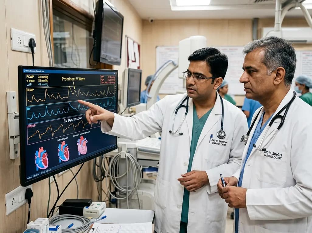
Left atrial stiffening generates prominent V waves that pressurize the pulmonary circulation and augment right ventricular afterload independently of intrinsic pulmonary vascular disease in patients with heart failure with preserved ejection fraction.
Today

A meta-analysis demonstrates that secondhand smoke exposure significantly elevates fetal loss risk by 21% among nonsmoking pregnant women. Clinicians must implement household screening and family-centered cessation interventions to prevent avoidable miscarriages and stillbirths.
Today
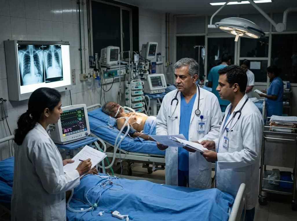
A recent cohort study reveals that postoperative infections occur in 17.3% of cardiac surgery patients using cardiopulmonary bypass. Routine inflammatory biomarkers like CRP and WBC show poor diagnostic accuracy due to bypass-induced systemic inflammation, demanding multi-parameter clinical vigilance.
Today

Explore the emerging role of glucose metabolic reprogramming in exercise-attenuated osteoporosis, examining how physical activity influences osteoblast glycolysis, osteoclast bioenergetics, and skeletal remodeling pathways to improve bone mineral density.
Today

A systematic review and meta-analysis of 33 studies evaluates the effectiveness of LDL apheresis techniques in familial hypercholesterolemia. Double-filtration plasmapheresis demonstrated the greatest reductions in total cholesterol and LDL-C, while safety and clinical feasibility guide individual therapy.
Today
