.webp&w=384&q=75)

Loading, please wait...
Loading, please wait...
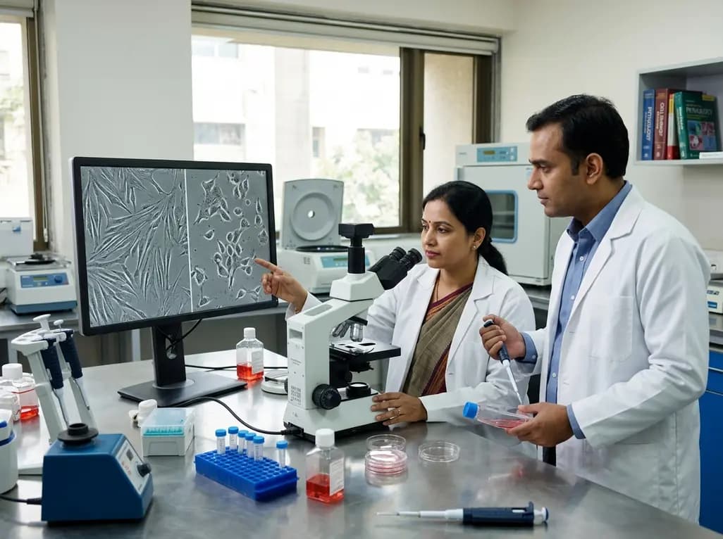
Human metapneumovirus (HMPV) is a primary cause of acute respiratory infections globally. While clinicians often associate it with childhood pneumonia, research indicates its impact extends to mesenchymal stem cells (MSCs). Human Metapneumovirus MSC infection significantly alters the structural and secretory landscape of these essential cells, potentially hindering their regenerative capabilities.
The study observed that HMPV reorganizes the distribution of actin and tubulin within MSCs. This structural shift likely facilitates viral entry and egress. Additionally, researchers noted an increase in Annexin V levels. This elevation suggests intense membrane modifications during viral fusion and budding. Consequently, the virus hijacks the cellular machinery to ensure its propagation.
HMPV significantly disrupts the secretion profile of growth factors. Specifically, infected MSCs show a marked decrease in EGF and VEGF levels. In contrast, Ang-2 and PDGF-AA levels rise significantly. These alterations create a precarious balance between ongoing cellular damage and the cells' remaining potential for tissue repair. Furthermore, despite these changes, the MSCs retain their ability to differentiate into various tissue types.
Understanding these interactions is vital for developing new therapeutic strategies. Because HMPV targets various cell types, including stem cells, its pathogenesis is more complex than previously thought. Future research must determine if these growth factor imbalances translate to impaired clinical recovery in patients with severe respiratory distress. However, the preservation of differentiation capacity provides a glimmer of hope for future stem-cell-based interventions.
No, the study indicates that despite structural and secretory changes, infected mesenchymal stem cells retain their ability to differentiate into various tissue types.
HMPV infection leads to a significant decrease in the secretion of VEGF and EGF, which are crucial for vascular health and cellular growth, while increasing levels of Ang-2 and PDGF-AA.
Disclaimer: This content is for informational and educational purposes only. It does not constitute medical advice or a substitute for professional healthcare consultation. Refer to the latest local and national guidelines for clinical practice.
References

Read summarized clinical updates, watch expert medical content, and earn CME certifications right from your smartphone.
Open your smartphone camera to scan and install.





Study reveals how HMPV infection disrupts growth factor secretion and cytoskeletal architecture in MSCs, while maintaining their differentiation potential....
4 months ago

Novel diimide and hydrazone scaffolds, particularly compound #C4, synergistically reactivate latent HIV-1 when combined with JQ1 or PKC activators. Demonstrating donor-matched proviral transcription ex vivo and non-canonical signaling, these findings advance shock and kill strategies toward reservoir eradication.
Today
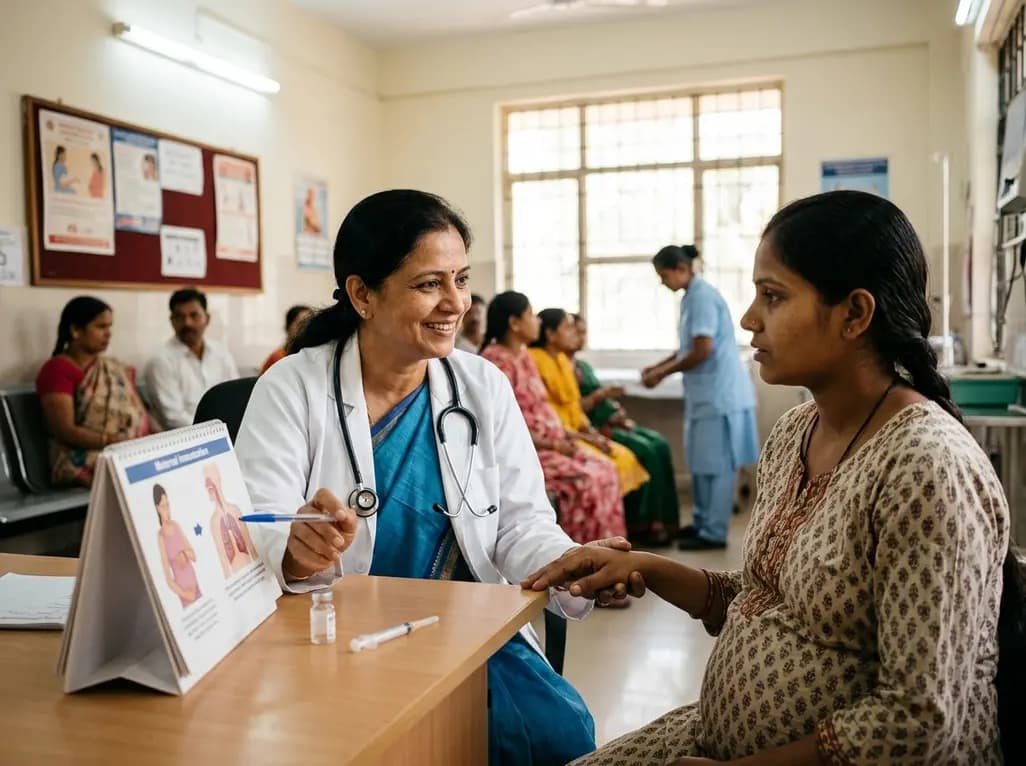
A cross-sectional study reveals that maternal RSV vaccination coverage reached only 43.7% in Argentina, with maternal vaccine knowledge being the sole independent predictor of uptake. Clinicians play a crucial role in eliminating informational and structural barriers to optimize infant health.
Today

Review how tailored exercise programs benefit frail older patients with heart failure. Explore pathophysiological mechanisms, individualized training prescriptions, and strategies to overcome adherence barriers in clinical practice.
Yesterday

This comparative study evaluates posterior labral tears with and without glenohumeral instability, highlighting distinct demographic patterns, MRI underestimation in stable shoulders, and successful arthroscopic repair outcomes.
Yesterday

A clinical study demonstrates that early intervention with tocilizumab, either as monotherapy or bridged with corticosteroids, achieves significant proptosis reduction and resolves active inflammation in treatment-naive thyroid eye disease.
Yesterday
