.webp&w=384&q=75)

Loading, please wait...
Loading, please wait...
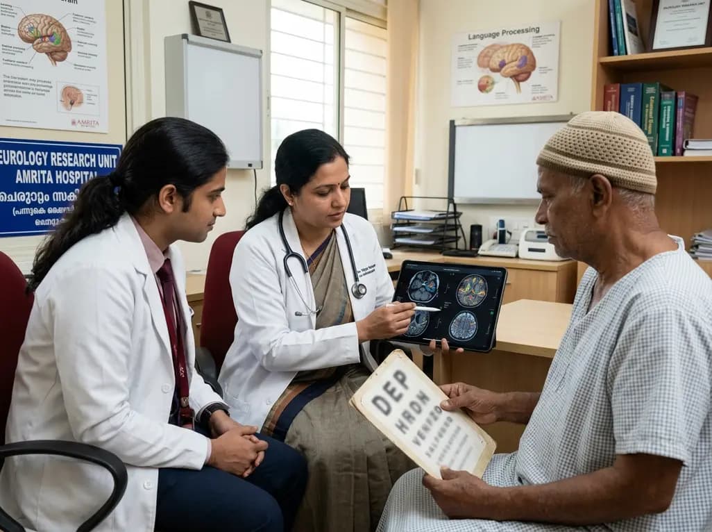
Pure alexia represents a classic neurological disconnection syndrome where stroke isolates reading comprehension from preserved speech and writing. Clinicians frequently encounter this condition following left posterior cerebral artery infarction. However, the exact neural pathways facilitating recovery from pure alexia remain poorly understood. A longitudinal study of a Korean stroke patient now provides vital insights into this process. Specifically, researchers tracked structural and functional brain connectivity over six months of behavioral improvement. Their findings demonstrate that distributed functional reorganization compensates for severe anatomical tract injury.
Pure alexia typically arises when an ischemic stroke damages the dominant occipital cortex and the splenium of the corpus callosum. Consequently, visual information cannot travel from either visual field to left-hemisphere language networks. Patients present with preserved verbal fluency, auditory comprehension, and intact handwriting. Nevertheless, they remain completely unable to read words, including sentences they wrote moments earlier.
In this recent report, clinicians evaluated a native Korean patient who developed pure alexia following an acute ischemic infarction. The patient displayed severe reading impairment for Hangul, the Korean syllabic script. Hangul combines alphabetic letters into two-dimensional syllabic blocks. Therefore, fluent reading demands seamless integration of visual form recognition and phonological conversion. Despite severe initial deficits, the patient achieved significant sentence-level reading recovery within six months.
Furthermore, this presentation emphasizes the importance of thorough bedside cognitive testing. Physicians frequently miss mild alexia because patients converse normally during standard clinical rounds. Therefore, structured reading assessments are essential in every posterior circulation stroke evaluation.
Advanced diffusion tensor tractography offers precise visualization of subcortical white matter pathways following cerebrovascular injury. In this investigation, tractography confirmed extensive structural damage caused by the posterior cerebral artery occlusion. Specifically, researchers identified severe degeneration within three vital tracts: the forceps major, the inferior longitudinal fasciculus, and the inferior fronto-occipital fasciculus.
The forceps major normally connects the bilateral visual cortices through the splenium of the corpus callosum. Meanwhile, the inferior longitudinal fasciculus links occipital visual hubs directly to the visual word form area. Finally, the inferior fronto-occipital fasciculus provides long-range structural connections to frontal language regions.
Importantly, serial evaluations demonstrated persistent structural disruption rather than axonal regeneration across the follow-up period. The damaged tracts showed no evidence of structural reconstitution at six months. Thus, behavioral improvement occurred despite permanent anatomical disconnection. This crucial finding confirms that structural tract integrity is not strictly necessary for meaningful clinical recovery after stroke.
Because structural pathways remained permanently disrupted, researchers evaluated whole-brain resting-state functional connectivity. Functional magnetic resonance imaging captured dynamic shifts in neural correlations across perilesional and distant cortical regions. Initially, the patient exhibited substantially diminished interhemispheric functional connectivity compared to healthy controls. The lesion effectively severed visual synchronization between the two cerebral hemispheres.
However, serial functional imaging demonstrated progressive intrahemispheric reorganization as reading ability improved. Perilesional visual areas formed strengthened functional correlations with associative language regions. In addition, the contralesional right hemisphere displayed adaptive activity changes, supporting compensatory processing. Although interhemispheric functional connectivity remained lower than normal, perilesional reorganization expanded steadily.
Consequently, these observations highlight the central role of functional neuroplasticity in stroke rehabilitation. Neural networks can dynamically reroute information through surviving cortical nodes to compensate for destroyed tracts. Therefore, recovery from pure alexia relies primarily on functional network plasticity rather than white matter tract regeneration.
Among all evaluated regions, the left angular gyrus demonstrated the most remarkable functional connectivity changes during behavioral recovery. The angular gyrus occupies an essential anatomical crossroad within the inferior parietal lobule. Traditionally, neuroscientists recognize this hub as a multisensory convergence zone linking orthographic, phonological, and semantic representations.
In this patient, functional connectivity between the left angular gyrus and perilesional occipital cortex changed significantly over time. Specifically, these connectivity surges coincided precisely with time points of rapid reading improvement. When ventral occipitotemporal pathways sustained damage, the angular gyrus actively recruited alternative processing streams. Thus, the parietal network compensated for the loss of direct ventral reading inputs.
Furthermore, this functional reconfiguration enabled the patient to implement alternative decoding strategies. By engaging dorsal phonological routes, the patient converted visual symbols into sounds sequentially. As a result, the angular gyrus served as an adaptive bridge, restoring sentence reading comprehension despite persistent ventral tract disruption.
The linguistic characteristics of Hangul provide valuable insights into orthographic processing. Because Hangul groups letters into discrete syllabic blocks, reading requires both holistic pattern recognition and linear phonological parsing. Initially, the patient struggled with single-block identification due to visual pathway injury. However, as compensatory networks matured, sentence-level reading recovered substantially.
Moreover, these neuroimaging insights carry direct implications for multilingual settings, including clinical practice across India. Indian scripts, such as Devanagari, Tamil, and Bengali, also combine complex phonetic and visual properties. Therefore, functional recruitment of dorsal parietal networks represents a universal compensatory mechanism across diverse writing systems.
Consequently, clinicians should design stroke rehabilitation programs that actively engage alternative pathways. Speech-language therapists can utilize tactile-kinesthetic tracing, multiple oral re-reading, and phonological assembly exercises to stimulate parietal circuits. Additionally, future therapies may employ non-invasive brain stimulation over the angular gyrus to enhance connectivity. Ultimately, targeted rehabilitation leverages functional neuroplasticity to restore functional literacy and patient independence.
Pure alexia presents as an isolated impairment in reading comprehension while spontaneous speech, verbal repetition, and written expression remain completely preserved. In contrast, standard aphasias involve broader language deficits that typically impair speech production, auditory comprehension, or grammatical formulation. Pure alexia occurs specifically when damage disconnects visual input from dominant language centers without injuring the primary language cortex. Consequently, affected individuals can write coherent sentences spontaneously yet cannot read what they just wrote.
Advanced neuroimaging modalities evaluate both structural tract continuity and dynamic functional correlations across surviving brain regions. While structural diffusion imaging reveals the anatomical extent of white matter damage, resting-state functional magnetic resonance imaging uncovers active network reorganization. Longitudinal increases in functional connectivity between perilesional hubs and associative areas, such as the angular gyrus, frequently signal adaptive neuroplasticity. Therefore, functional imaging helps clinicians forecast compensatory potential even when primary white matter pathways experience irreversible disruption.
Rehabilitation protocols typically combine multiple oral re-reading techniques, tactile-kinesthetic training, and phonological decoding exercises. In tactile-kinesthetic training, patients trace letters to access motor memory pathways, bypassing damaged visual routes. Multiple oral re-reading involves reading passages repeatedly to improve reading speed and accuracy through contextual cues. Furthermore, emerging evidence suggests that non-invasive brain stimulation targeting parietal compensatory nodes, such as the left angular gyrus, may enhance functional connectivity and accelerate meaningful behavioral gains during therapy.
Disclaimer: This content is for informational and educational purposes only and should not be considered medical advice. It is not intended to replace consultation with a qualified healthcare professional. While we strive to provide accurate and up-to-date information, medical knowledge is constantly evolving, and individual cases may vary. Readers are encouraged to confirm information with other sources and consult a healthcare provider for specific medical guidance. Refer to the latest local and national guidelines for clinical practice.
References

Read summarized clinical updates, watch expert medical content, and earn CME certifications right from your smartphone.
Open your smartphone camera to scan and install.





A new case study illuminates how functional connectivity changes drive recovery from pure alexia following posterior cerebral artery infarction. Functional reorganization supports reading recovery despite permanent structural tract disconnection, highlighting key implications for stroke rehabilitation.
last month

Explore why official drug package inserts fail to reflect modern evidence for clozapine prescribing. Learn how legal standards of care, case law, slow titration, and vital adverse event monitoring empower clinicians to prioritize evidence-based medical practice over outdated manufacturer labeling.
Today
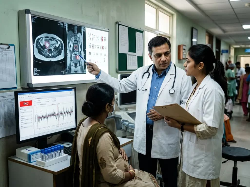
Occult maternal malignancy accounts for 3.82% of persistent unreportable NIPT results with genomic noise. Multiple chromosomal aberrations, notably on chromosome 18, warrant thorough oncological evaluation and whole-body MRI to ensure timely maternal diagnosis while preserving fetal care.
Today
This article reviews the forensic and clinical complexities of acute myocarditis arising on left ventricular hypertrophy in children, highlighting early diagnostic red flags and multidisciplinary postmortem autopsy protocols.
Today
A randomized controlled trial demonstrates that an eight-week dual-task training intervention significantly improves static and dynamic postural control over standard balance exercises in individuals with chronic ankle instability, addressing critical neurocognitive deficits during athletic and daily activities.
Today
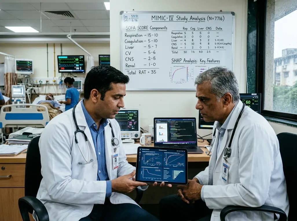
A retrospective cohort study of 7,716 ICU patients from the MIMIC-IV database demonstrates that Gradient Boosting Decision Trees significantly outperform traditional SOFA scores in predicting in-hospital mortality among patients with sepsis and diabetes, with SHAP analysis providing clinical interpretability.
Today
