.webp&w=384&q=75)

Loading, please wait...
Loading, please wait...
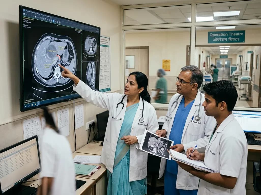
Intrahepatic cholangiocarcinoma arising from the caudate lobe presents unique anatomical and surgical challenges due to its deep spatial location and intimate association with major retrohepatic vascular structures. Recently, hepatobiliary surgeons identified a previously undescribed pathway of locoregional spread termed hepatocaval ligament dissemination in a patient with caudate-origin intrahepatic cholangiocarcinoma. This novel clinical observation highlights how fibrous retrohepatic ligamentous structures can serve as dedicated conduits for localized malignant expansion directly posterior to the inferior vena cava. Understanding this pathway is vital for surgical oncologists striving for margin-negative resections.
The caudate lobe, designated as segment I in Couinaud's liver anatomy, occupies a deep and central position within the posterior aspect of the liver. It sits directly anterior to the infrahepatic and retrohepatic inferior vena cava while remaining bounded anteriorly by the main portal vein bifurcation and primary hepatic ducts. Because of its complex dual vascular supply and intricate venous drainage through multiple short hepatic veins directly into the vena cava, primary caudate malignancies are extraordinarily difficult to manage surgically. Intrahepatic cholangiocarcinoma originating in segment I frequently invades adjacent portal triads, right hepatic arteries, and hepatic veins early in its development. Furthermore, the lymphatic drainage pathways from the caudate lobe flow bilaterally into retrocaval, paracaval, and superior pancreatoduodenal lymph nodes. Surgical resection represents the only potentially curative intervention for localized biliary tract tumors, yet achieving clear microscopic margins requires high-level hepatobiliary expertise. Anatomical variations in the caudate process and Spiegel lobe further complicate surgical dissection during radical hepatic procedures. When tumors involve both the paracaval portion and caudate process, complete extirpation typically requires combining caudate lobectomy with major hepatectomy. Consequently, surgical teams must carefully evaluate retrohepatic spatial relationships during preoperative planning to ensure procedural safety and complete tumor removal.
Intrahepatic cholangiocarcinoma typically disseminates through established anatomical pathways, including direct intraparenchymal infiltration, perineural sheaths, lymphatic networks, and portal venous channels. However, hepatocaval ligament dissemination represents an atypical retrohepatic pathway that extends tumor spread beyond standard anatomical boundaries. The hepatocaval ligament is a fibrous band or liver tissue bridge that spans between segment VII or segment I and the posterior wall of the inferior vena cava. In this newly described mechanism, malignant cells track directly along this fibrous connective tissue plane behind the main vena caval trunk. Consequently, retrocaval tumor expansion occurs without direct intravascular invasion into the vessel lumen itself. Surgical oncologists managing caudate tumors must realize that tumor cells can travel along these subtle ligamentous structures into otherwise unsuspected anatomic spaces. Dissection within the retrocaval space requires meticulous technique to avoid catastrophic avulsion of short hepatic veins or tearing of the vena caval wall. Moreover, incomplete resection of tissue within the hepatocaval ligament inevitably leaves behind microscopic residual disease, leading to early local recurrence. Identifying hepatocaval ligament dissemination allows surgeons to execute complete en bloc resections that thoroughly clear retrohepatic connective tissue planes.
Locally advanced caudate intrahepatic cholangiocarcinoma presenting with bulky regional lymphadenopathy historically carried an extremely poor clinical prognosis and low resectability rate. Contemporary gastrointestinal oncology increasingly relies on aggressive neoadjuvant therapy to downstage locally advanced biliary tract malignancies prior to operative intervention. Combining systemic gemcitabine and cisplatin with the anti-PD-1 checkpoint inhibitor pembrolizumab has established a potent therapeutic standard for advanced biliary carcinomas. Neoadjuvant chemoimmunotherapy effectively shrinks primary tumor volume, controls regional micrometastatic disease, and alleviates malignant biliary tree compression. In patients presenting with severe obstructive jaundice and extensive nodal burdens, systemic therapy can convert previously unresectable disease into a surgically resectable state. Furthermore, a robust histological response following systemic treatment facilitates achieving negative surgical margins during complex resections. Multidisciplinary tumor boards evaluate serial radiological imaging to track treatment response and optimize the timing of surgical intervention. Patients demonstrating favorable clinical response or stable disease proceed to radical resection, such as open right hepatectomy with total caudate lobectomy. Integrating state-of-the-art neoadjuvant regimens with radical surgical management provides patients with advanced caudate cholangiocarcinoma the highest likelihood of achieving durable long-term disease control.
Achieving negative surgical margins in caudate-origin intrahepatic cholangiocarcinoma demands detailed preoperative strategy and precise intraoperative execution. Surgical teams routinely perform an open right hepatectomy combined with complete caudate lobectomy and comprehensive regional lymphadenectomy. During the operation, surgeons mobilize the right hepatic lobe extensively, exposing the entire retrohepatic inferior vena cava from the infrahepatic level to the diaphragm. Mobilization provides direct visualization of the short hepatic veins that drain the caudate lobe directly into the anterior and lateral walls of the vena cava. Careful dissection within the retrocaval space allows the surgical team to identify unexpected tumor extension, specifically tracking along the hepatocaval ligament. Performing an en bloc resection of the primary caudate tumor along with the fibrous hepatocaval ligament ensures complete clearing of retrohepatic tissue planes. Surgeons frequently utilize intraoperative ultrasound to map tumor margins, verify vascular patency, and confirm adequate clearance from major hepatic veins in real time. Meticulous double-ligation or vascular stapling of short hepatic veins prevents major intraoperative hemorrhage during caudate detachment. Additionally, systematic lymphadenectomy encompassing the hepatoduodenal ligament, retropancreatic area, and retrocaval space ensures accurate pathological staging and reduces local recurrence risks.
Accurate preoperative assessment of caudate intrahepatic cholangiocarcinoma is essential for establishing resectability and planning complex surgical strategies. High-resolution multiphasic contrast-enhanced computed tomography and magnetic resonance cholangiopancreatography serve as the primary diagnostic modalities. These advanced imaging studies delineate primary tumor dimensions, evaluate biliary tree obstruction, and identify vascular invasion into portal or hepatic veins. However, subtle retrohepatic ligamentous extension along the hepatocaval ligament can easily escape detection on standard cross-sectional axial imaging. Diagnostic radiologists and hepatobiliary surgeons must scrutinize the retrocaval space specifically, evaluating soft tissue stranding or thickening behind the inferior vena cava. Advanced three-dimensional anatomical visualization tools enable surgical teams to reconstruct retrohepatic vascular relationships and model resection boundaries before surgery. Preoperative biliary drainage using endoscopic or percutaneous transhepatic biliary stents may be required if bulky lymphadenopathy induces severe obstructive jaundice prior to initiating neoadjuvant therapy. Rigorous preoperative staging prevents non-therapeutic exploratory laparotomies and helps select ideal candidates for systemic chemoimmunotherapy protocols. Consequently, close collaboration between dedicated gastrointestinal radiologists and hepatobiliary surgeons optimizes patient selection and improves overall surgical success.
The discovery of hepatocaval ligament dissemination underscores the rapidly evolving landscape of hepatobiliary surgical oncology. Managing complex caudate intrahepatic cholangiocarcinoma requires close coordination among surgical oncologists, medical oncologists, diagnostic radiologists, and gastroenterologists within a multidisciplinary framework. Modern neoadjuvant regimens incorporating immunotherapy have transformed treatment paradigms, enabling successful resections in complex presentations that were previously considered surgically incurable. Surgeons must maintain heightened vigilance during retrohepatic mobilization to recognize unexpected ligamentous tumor spread behind major vascular trunks. Educational documentation of these uncommon dissemination pathways through surgical case reports and instructional videos provides valuable insights for the international surgical community. Combining comprehensive preoperative imaging, effective neoadjuvant chemoimmunotherapy, and precise retrohepatic surgical dissection represents the current gold standard for maximizing survival in patients with challenging caudate biliary malignancies.
Hepatocaval ligament dissemination represents a novel retrohepatic pathway where caudate-origin intrahepatic cholangiocarcinoma extends along fibrous tissue behind the inferior vena cava. Recognizing this route is vital for hepatobiliary surgeons because incomplete removal of retrohepatic ligamentous tissue leads to positive surgical margins and local tumor recurrence. Identifying this specific pathway ensures complete en bloc surgical resection during complex hepatectomy procedures.
Neoadjuvant chemoimmunotherapy, combining gemcitabine, cisplatin, and pembrolizumab, significantly helps downstage locally advanced intrahepatic cholangiocarcinoma. This systemic regimen reduces primary tumor volume, clears bulky regional lymph node disease, and relieves severe biliary obstruction. Consequently, systemic treatment converts previously borderline or unresectable caudate tumors into resectable lesions, allowing complete margin-negative surgical resections and improving long-term patient outcomes.
Caudate lobe resections are technically demanding due to the deep anatomical location of segment I between the portal vein bifurcation and the inferior vena cava. Numerous short hepatic veins drain directly from the caudate lobe into the vena cava. Surgeons must carefully dissect these short vessels without causing severe intraoperative hemorrhage while achieving clear resection margins around retrohepatic fibrous structures.
Disclaimer: This content is for informational and educational purposes only, and should not be used as medical advice or replace consultation with a qualified healthcare professional. Refer to the latest local and national guidelines for clinical practice.
References

Read summarized clinical updates, watch expert medical content, and earn CME certifications right from your smartphone.
Open your smartphone camera to scan and install.





A case report highlights a novel dissemination pathway along the hepatocaval ligament in caudate-origin intrahepatic cholangiocarcinoma. Successful management involved neoadjuvant gemcitabine, cisplatin, and pembrolizumab followed by open right hepatectomy with caudate lobectomy and negative margins.
last month

Clinical guidelines rely heavily on isolated biomarkers like IGF-1 and HbA1c. However, portal insulin delivery fundamentally gates hepatic growth hormone sensitivity. This physiological continuum unites type 1 and type 2 diabetes, obesity, cirrhosis, and acromegaly, challenging conventional treatment strategies.
Today
The ClinGen Prenatal Gene Curation Expert Panel evaluated 63 disease relationships across 61 genes, establishing clinical validity for severe fetal phenotypes like hydrops and stillbirth to enhance prenatal genomic interpretation and clinical care.
Today
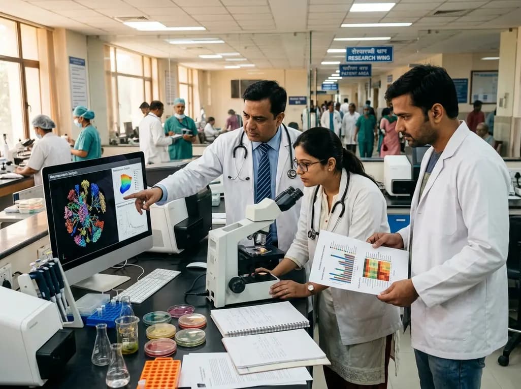
A metataxonomic study reveals distinct gut bacteriome biomarkers in type 2 diabetes, obesity, and cardiovascular complications, identifying specific bacterial shifts that pave the way for precision metabolic medicine.
Today
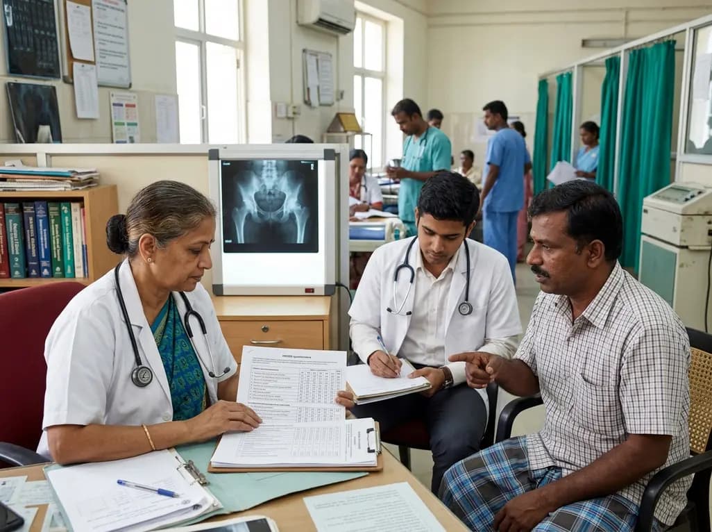
A psychometric validation study confirms that the Turkish Copenhagen Hip and Groin Outcome Score (HAGOS-T) offers excellent reliability, construct validity, and interpretability for patients with hip osteoarthritis, providing clinicians with a robust tool for functional assessment.
Today
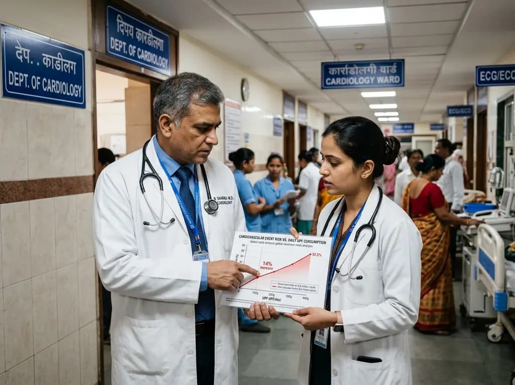
A global dose-response meta-analysis of 8.8 million adults reveals that consuming an extra 100 grams of processed food daily raises cardiovascular event risk by 14%. Explore how industrial additives, systemic inflammation, and metabolic disruption heighten long-term multimorbidity and cardiometabolic risks.
Today
