.webp&w=384&q=75)

Loading, please wait...
Loading, please wait...

Anterior cervical discectomy and fusion remains the gold standard surgical procedure for treating cervical radiculopathy and degenerative myelopathy. However, clinicians around the world encounter significant ambiguity when evaluating arthrodesis success. A recent international Delphi-informed investigation analyzed contemporary clinical practices among high-impact cervical spine specialists worldwide. The investigators discovered profound heterogeneity regarding imaging strategies, implant choices, and diagnostic criteria. Therefore, establishing a reliable standard for ACDF fusion assessment has become an urgent clinical priority. Spine surgeons require unified, evidence-based guidelines to interpret osseous bridging accurately and optimize postoperative management.
Surgeons historically debate the optimal implant choices for achieving cervical arthrodesis. According to recent survey data, most spine surgeons globally prefer anterior plate fixation over stand-alone constructs. Specifically, 71% of surveyed experts utilize supplemental anterior plating for one- to two-level procedures. Furthermore, this preference rises sharply to 92% when surgeons perform three- to four-level reconstructions. Anterior cervical plates provide rigid biomechanical stabilization, which minimizes shear forces across graft interfaces. Meanwhile, interbody device preferences reveal a persistent inclination toward metallic cages. For instance, metallic implants accounted for 40% of preferences in short constructs and 42% in multilevel reconstructions. Although polyetheretherketone (PEEK) and structural autografts remain viable alternatives, metallic implants provide immediate structural rigidity and enhanced osteointegration. Consequently, construct rigidity strongly influences the timeline and radiographic appearance of bony bridging. Spine specialists frequently select rigid internal stabilization to reduce construct failure risks in complex degenerative pathologies. As a result, stabilizing hardware dictates which radiographic modalities provide diagnostic accuracy during follow-up reviews.
Accurate ACDF fusion assessment demands reliable imaging techniques that differentiate solid arthrodesis from pseudarthrosis. Consequently, spine surgeons utilize diverse postoperative imaging protocols across international centers. Dynamic flexion-extension radiographs remain the most popular initial screening tool among surveyed spine practitioners. In addition, surgeons routinely deploy these functional radiographs to detect pathologic interspinous motion across reconstructed motion segments. However, flexion-extension plain radiographs carry substantial measurement error and often suffer from voluntary patient guarding. Therefore, experts increasingly view thin-cut computed tomography (CT) as the definitive diagnostic standard. Multidetector CT offers superior cross-sectional visualization of continuous interbody trabeculae and facet articulation integrity. Nevertheless, clinicians frequently limit routine CT utilization because radiation exposure and financial costs raise legitimate concerns. Surgeons often reserve CT evaluation for symptomatic patients or uncertain radiographic findings. Furthermore, metal artifacts from anterior plates and titanium cages occasionally obscure subtle trabecular bridging. Thus, clinicians must weigh the accessibility of dynamic radiographs against the high spatial resolution of cross-sectional imaging when monitoring cervical healing.
Defining true bony union requires strict qualitative and quantitative radiographic criteria. The global Delphi survey highlighted that surgeons assign varying diagnostic weight to different radiographic signs. Most notably, spine specialists endorsed continuous trabecular bridging across the intervertebral disc space as the single strongest criterion for successful arthrodesis. In addition, experts regard the complete absence of angular motion on dynamic radiographs as robust proof of spinal stability. Conversely, surgeons identify peri-implant radiolucency and halo formation as alarming signs of nonunion. However, subjective interpretations often create diagnostic discrepancies between radiologists and treating spine surgeons. For example, subtle radiolucent halos around screws might reflect normal settling rather than definite mechanical loosening. Moreover, inter-observer agreement remains modest when evaluating trabecular continuity inside dense metallic interbody cages. Accordingly, many specialists advocate for standardized scoring systems to minimize observer bias. Because pseudarthrosis directly influences patient-reported outcomes, clinicians must synthesize multiple imaging parameters rather than relying on a solitary metric. Comprehensive structural assessment ensures timely detection of biomechanical failure and prevents unnecessary revision surgeries.
Postoperative rehabilitation and immobilization strategies vary widely among international spinal surgery practices. While some surgeons mandate rigid cervical orthoses following multilevel fusions, others permit immediate mobilization without external support. Furthermore, surgical construct stiffness largely dictates bracing decisions. For instance, rigid anterior plating provides sufficient mechanical stability, which allows surgeons to minimize external collar duration safely. In contrast, uninstrumented or multilevel reconstructions frequently prompt surgeons to prescribe external immobilization for four to eight weeks. Beyond external bracing, the duration of outpatient clinical monitoring exhibits noticeable divergence. The survey revealed that patient-reported satisfaction and definitive radiographic fusion status serve as the two primary drivers of follow-up cessation. Therefore, asymptomatic patients demonstrating solid trabecular bridging typically discontinue formal follow-up between six and twelve months postoperatively. Conversely, persistent axial pain or persistent radicular symptoms prolong clinical observation considerably. Clinicians carefully observe symptomatic patients to distinguish transient muscle pain from symptomatic hardware loosening. Consequently, longitudinal follow-up protocols balance clinical efficiency against the risk of overlooking delayed pseudoarthrosis.
The observed worldwide divergence in spinal assessment holds critical implications for surgical practice in India. In Indian clinical centers, cost constraints and disparate healthcare access deeply influence postoperative surveillance. For example, private tertiary hospitals easily access advanced multi-detector CT scanners, whereas rural institutions rely strictly on serial plain radiography. Moreover, out-of-pocket healthcare financing makes repeated advanced imaging challenging for many patients. Consequently, Indian spine specialists must adopt pragmatic, stepwise assessment protocols. Routine follow-up should initially utilize properly positioned lateral and dynamic flexion-extension radiographs. Furthermore, clinicians should preserve CT scans specifically for patients who experience persistent mechanical axial pain or questionable radiographic lucency. Additionally, selecting cost-effective implant constructs remains essential. Given that anterior plating and metallic cages demonstrate reliable clinical fusion worldwide, Indian surgeons can achieve reproducible outcomes through standardized surgical instrumentation. Ultimately, bridging the gap between global consensus and resource-conscious healthcare delivery will elevate patient safety and standardize cervical spine care throughout developing regions.
Computed tomography remains the gold standard modality for assessing bony fusion after cervical reconstruction. Multidetector thin-cut CT scans clearly visualize continuous trabecular bridging across the intervertebral disc space and evaluate facet joint ankylosis accurately. Although flexion-extension dynamic radiographs offer an accessible, low-radiation screening option, they lack precision due to voluntary patient guarding. Therefore, clinicians reserve CT imaging for symptomatic patients or ambiguous plain radiographs to confirm definitive arthrodesis.
Surgeons overwhelmingly prefer supplemental anterior plating in multilevel cervical procedures because multiple operative segments experience higher biomechanical shear stress and construct instability. Anterior plates enhance structural rigidity, minimize micromotion at graft interfaces, and lower nonunion rates considerably. Additionally, plate fixation preserves cervical lordosis and prevents early cage dislodgement or subsidence. Consequently, international guidelines and clinical consensus strongly recommend supplemental plate stabilization when surgeons perform three- or four-level cervical disc fusions.
Clinicians evaluate clinical symptomatology and dynamic radiographic findings concurrently to differentiate pseudarthrosis from healing bone. Successful arthrodesis demonstrates uninterrupted trabecular bridging across endplates and stable interspinous distance on flexion-extension imaging. Conversely, pseudarthrosis often exhibits radiolucent halos around screws, visible interspace gaps, or dynamic segment motion exceeding two millimeters. If patients remain completely asymptomatic despite ambiguous radiographs, surgeons frequently opt for serial radiological surveillance rather than recommending immediate surgical revision.
Disclaimer: This content is for informational and educational purposes only and does not constitute formal medical advice. Healthcare professionals must exercise independent clinical judgment when diagnosing and treating individual patients. Refer to the latest local and national guidelines for clinical practice.
References

Read summarized clinical updates, watch expert medical content, and earn CME certifications right from your smartphone.
Open your smartphone camera to scan and install.





A global cross-sectional survey of cervical spine experts reveals significant variation in ACDF fusion assessment practices, imaging modalities, and fixation preferences, underscoring the need for standardized post-surgical protocols.
Today
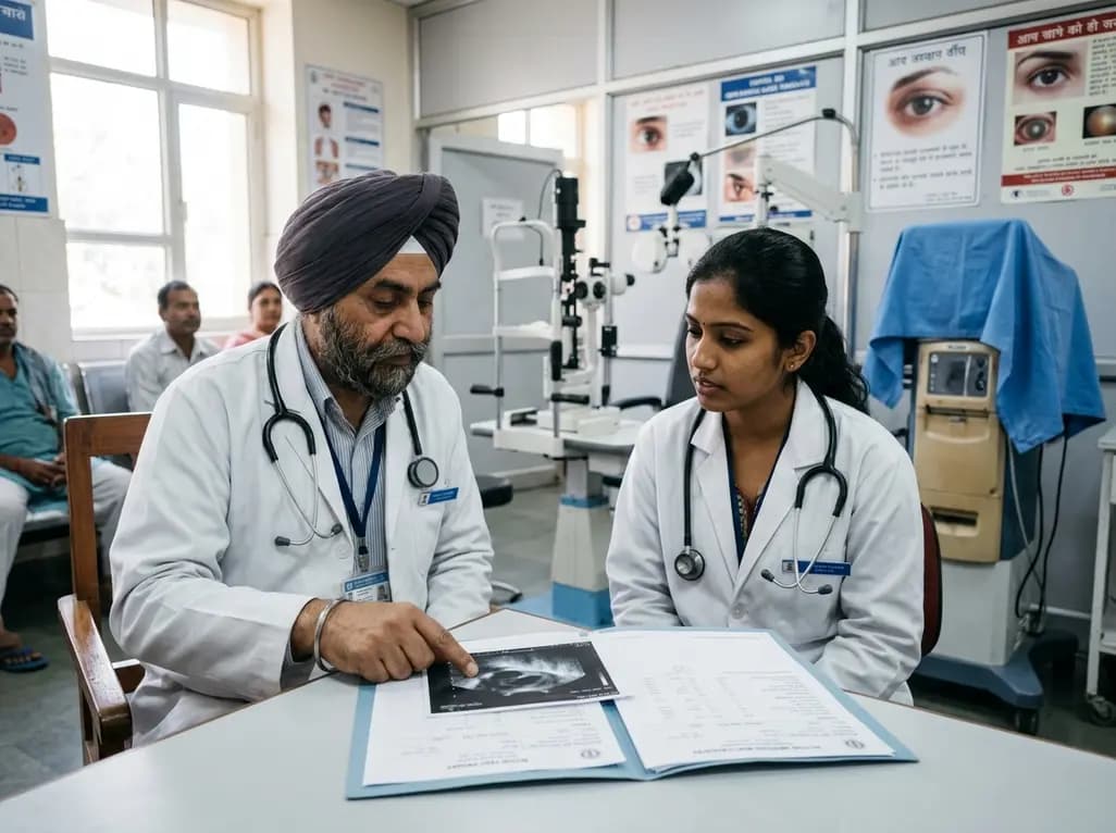
A multicenter study reveals that a significant proportion of HCV antibody-positive ophthalmic surgical patients remain untreated and eligible for direct-acting antivirals. Stratifying antibody titers and streamlining referral pathways can optimize hepatitis C micro-elimination efforts.
Today
Robotic single-port totally extraperitoneal (TEP) repair enables simultaneous excision of a canal of Nuck cyst and indirect hernia repair in women. Using the da Vinci SP platform and self-adhesive mesh, this approach offers minimal tissue trauma, low operative times, and rapid discharge without complications.
Today
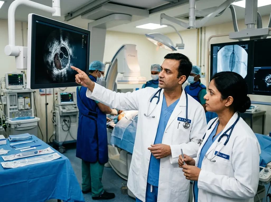
A multicenter REAL-LEAD registry sub-analysis evaluates how intravascular ultrasound-defined circumferential calcification and post-procedural minimum lumen area govern 1-year primary patency after drug-coated balloon angioplasty for femoropopliteal lesions.
Today

A multicenter real-world prospective study in Taiwan demonstrates that liraglutide effectively reduces body weight and BMI standard deviation score in adolescents with obesity. The therapy shows a favorable safety profile with mild gastrointestinal adverse events and zero treatment discontinuations due to intolerance.
Today
