.webp&w=384&q=75)

Loading, please wait...
Loading, please wait...
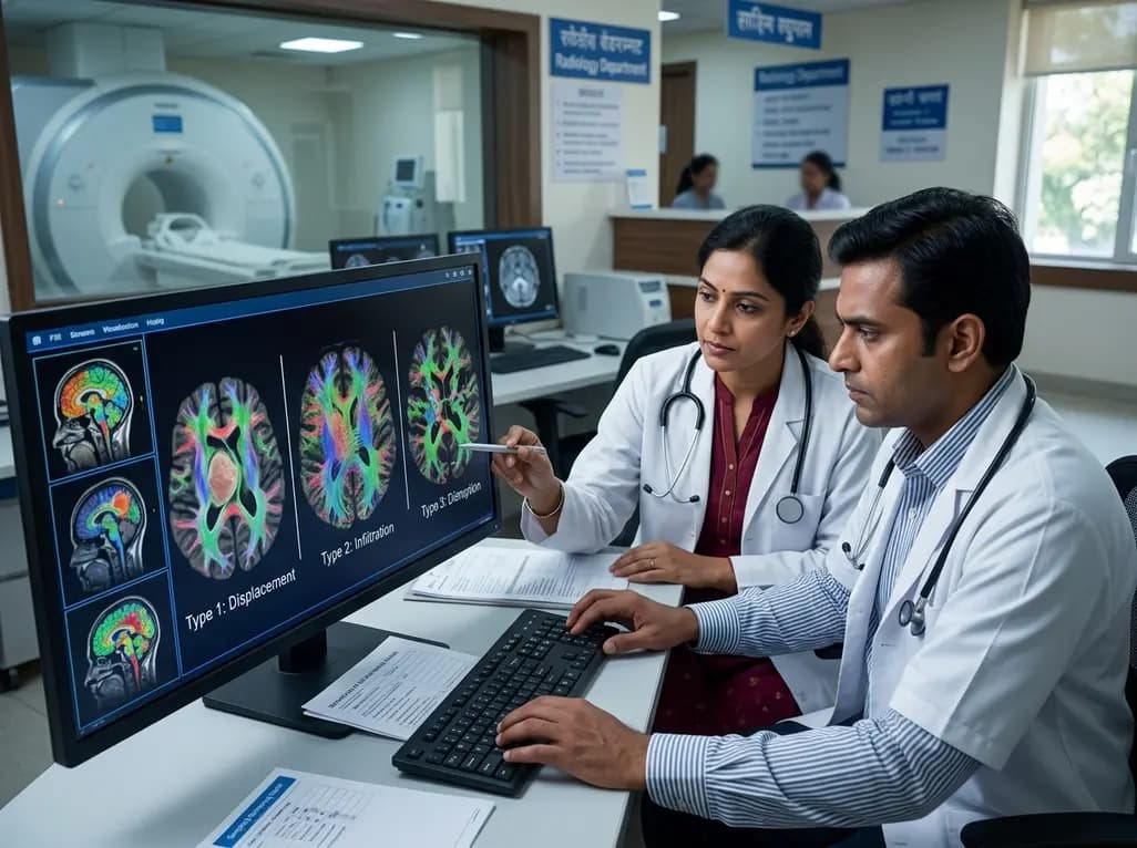
Surgical management of diffuse gliomas requires a delicate balance between maximizing tumor resection and preserving eloquent neurological pathways. Recent advances in neuroimaging provide deeper insights into structural connectivity. Specifically, characterizing glioma tract interactions helps neurosurgeons anticipate tumor invasiveness and functional risks prior to craniotomy. Diffuse intrinsic tumors frequently distort, invade, or sever subcortical neural bundles, which complicates intraoperative navigation. Consequently, clinicians need objective imaging criteria to evaluate these subcortical relationships. A groundbreaking study published in Neuro-Oncology established a standardized 3-tier classification system utilizing diffusion magnetic resonance imaging and automated fiber tractography. By analyzing preoperative scans from 360 surgical patients, investigators mapped how gliomas disrupt adjacent white matter architecture. Furthermore, this framework bridges advanced tractography with molecular neuropathology and surgical outcomes. Clinicians can now evaluate whether a tract suffers pure displacement, active infiltration, or structural disruption before entering the operating theater. Therefore, this paradigm shift enhances individualized surgical planning, improves risk stratification, and protects patient quality of life.
Automated tractography reconstructs essential subcortical pathways, including the corticospinal tract, arcuate fasciculus, and optic radiation. The proposed 3-tier classification categorizes glioma tract interactions into displacement, infiltration, and disruption. In displacement-type interactions, the expanding tumor mass pushes intact white matter bundles aside without penetrating axonal sheaths. Accordingly, fiber continuity remains entirely preserved on diffusion imaging metrics. In contrast, infiltration-type interactions demonstrate neoplastic cells permeating through the interstitial spaces of the tract, causing signal heterogeneity while maintaining structural fiber orientation. Disruption-type interactions represent the most destructive pattern, characterized by complete loss of tract integrity and severe axonal breakdown. Among the cohort of 360 patients, infiltration was the most common presentation, occurring in 78.6% of cases. Meanwhile, displacement appeared in 9.7% of patients, and disruption occurred in 11.7%. Moreover, automated fiber tracking accurately distinguished these categories with high reproducibility. Hence, neuro-radiologists and surgeons gain a reliable anatomical roadmap that standardizes radiological reporting and guides surgical boundaries.
To confirm the biological validity of the imaging classification, researchers conducted double immunohistochemical staining on neuronavigation-guided tissue samples. They stained biopsy specimens for isocitrate dehydrogenase mutations and myelin basic protein to verify axonal architecture at microscopic margins. Histological analysis strongly corroborated the diffusion imaging categories. Displacement zones showed compressed, intact myelin sheaths devoid of tumor infiltration. Conversely, infiltrated tracts contained scattered glioma cells navigating along preserved myelin fibers, whereas disrupted tracts revealed extensive myelin disintegration. Crucially, the 3-tier imaging classification demonstrated remarkable alignment with tumor molecular status. Disruption-type tracts correlated predominantly with aggressive isocitrate dehydrogenase wild-type gliomas, accounting for 87.2% of such lesions. In contrast, IDH-mutant gliomas more frequently exhibited displacement and infiltration patterns. Furthermore, IDH wild-type status drives aggressive microenvironmental invasion, which explains the high frequency of axonal destruction. Thus, preoperative diffusion imaging acts as a non-invasive surrogate biomarker reflecting underlying tumor biology and molecular aggression.
Achieving supratotal or gross total resection remains a cornerstone of glioma therapy, yet preservation of motor function is paramount. The study revealed that tract classification significantly influenced surgical resectability and postoperative outcomes. Specifically, patients with displacement-type tracts achieved significantly higher rates of gross total resection compared to those with infiltration-type tumors. Because displacement preserves a distinct anatomical cleavage plane, surgeons can safely resect neoplastic tissue without violating functional fiber bundles. In cases involving the corticospinal tract, displacement-type tumors showed preserved motor strength following surgery without new neurological deficits. Conversely, infiltration and disruption types demonstrated strong correlations with postoperative motor deterioration. When tumors infiltrate functional pathways, complete surgical eradication often risks permanent paralysis. Therefore, identifying infiltration or disruption preoperatively alerts the surgical team to employ intraoperative neuromonitoring, awake mapping techniques, and subcortical stimulation. Consequently, surgeons can tailor aggressive debulking while establishing realistic functional expectations for patients and caregivers.
Beyond guiding surgical extent and functional preservation, tract interaction patterns offer vital prognostic information. Overall survival and progression-free survival differ markedly across the three classifications. Patients presenting with displacement-type tumors demonstrated superior median survival times, reflecting both higher resectability rates and favorable IDH mutation status. In stark contrast, patients with disruption-type tracts experienced significantly shorter progression-free and overall survival. Multivariable survival analyses confirmed that white matter tract disruption serves as an independent predictor of poor clinical outcome. Moreover, axonal disruption indicates extensive neoplastic infiltration into deep brain structures, which limits the efficacy of adjuvant radiotherapy and chemotherapy. Infiltration-type cases occupied an intermediate prognostic category, where survival heavily depended on achieving maximal safe cytoreduction. Thus, integrating diffusion tractography metrics into standard prognostic risk scoring models refines clinical staging. As a result, multidisciplinary neuro-oncology tumor boards can better stratify patients for clinical trials and aggressive adjuvant protocols.
In India, neurosurgical centers manage high volumes of complex glioma cases under varied resource settings. Incorporating advanced diffusion magnetic resonance tractography into routine presurgical workups offers immense value across tertiary cancer centers and regional hospitals. While advanced intraoperative MRI remains limited to premier institutions, preoperative tractography is widely accessible on modern 1.5T and 3T MRI platforms. Indian neurosurgeons can readily implement this 3-tier classification to categorize tract displacement, infiltration, and disruption during multidisciplinary tumor board discussions. Furthermore, establishing these tract interactions optimizes intraoperative resources, such as direct cortical and subcortical electrical stimulation and neuronavigation systems. Preoperative counseling also benefits immensely, as clinicians can provide families with objective assessments regarding neurological risk and functional recovery. Moreover, training postgraduate trainees in tractography interpretation enhances overall neuro-oncological care. Therefore, adopting this standardized imaging classification strengthens surgical precision, optimizes oncological outcomes, and elevates the standard of neuro-oncology care throughout India.
The classification categorizes interactions into displacement, infiltration, and disruption using diffusion MRI tractography. Displacement indicates intact fibers pushed aside by tumor mass. Infiltration represents neoplastic cells spreading along intact myelin sheaths. Disruption denotes severe axonal destruction and loss of fiber continuity, commonly seen in aggressive isocitrate dehydrogenase wild-type gliomas.
Displacement-type tracts allow higher rates of gross total resection because tumor margins remain distinct from intact neural pathways. Conversely, infiltration and disruption patterns signal high risk for postoperative motor deficits. Surgeons manage these complex patterns by utilizing intraoperative neuromonitoring, awake brain mapping, and subcortical electrical stimulation to preserve neurological function.
Tract disruption strongly correlates with isocitrate dehydrogenase wild-type biology and aggressive microstructural invasion. This destructive phenotype reflects rapid tumor proliferation, deep brain infiltration, and lower rates of complete resection. Consequently, patients with disrupted tracts face earlier disease progression and significantly shorter overall survival despite standard adjuvant chemoradiotherapy.
Disclaimer: This content is for informational and educational purposes only. It is not intended as a substitute for professional medical advice, diagnosis, or treatment. Always seek the advice of your physician or other qualified health provider with any questions you may have regarding a medical condition. Never disregard professional medical advice or delay in seeking it because of something you have read here. Clinical decisions should not be based solely on this information. Refer to the latest local and national guidelines for clinical practice.
References

Read summarized clinical updates, watch expert medical content, and earn CME certifications right from your smartphone.
Open your smartphone camera to scan and install.





A novel 3-tier diffusion MRI classification system categorizes glioma-white matter tract interactions into displacement, infiltration, and disruption, correlating directly with IDH mutation status, gross total resection rates, postoperative motor preservation, and overall patient survival.
last month

Discover how oxidative stress drives inflammatory bowel disease (IBD) pathogenesis. Explore cutting-edge insights on Nrf2 signaling, ferroptosis suppression, microbiome interactions, and emerging targeted redox therapies.
Today

A pharmacometric and machine learning study reveals that specific vaginal bacteria alter cervical antiretroviral exposure, highlighting the need for precision HIV PrEP strategies.
Yesterday

Abdominal aortic aneurysm rupture remains a catastrophic vascular emergency. Groundbreaking research reveals that USP53 accelerates aneurysm progression by reprogramming smooth muscle cell metabolism toward aerobic glycolysis, highlighting a promising target beyond conventional diameter-based surveillance.
Today

A breakthrough study leverages natural language processing to accurately identify incident medication-related osteonecrosis of the jaw and track antiresorptive drug holidays from clinical narratives in electronic health records, advancing osteoporosis pharmacovigilance and dental safety.
Today

Recent research defines ferro-aging as a conserved iron-lipid axis driving organ decline across primates. Age-related iron dyshomeostasis stimulates ACSL4-mediated lipid peroxidation, promoting cellular senescence. Notably, vitamin C directly inhibits ACSL4, suppressing phospholipid oxidation and multi-organ decay.
Yesterday
