.webp&w=384&q=75)

Loading, please wait...
Loading, please wait...

Primary cardiac tumors represent an extraordinary diagnostic challenge in contemporary cardiovascular medicine. Although benign histologically, a giant left atrial myxoma can produce severe mechanical obstruction and profound hemodynamic compromise. When these tumors prolapse across the mitral valve orifice during diastole, they frequently mimic severe rheumatic mitral valve stenosis. Consequently, affected patients develop marked secondary pulmonary hypertension, exercise intolerance, and severe heart failure symptoms. Early identification and prompt intervention are imperative to prevent sudden cardiac death, acute embolic events, or irreversible pulmonary vascular remodeling. Modern advances in cardiothoracic surgery now allow selected patients to undergo successful resection via minimally invasive techniques, avoiding traditional median sternotomy while ensuring complete tumor removal and excellent clinical outcomes.
A 57-year-old woman presented with progressive exertional dyspnea corresponding to New York Heart Association class III status, accompanied by persistent palpitations. Her past medical history was notable for cardiovascular risk factors, specifically hypercholesterolemia and obesity. Initial physical examination revealed signs consistent with elevated left-sided cardiac filling pressures. Therefore, clinicians promptly performed first-line transthoracic echocardiography to evaluate her structural heart function. Diagnostic imaging demonstrated a giant, highly mobile left atrial mass measuring 4.7 by 4.8 centimeters. The lesion was attached securely to the basal third of the interatrial septum. During cardiac diastole, the large tumor prolapsed prominently across the mitral valve annulus into the left ventricle.
This dynamic diastolic prolapse resulted in severe left ventricular inflow tract obstruction. Echocardiography measured a mean transmitral pressure gradient of 16 mmHg, confirming severe functional mitral valve stenosis. Furthermore, Doppler imaging revealed severe secondary pulmonary hypertension, with an estimated pulmonary artery systolic pressure reaching approximately 80 mmHg. Despite these severe hemodynamic abnormalities, the mitral valve leaflets remained structurally intact without primary intrinsic disease. The severe gradient stemmed entirely from mechanical obstruction by the mobile mass. This presentation underscores how an intracardiac mass can dramatically alter cardiac hemodynamics, requiring urgent clinical attention and definitive intervention.
Given the patient's age and established cardiovascular risk factors, non-invasive assessment of the coronary arteries was vital prior to surgical intervention. Clinicians performed preoperative coronary computed tomography angiography to evaluate for underlying obstructive coronary artery disease. This imaging modality served a dual purpose by characterizing the anatomic relationships of the intracardiac tumor while simultaneously excluding significant epicardial coronary stenosis. The computed tomography scan clearly highlighted widespread coronary atheromatosis but confirmed the absence of hemodynamically significant vessel luminal narrowing, thereby eliminating the need for concomitant coronary artery bypass surgery.
Following imaging, a multidisciplinary heart team meeting took place to formulate a personalized management strategy. The team reviewed the echocardiographic parameters, anatomical features, and surgical risks. Given the tumor size and mobile nature, surgical resection was scheduled within two weeks of initial diagnosis. To minimize embolic risk prior to surgery, clinicians implemented strict physical activity restrictions and strict clinical monitoring. Preoperative systemic anticoagulation was intentionally omitted, as unmanaged anticoagulation carries a risk of tumor hemorrhage or mechanical dislodgement without clear evidence of benefit. The heart team unanimously selected a minimally invasive thoracoscopic approach with femoro-femoral cardiopulmonary bypass, balancing operative safety with enhanced postoperative recovery advantages.
The surgical team performed complete tumor excision utilizing a right minimally invasive video-assisted thoracoscopic approach. Peripheral vascular access was established via femoro-femoral cannulation for cardiopulmonary bypass management. Minimally invasive cardiac surgery offers significant advantages over conventional median sternotomy, including reduced surgical trauma, less postoperative pain, decreased blood loss, and faster return to functional baseline. Through a small right mini-thoracotomy, the surgeon achieved excellent visualization of the left atrium and interatrial septum. Careful handling of the mass was prioritized throughout the procedure to prevent tumor fragmentation and catastrophic systemic embolization.
The surgeon resected the giant tumor en bloc along with its septal attachment, ensuring clear surgical margins while preserving the native mitral valve apparatus. Surgical inspection confirmed that the underlying mitral valve leaflets and subvalvular structures were anatomically normal and functionally undamaged. Following copious irrigation of the cardiac chambers to remove cellular debris, the atrial septal defect was closed systematically. The patient was successfully weaned from cardiopulmonary bypass without complication. The entire intraoperative course proceeded smoothly, demonstrating that complex intracardiac mass resections can be performed safely via thoracoscopic techniques in high-volume expert centers.
The patient experienced an uncomplicated postoperative recovery period in the intensive care unit. Thanks to the minimally invasive approach, early extubation was achieved shortly after surgery, followed by rapid mobilization on the first postoperative day. Pathological evaluation of the resected surgical specimen confirmed the definitive diagnosis. Histopathological examination revealed a well-circumscribed, non-encapsulated mass characterized by a smooth surface. Microscopic analysis demonstrated a hypocellular proliferation of spindle-to-stellate cells suspended within a rich, abundant myxoid stroma. Importantly, there was no evidence of cellular atypia, mitotic activity, or tumor necrosis. Diagnostic immunohistochemistry revealed positive staining for calretinin, confirming a classic benign cardiac myxoma.
At the one-month outpatient follow-up evaluation, the patient demonstrated complete clinical symptom resolution, reporting excellent exercise tolerance. Follow-up transthoracic echocardiography confirmed complete normalization of cardiac hemodynamics. The transmitral pressure gradient had dropped to normal levels, and the estimated pulmonary artery systolic pressure had fully normalized. Echocardiography identified only mild, stable residual mitral regurgitation that required no immediate intervention. This remarkable hemodynamic recovery highlights the complete reversibility of functional mitral stenosis and secondary pulmonary hypertension once mechanical outflow tract obstruction is surgically eliminated.
Managing large left atrial tumors requires rapid clinical decision-making and clear interdisciplinary collaboration. First, clinicians must maintain a high index of suspicion for intracardiac masses when evaluating patients with sudden onset heart failure or atypical mitral valve murmur. Second, echocardiography remains the primary diagnostic standard, but cross-sectional imaging with coronary computed tomography angiography provides valuable structural and vascular insights without invasive risk. Third, functional mitral stenosis caused by a myxoma can lead to profound pulmonary hypertension, which typically resolves rapidly following tumor removal. Fourth, surgical excision should be performed expeditiously to eliminate the constant threat of systemic arterial embolization or sudden hemodynamic collapse. Finally, minimally invasive thoracoscopic approaches represent a safe, highly effective alternative to traditional sternotomy in carefully selected patients managed by expert multidisciplinary teams.
A left atrial myxoma is the most common primary benign heart tumor. It typically arises from the interatrial septum within the left atrium. As the mass increases in size, its mobile structure can physically prolapse through the mitral valve orifice during diastole. This mechanical blockage restricts blood flow from the left atrium into the left ventricle, creating severe functional mitral stenosis and elevated heart pressures.
Minimally invasive thoracoscopic resection offers substantial advantages over standard median sternotomy for treating cardiac tumors. By utilizing smaller lateral incisions and video assistance, this surgical technique reduces overall operative trauma, minimizes perioperative blood loss, decreases postoperative pain, and shortens hospital stay. Patients generally experience faster functional recovery and rapid return to daily activities while achieving full, safe tumor resection.
Yes, pulmonary hypertension secondary to a left atrial tumor frequently resolves following complete surgical excision. When a myxoma obstructs the mitral valve, upstream pressures rise rapidly, transmitting severe pressure into the pulmonary vasculature. Once the tumor is successfully resected and mechanical obstruction is cleared, left atrial pressures fall immediately, leading to prompt normalization of pulmonary artery pressures and complete relief of clinical symptoms.
Disclaimer: This content is for informational and educational purposes only and does not constitute medical advice, diagnosis, or treatment. Always seek the direct advice of a qualified healthcare provider regarding any clinical condition or surgical decision. Refer to the latest local and national guidelines for clinical practice.
References
Daher A et al. Giant Left Atrial Myxoma Causing Functional Mitral Stenosis: A Minimally Invasive Approach. Cureus. 2026 Aug undefined. doi: 10.7759/cureus.114155. PMID: 42571329.

Read summarized clinical updates, watch expert medical content, and earn CME certifications right from your smartphone.
Open your smartphone camera to scan and install.





A 57-year-old woman presented with severe dyspnea caused by a giant left atrial myxoma prolapsing into the mitral valve, creating functional mitral stenosis and pulmonary hypertension. She underwent successful minimally invasive thoracoscopic resection with complete symptom resolution and normalized pressures.
last month

A metataxonomic study reveals distinct gut bacteriome biomarkers in type 2 diabetes, obesity, and cardiovascular complications, identifying specific bacterial shifts that pave the way for precision metabolic medicine.
Today

Researchers have engineered HER2-targeted AIE nanotheranostics to overcome SHR-A1811 resistance in breast cancer. By inducing immunogenic pyroptosis and reprogramming tumor-associated macrophages, this multimodal nanoplatform achieves near-complete tumor regression in refractory preclinical models.
Today

A global dose-response meta-analysis of 8.8 million adults reveals that consuming an extra 100 grams of processed food daily raises cardiovascular event risk by 14%. Explore how industrial additives, systemic inflammation, and metabolic disruption heighten long-term multimorbidity and cardiometabolic risks.
Today

A psychometric validation study confirms that the Turkish Copenhagen Hip and Groin Outcome Score (HAGOS-T) offers excellent reliability, construct validity, and interpretability for patients with hip osteoarthritis, providing clinicians with a robust tool for functional assessment.
Today
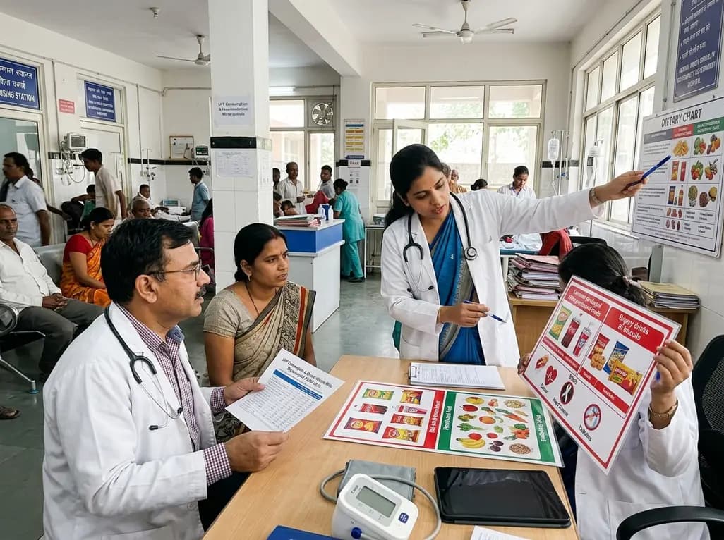
A landmark dose-response meta-analysis of 8.8 million adults reveals that high ultra-processed food intake significantly elevates risks for cardiovascular disease, cancer, diabetes, depression, and all-cause mortality, highlighting urgent needs for clinical lifestyle interventions.
Today
