

Loading, please wait...
Loading, please wait...
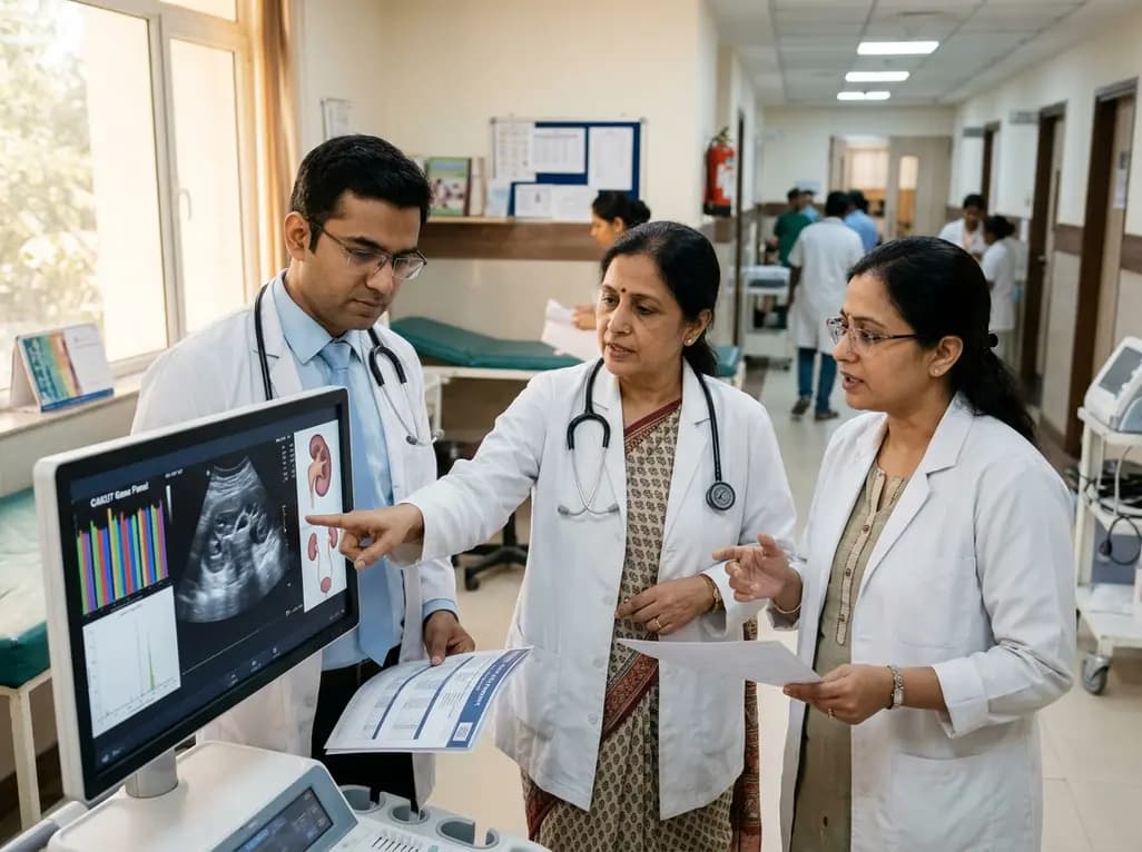
Congenital anomalies of the kidney and urinary tract represent the leading cause of pediatric end-stage kidney failure worldwide. Clinicians now recognize that understanding genetics in CAKUT provides vital insights into disease pathogenesis, patient risk, and familial counseling. Although anatomical malformations frequently require urgent surgical evaluation, underlying genomic variants govern long-term parenchymal survival and extrarenal morbidity. Consequently, pediatric and adult urologists must integrate modern molecular findings into routine practice to optimize patient care.
Congenital renal anomalies display an extraordinary spectrum of anatomical diversity. For example, clinical presentations range from isolated vesicoureteral reflux to bilateral renal agenesis and severe multicystic dysplasia. Historically, clinicians viewed these malformations purely as isolated mechanical disruptions during embryogenesis. However, recent genomic discoveries confirm that disrupted developmental pathways frequently cause abnormal ureteric bud outgrowth and defective metanephric differentiation.
Importantly, monogenic mutations account for nearly twenty percent of developmental urinary disorders. In addition, large copy number variations frequently trigger complex urinary phenotypes alongside multi-organ defects. Nevertheless, variable expressivity and incomplete penetrance complicate clinical interpretation. For instance, relatives carrying identical pathogenic mutations often exhibit widely discordant clinical courses. While one family member exhibits mild unilateral anomalies, another might progress to end-stage kidney disease during early childhood. Furthermore, maternal metabolic factors and epigenetic mechanisms modify structural gene expression during fetal organogenesis. Therefore, urologists must appreciate this biological complexity when counseling families and arranging genetic investigations.
Selecting appropriate candidates for genomic evaluation represents a critical clinical responsibility. Because genetic testing involves substantial financial expense, urologists must apply clear diagnostic criteria. In particular, diagnostic yields rise dramatically in patients presenting with bilateral parenchymal malformations or severe renal hypodysplasia. When congenital defects affect both kidneys, monogenic causes occur much more frequently.
Furthermore, clinicians should initiate genetic evaluation whenever marked discrepancies emerge between anatomical abnormalities and biochemical renal function. If a child with mild hydronephrosis develops severe, progressive renal impairment, an underlying developmental nephropathy likely exists. Additionally, consanguineous parentage and positive family histories strongly favor targeted genomic testing. For instance, identifying urinary defects across multiple generations confirms an inherited mendelian trait. Conversely, isolated unilateral anomalies such as unilateral pelviureteric obstruction rarely stem from monogenic variants. In these sporadic cases, corrective surgery successfully resolves the obstruction without extensive genomic screening. Thus, prioritizing bilateral, severe, or familial disease maximizes diagnostic utility and optimizes healthcare resources.
A confirmed genetic diagnosis provides significant clinical value by uncovering unrecognized extrarenal abnormalities. Notably, structural renal defects often represent the earliest manifestation of multi-system developmental syndromes. For example, pathogenic mutations in the HNF1B gene cause renal cysts alongside diabetes syndrome, pancreatic atrophy, and abnormal liver enzymes. Early molecular diagnosis allows clinicians to initiate proactive endocrine monitoring before symptomatic hyperglycemia develops.
Similarly, mutations in PAX2 cause renal coloboma syndrome, producing optic nerve dysplasia and progressive renal hypoplasia. Therefore, an urgent ophthalmological examination becomes essential whenever testing identifies a PAX2 mutation. Furthermore, branchio-oto-renal syndrome pairs branchial fistulae and hearing impairment with urinary malformations. Because these syndromic conditions present with subtle extrarenal signs, urologists may overlook them during focused surgical evaluations. However, discovering a systemic genetic variant prompts immediate multidisciplinary referrals to audiology, ophthalmology, and medical genetics specialists. Consequently, genomic testing transforms isolated surgical therapy into comprehensive systemic healthcare for complex pediatric patients.
Although genomic testing offers vital diagnostic data, genetic results must never replace standard surgical indications. Indeed, mechanical obstruction and significant urinary stasis require urgent anatomical correction regardless of underlying genetic findings. For instance, severe posterior urethral valves cause rapid hydrostatic bladder injury and renal parenchymal destruction. Urologists must ablate these valves immediately to preserve renal tissue, even if pathogenic mutations exist.
Similarly, high-grade pelviureteric junction obstruction causing functional decline demands prompt pyeloplasty. Surgical intervention successfully resolves urinary stasis, prevents recurrent pyelonephritis, and protects surviving nephrons. However, surgery cannot restore congenitally deficient nephron numbers or reverse intrinsic parenchymal dysplasia. Consequently, successful structural reconstruction does not guarantee normal long-term renal function in dysplastic kidneys. Clinicians must clearly explain this distinction to parents before performing operative procedures. By separating mechanical repair from genetic nephron loss, surgeons establish realistic long-term expectations. Thus, molecular insights complement rather than replace expert surgical management in everyday clinical practice.
Genomic insights substantially improve long-term prognostic counseling for pediatric and adult urology patients. Specifically, identifying causative genetic variants helps clinicians anticipate the trajectory of renal functional decline. While children with isolated obstructive lesions often achieve stable kidney function after surgery, patients harboring monogenic defects face progressive renal failure. Therefore, pediatric nephrologists and urologists must establish personalized surveillance protocols based on underlying genomic findings.
Furthermore, genetic diagnoses guide accurate reproductive counseling and familial risk stratification. For example, parents receive reliable recurrence figures for subsequent pregnancies after geneticists differentiate de novo mutations from inherited variants. In addition, adult urologists frequently encounter mature patients who transition from pediatric care. Many adults with congenital renal hypoplasia present with sudden hypertension or proteinuria during routine medical visits. Identifying genetic anomalies in these adult cohorts informs cardiovascular risk reduction and avoids nephrotoxic exposures. Ultimately, close collaboration among urologists, geneticists, and nephrologists ensures optimal lifespan care across diverse clinical settings.
Genetic testing provides high diagnostic utility in patients presenting with bilateral parenchymal malformations, severe hypodysplasia, or multi-organ syndromic features. Furthermore, clinicians should evaluate children with positive family histories of developmental renal anomalies or consanguineous parentage. Conversely, isolated unilateral anomalies rarely yield pathogenic variants. Therefore, targeted genomic screening optimizes diagnostic success, clarifies familial recurrence risks, informs clinical prognosis, and avoids unnecessary financial expenditure for affected families.
Genetic testing never replaces indicated surgical interventions for structural anomalies. When progressive obstruction or severe vesicoureteral reflux threatens renal function, urologists must perform prompt surgical decompression to prevent irreversible tissue damage. However, genetic evaluations complement surgery by identifying intrinsic nephron deficiencies that surgery cannot resolve. Consequently, molecular data guide medical surveillance and family counseling without altering established indications for necessary operative repair.
A confirmed genetic diagnosis allows clinicians to calculate precise recurrence risks and offer targeted carrier screening to relatives. Furthermore, identifying specific pathogenic mutations alerts physicians to unsuspected extrarenal complications, including sensorineural hearing loss, early-onset diabetes, and ocular colobomas. With this vital information, medical teams implement proactive surveillance protocols that detect secondary manifestations early, improving overall patient safety and long-term quality of care.
Disclaimer: This content is for informational and educational purposes only... Refer to the latest local and national guidelines for clinical practice.
References

Read summarized clinical updates, watch expert medical content, and earn CME certifications right from your smartphone.
Open your smartphone camera to scan and install.





Explore the critical role of genetics in CAKUT for pediatric and adult urologists. Learn when to test, how molecular results guide prognosis, and why surgery remains vital.
Today
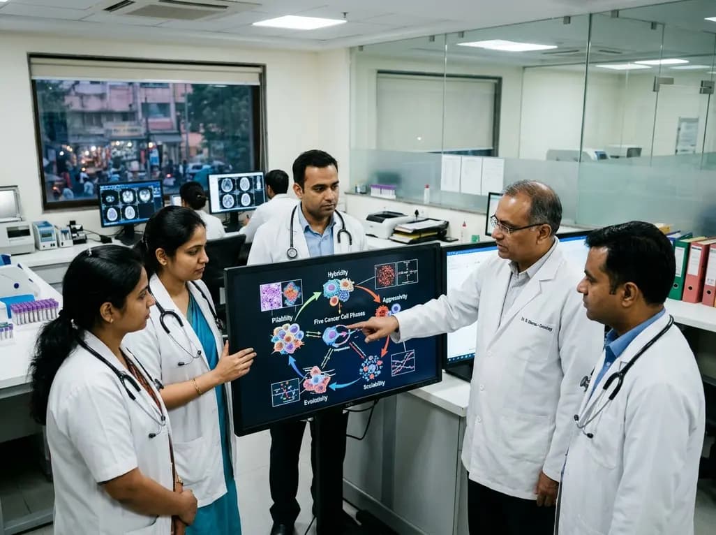
The PHASE framework conceptualizes cancer biology through five interacting processes: Pliability, Hybridity, Aggressivity, Sociability, and Evolutivity. This regulatory circuit offers a systems-level perspective on tumor heterogeneity, disease progression, and interaction-guided therapeutic strategies.
Today

Maternal cardiovascular risk factors such as hypertension, obesity, and diabetes significantly elevate offspring cardiovascular disease risk through placental dysfunction and fetal programming. Enhancing preconception screening and antenatal care offers clinicians a powerful strategy to protect long-term heart health.
Yesterday
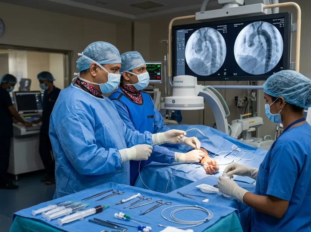
The novel CAST technique utilizes the indwelling Safari XS guidewire as a tracking rail to reposition a radial pigtail catheter into the descending aorta during minimalist TAVI, overcoming challenging vascular anatomy without requiring secondary femoral access or specialized equipment.
Today

A retrospective cohort study reveals that lumbar spine bone mineral density does not distinguish extremity long bone fracture status in adults with severe motor and intellectual disabilities. Because age drove fracture risk rather than spinal density, clinicians must adopt comprehensive assessment tools.
Yesterday

A case report details how reusing single-use insulin cartridges caused device failure and diabetic ketoacidosis in a type 1 diabetes patient using an advanced hybrid closed-loop system. The report highlights technological blindspots, mechanical degradation, and safety protocols during supply shortages.
Today
