

Loading, please wait...
Loading, please wait...

Pediatric craniofacial reconstruction demands an intricate balance between structural stability and dynamic cranial growth. Primary fronto-orbital advancement remains the benchmark surgical intervention for nonsyndromic unilateral coronal craniosynostosis. However, reconstructive surgeons continually refine fixation modalities to mitigate secondary relapse and growth restriction. A recent cohort study published in the Journal of Craniofacial Surgery evaluates the efficacy of a hybrid fixation technique that integrates resorbable plates with absorbable sutures. This detailed analysis examines how this tailored strategy achieves remarkable cosmetic outcomes while preserving perioperative safety.
Unilateral coronal craniosynostosis occurs when a single coronal suture fuses prematurely in infancy. Consequently, this premature fusion restricts anterior-posterior skull growth on the affected side while driving compensatory calvarial expansion elsewhere. Clinicians identify a distinct phenotypic presentation termed anterior plagiocephaly. This deformity features ipsilateral forehead flattening, superior and posterior recession of the supraorbital rim, and contralateral frontal bossing. Additionally, patients frequently present with the characteristic Harlequin eye sign on radiographic imaging, marked by elevation of the lesser sphenoid wing.
Beyond aesthetic asymmetry, uncorrected coronal fusion poses significant clinical risks. Unrelieved calvarial constraint can elevate intracranial pressure and compromise neurocognitive development. Furthermore, secondary orbital dysmorphology frequently provokes ocular motility disorders, including vertical strabismus and astigmatism. When pediatric patients present past the optimal early window for minimally invasive endoscopic strip craniectomy, open reconstruction becomes imperative. Therefore, surgical teams employ cranial vault remodeling to re-establish intracranial volume, protect developing brain parenchyma, and restore facial symmetry before permanent osseous consolidation occurs.
Historically, craniofacial surgeons secured osteotomized cranial fragments using stainless steel wires or titanium microplates. Although metallic fixation provides rigid structural stability, it introduces notable long-term clinical complications. Pediatric skull expansion often causes rigid metallic hardware to translocate intracranially or erode through thin infant skin. Moreover, metallic plates can restrict physiological calvarial growth, creating secondary contour irregularities. To address these limitations, surgeons adopted biodegradable polymer plates made from poly-L-lactic and polyglycolic acids.
However, pure resorbable plating systems also present distinct surgical challenges. Applying extensive rigid plates across multiple infant bone segments requires substantial foreign material. Consequently, this foreign mass increases operative expense and may incite localized foreign-body inflammatory reactions. In response, modern craniofacial teams developed hybrid fixation. This balanced concept deploys biodegradable plates solely at crucial load-bearing structural transitions, such as the lateral orbital rim and temporal tenon extensions. Meanwhile, surgeons secure individual remodeled calvarial bone fragments using flexible absorbable sutures. Thus, hybrid fixation provides immediate stability while reducing foreign implant mass and preserving bone compliance.
A dedicated surgical team at the JSC National Center for Neurosurgery in Astana, Kazakhstan, conducted a retrospective cohort investigation. The investigators evaluated 14 consecutive pediatric patients treated between January 2023 and December 2024. Every enrolled patient had computed tomography confirmation of primary, nonsyndromic unilateral coronal craniosynostosis. The cohort presented at a mean surgical age of 16.7 months, representing children who had progressed beyond the timeframe for endoscopic suturectomy.
Surgeons executed an extended fronto-orbital advancement combined with meticulous cranial vault remodeling. Through a coronal zig-zag incision, the team performed bifrontal craniotomies and mobilized the supraorbital bandeau. They carefully reshaped the recessed supraorbital bar, advanced the affected side, and repositioned the remodeled frontal bone flaps. Crucially, the reconstructive protocol utilized hybrid fixation. Biodegradable plates established firm stabilization at key structural anchor points across the supraorbital rim. Simultaneously, surgeons reapproximated the reshaped calvarial segments using high-tensile absorbable braided sutures. The team monitored clinical progress over a mean postoperative follow-up interval of 16 months.
The clinical results revealed exceptional aesthetic success across the entire patient cohort. Independent evaluators graded surgical outcomes using the validated Whitaker classification system. Remarkably, 92.9% of patients achieved a Whitaker Class I outcome, signifying complete aesthetic restoration requiring no revision surgery. The remaining 7.1% achieved Class II status, requiring only minor soft-tissue adjustments. Notably, no patient demonstrated Class III or Class IV deformities, and zero cases required major revision osteotomies.
Furthermore, an independent panel of ten clinician assessors scored postoperative aesthetics using a Visual Analog Cosmetic Score. The median cosmetic score reached 9.5 out of 10, highlighting robust aesthetic harmony. Objective morphometric imaging confirmed these clinical observations. Specifically, the Frontal Asymmetry Index decreased significantly in 10 of the 14 patients, proving substantial frontal realignment. Although Cephalic Index values decreased in six children, overall cranial proportions did not completely normalize. Additionally, nasal deviation angles showed no statistically significant directional shift. These objective findings emphasize that while hybrid fronto-orbital advancement restores regional frontal symmetry effectively, global skull dysmorphology may partially persist.
Extensive pediatric calvarial reconstruction entails inherent perioperative challenges, particularly regarding hemodynamic stability. Scalp elevation, cranial osteotomies, and dural dissection frequently precipitate significant intraoperative blood loss. In this series, all 14 patients received intraoperative autologous blood reinfusion alongside allogeneic packed red blood cell transfusions. Cell salvage strategies minimized donor blood exposure, stabilized intraoperative hemodynamics, and preserved adequate oxygen delivery.
Importantly, the study demonstrated an exemplary safety profile throughout the perioperative timeline. The surgical team recorded zero instances of postoperative cerebrospinal fluid leaks, dural tears, or surgical-site infections. Furthermore, clinicians detected no hardware infections, plate expulsions, or secondary bone flap resorption during follow-up. The mean total hospital stay spanned 18.9 days, encompassing comprehensive preoperative staging and cautious postoperative convalescence. Although extended hospitalization reflects conservative inpatient monitoring common in regional tertiary centers, early clinical stability remained universal. Consequently, these safety metrics validate the procedural viability of hybrid stabilization in complex pediatric cranial reconstruction.
This single-center experience highlights the therapeutic promise of hybrid fixation for unilateral coronal craniosynostosis. By combining bioabsorbable plates at key structural points with absorbable sutures for calvarial reshaping, surgeons achieve outstanding frontal symmetry and robust structural integrity. Moreover, this approach minimizes foreign material load, curtailing procedural costs and potential biological reactivity. The remarkable absence of early relapses or reoperations supports the clinical reliability of this technique.
Nevertheless, craniofacial specialists must interpret these findings within their clinical context. The authors emphasize that this retrospective cohort cannot isolate the precise mechanical contribution of hybrid fixation versus the comprehensive fronto-orbital osteotomy itself. Furthermore, open reconstruction beyond 12 months requires diligent blood management and coordinated multidisciplinary neurosurgical collaboration. Future multicenter investigations featuring larger cohorts and extended follow-up into skeletal maturity remain essential. Thus, prospective comparative trials will clarify long-term vector stability, cranial growth dynamics, and neurocognitive development following hybrid craniofacial reconstruction.
Hybrid fixation combines biodegradable plates at high-stress structural anchor points with flexible absorbable sutures between remodeled calvarial fragments. Consequently, this balanced construct secures immediate cranial stability while avoiding rigid circumferential encasement. Surgeons effectively reduce foreign material burden, lower localized bone resorption risks, and minimize restrictions on natural brain expansion. Furthermore, this dynamic technique decreases the likelihood of hardware palpability and late plate migration, supporting physiological pediatric skull growth during rapid developmental phases.
The Whitaker classification categorizes aesthetic and structural outcomes after craniofacial reconstruction into four clinical tiers based on the necessity for surgical revision. Category I represents an ideal outcome requiring no revision. Category II denotes minor contour irregularities treatable with localized soft-tissue adjustments. In contrast, Category III necessitates significant osteotomies or bone grafting, while Category IV requires a complete reoperation. Thus, achieving Category I or II reflects favorable procedural stability and predictable long-term cosmetic harmony.
Cranial vault reconstruction in young infants carries substantial risks of rapid hemorrhage due to extensive scalp vascularity, bone marrow bleeding, and dural venous stripping. Because infant blood volume is relatively low, even moderate blood loss quickly precipitates hemodynamic instability. Therefore, surgical teams utilize proactive blood-conservation protocols, including autologous cell salvage, meticulous bipolar electrocautery, and timely allogeneic transfusion. Consequently, maintaining tight physiological control prevents coagulopathy, supports tissue perfusion, and protects neurological function throughout these lengthy reconstructive procedures.
Disclaimer: This content is for informational and educational purposes only and should not be taken as professional medical advice. Always consult a qualified healthcare provider for diagnosis and treatment of medical conditions. Refer to the latest local and national guidelines for clinical practice.
References

Read summarized clinical updates, watch expert medical content, and earn CME certifications right from your smartphone.
Open your smartphone camera to scan and install.





A cohort study evaluates fronto-orbital advancement with hybrid fixation in pediatric unilateral coronal craniosynostosis. The technique achieved favorable Whitaker class I outcomes and significant symmetry correction without major complications.
Today

A master diving instructor suffered severe decompression sickness with hemiplegia and hepatic intravascular gas after rapid ascent. Emergency hyperbaric oxygen therapy via US Navy Treatment Table 6 achieved prompt, complete clinical recovery, underscoring essential dive safety and rapid recompression protocols.
Today

The GESS-AEEM consensus establishes a clear clinical implementation framework for testosterone therapy in postmenopausal women with hypoactive sexual desire disorder, focusing on transdermal dosing, precise laboratory monitoring, and safety communication.
Today

A Bayesian component network meta-analysis of 130 RCTs reveals that self-monitoring and gamification provide the clearest positive effects in mHealth physical activity interventions, while social comparison may have adverse psychological trade-offs.
Today
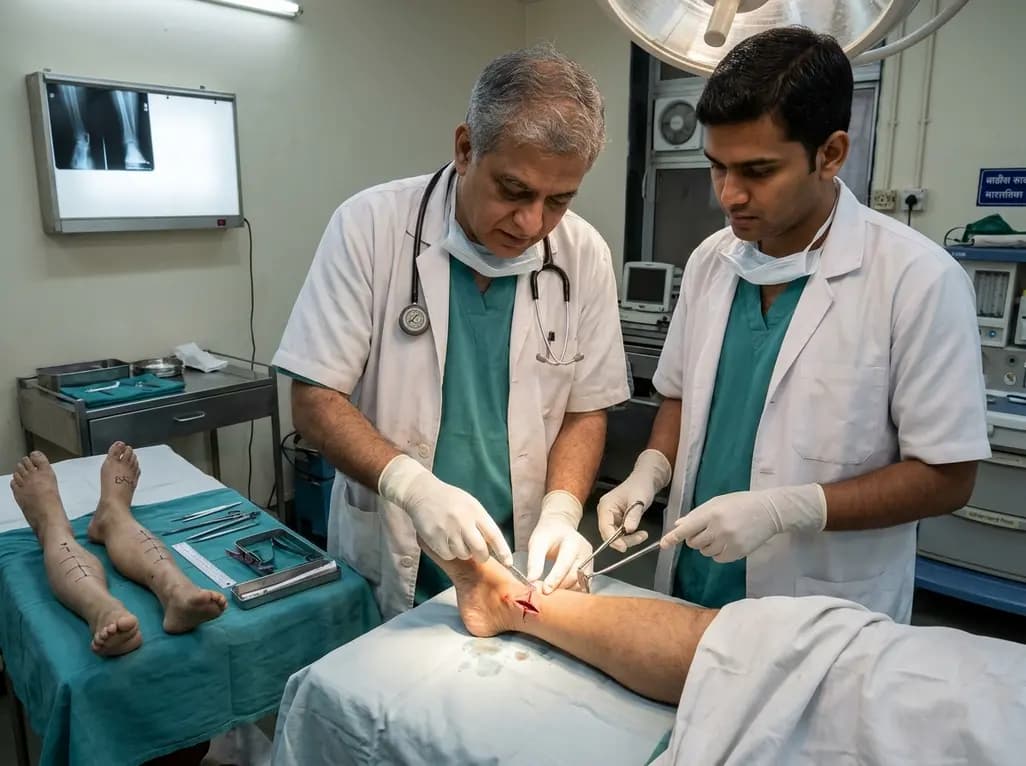
A paired cadaveric study compared triple and quintuple percutaneous Achilles tendon lengthening. While quintuple hemisection showed numerically greater elongation, differences were not statistically significant.
2 days back

With GLP-1 receptor agonists reducing daily caloric intake by up to 39%, Indian patients face a heightened risk of sarcopenia and micronutrient deficits. An expert consensus highlights structured dietary interventions, high-quality protein, and diabetes-specific nutrition to safeguard metabolic outcomes.
Today
