
Loading, please wait...
Loading, please wait...
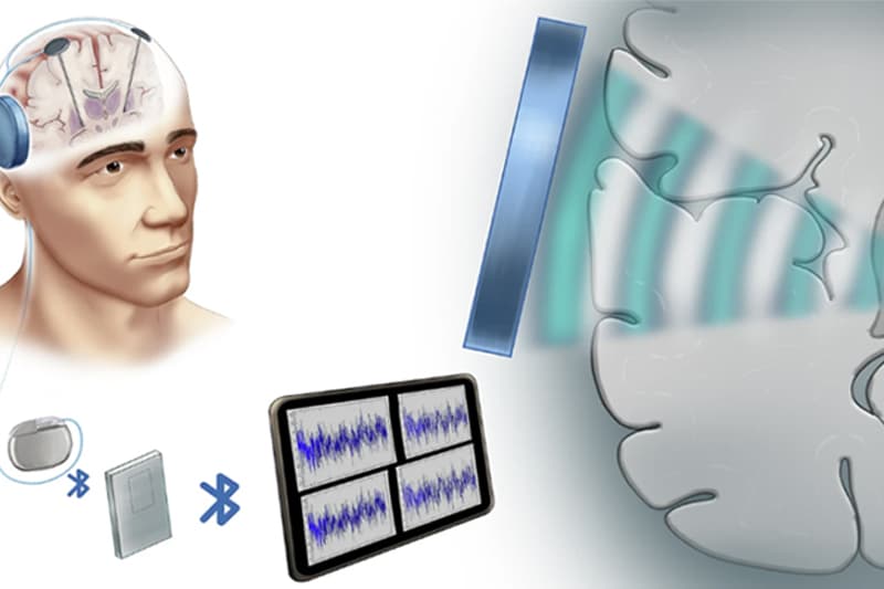
Focused ultrasound neuromodulation represents a revolutionary frontier for treating complex neurological disorders. This non-invasive technology uses acoustic energy to target deep brain structures precisely. However, most preclinical research relies on healthy animal models. Consequently, understanding how a diseased brain reacts differently than a healthy one is essential for clinical progress.
Recent research investigated how Parkinson’s disease (PD) impacts the efficacy of focused ultrasound neuromodulation. Scientists used a unilateral dopamine depletion model to compare responses between healthy and diseased mice. They specifically targeted the left external globus pallidus (GPe) using a wearable device. Interestingly, the results showed that the disease state completely changed the brain's response to the intervention.
In mice with Parkinson’s, the stimulation significantly inhibited GPe calcium activity for approximately three minutes. Furthermore, this physiological change led to dramatic motor improvements. The researchers observed a reduction in ipsilateral circling that persisted for at least 50 minutes after the session. In contrast, healthy mice showed no significant changes in calcium levels or rotational behavior.
One major concern with any deep-brain intervention is the potential for tissue damage. Fortunately, histological analysis confirmed that this specific protocol caused no neuronal damage or inflammation. There was no evidence of astrocytic activation or microglial proliferation in either group. These safety findings support the potential for translating this technology into human clinical trials.
Notably, the transient inhibition of the GPe appears to correct the pathological overactivity typical of the Parkinsonian state. This selective response highlights why evaluating neuromodulation strategies in relevant disease models is critical. Therefore, future therapies must account for the specific neurochemical environment of the patient’s brain to ensure maximum therapeutic benefit.
In this study, the inhibition of brain cell activity lasted for three minutes, while the improvements in motor movement lasted for 50 minutes after stimulation.
No. Histological exams revealed no signs of cell death, scarring, or immune system activation in the brain regions targeted by the ultrasound.
The ultrasound appears to correct specific imbalances caused by dopamine loss. Because healthy mice do not have these imbalances, the stimulation does not produce the same inhibitory or behavioral effects.
Disclaimer: This content is for informational and educational purposes only. It does not constitute medical advice and is not intended to replace professional judgment or treatment. Refer to the latest local and national guidelines for clinical practice.
References

Read summarized clinical updates, watch expert medical content, and earn CME certifications right from your smartphone.
Open your smartphone camera to scan and install.


New research shows focused ultrasound neuromodulation selectively inhibits GPe activity and improves motor function in Parkinson's mice, but not in healthy ...
5 months ago

Andhra Pradesh reported 10 new Covid-19 cases, taking the state tally to 49 while deaths remain at four. With 24 patients hospitalized and 16 under home isolation, the Health Department has intensified monitoring. Medical professionals should review regional distribution, diagnostic protocols, and management plans.
Today

An 11-year Swedish registry study of 618 uterine sarcoma patients found that minimally invasive surgery yielded survival comparable to open surgery in early stages. However, adjuvant chemotherapy conferred no survival benefit in localized or advanced disease, highlighting stage and histology as key outcomes.
3 days back

A cross-sectional study evaluates post-intensive care syndrome in cardiac patients 2-4 weeks post-ICU discharge, highlighting cognitive, psychological, and functional impairments and the need for structured multidisciplinary rehabilitation.
3 days back

Anterior cruciate ligament reconstruction failure lacks uniform definition. A narrative review proposes an integrative framework incorporating objective and subjective instability, persistent pain, restricted motion, graft rupture, and secondary meniscal injury to standardize clinical reporting.
3 days back

With World Obesity Atlas data warning that over 41 million Indian children are overweight or obese, ICMR and NIN have unveiled a 10-point policy roadmap. The initiative calls for mandatory front-of-pack labeling, HFSS taxes, strict marketing bans, and healthier school environments to curb non-communicable diseases.
Today
