.webp&w=384&q=75)

Loading, please wait...
Loading, please wait...
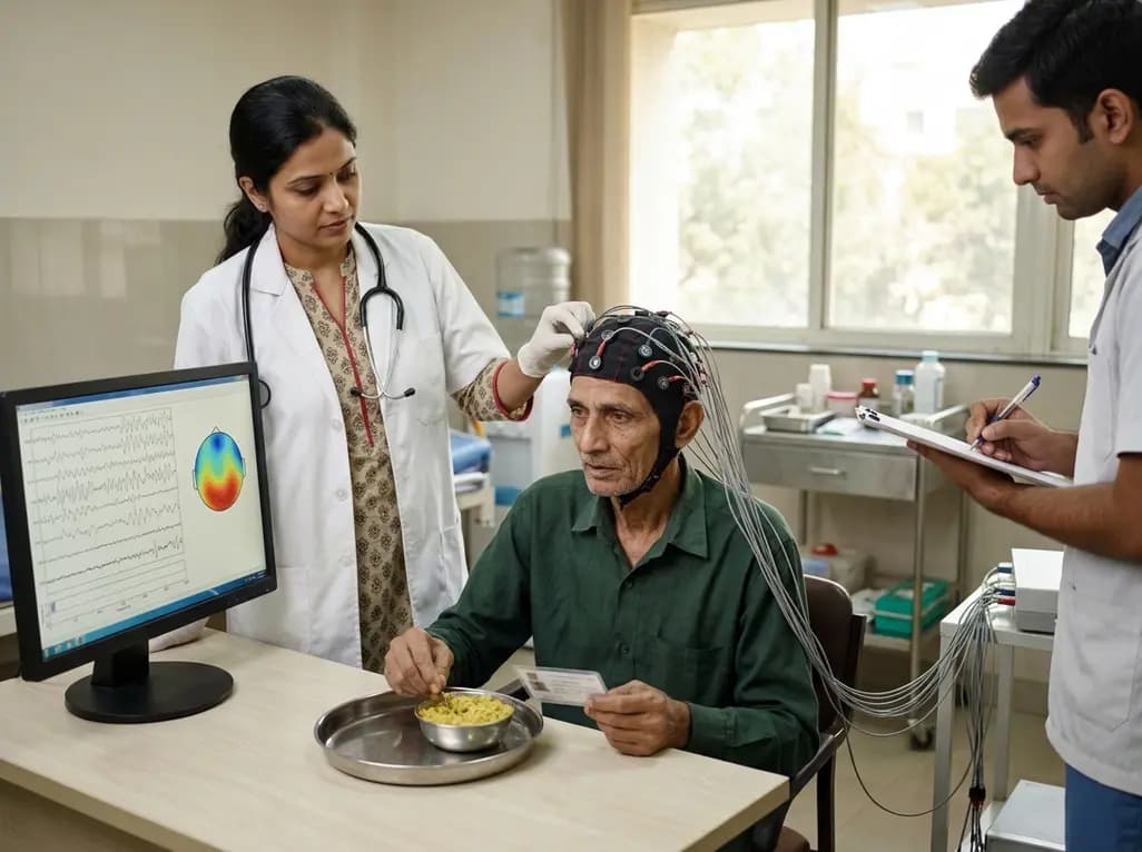
Geriatric anorexia and unexplained hyporexia represent major clinical challenges that directly impair nutritional status, physical recovery, and independent living in older adults. Even in the absence of overt dementia, many elderly individuals struggle to imagine or articulate their food desires, which severely complicates dietary interventions. Traditional appetite assessments depend heavily on subjective patient recall or visual analogue scales, both of which often fail in frailer populations. Consequently, developing an objective, non-invasive neuroimaging methodology has become an urgent clinical goal. Emerging exploratory research indicates that fNIRS appetite assessment targeting the medial and frontopolar prefrontal regions (MFPR) provides measurable, real-time hemodynamic signals during active meal consumption. By examining how cortical activation shifts in response to food preference and gender, clinicians may soon possess practical diagnostic biosensors to quantify eating motivation and tailor nutritional therapy effectively.
Appetite decline among aging populations arises from complex, overlapping pathophysiological mechanisms. Age-related changes in chemosensory perception, polypharmacy, gastrointestinal dysmotility, and altered neuroendocrine signaling all reduce baseline hunger cues. Furthermore, subclinical depressive symptoms and dental or swallowing difficulties frequently suppress eating desire. In hospital and long-term care environments, clinicians frequently encounter older patients who refuse meals but cannot clearly express their dietary preferences or food aversions.
Standard cognitive screening instruments and bedside dietary records lack the sensitivity required to evaluate hedonic food appraisal accurately. Moreover, conventional functional magnetic resonance imaging (fMRI) cannot readily capture the physiological mechanics of active chewing and swallowing due to severe motion artifacts and restrictive scanner geometry. Because nutritional deficits rapidly accelerate sarcopenia, immune compromise, and frailty, physicians need bedside-capable neuroimaging tools. Establishing objective cortical markers of meal appreciation in healthy cohorts therefore represents an essential stepping stone toward resolving diagnostic blind spots in geriatric nutrition and rehabilitation medicine.
Functional near-infrared spectroscopy offers a lightweight, portable, and motion-tolerant platform ideally suited for real-time nutritional neuroscience. In recent experimental protocols, researchers equipped participants with multichannel fNIRS head caps covering the medial and frontopolar prefrontal cortices to record continuous changes in oxygenated and deoxygenated hemoglobin levels during natural eating tasks.
Investigators evaluated healthy young volunteers across multiple test sessions under standardized nutritional states. Participants completed structured questionnaires detailing baseline food intake frequency, hedonic ratings, and specific dietary likes and dislikes. Subsequently, each participant attended distinct laboratory sessions to visually inspect and then consume either a standardized control dish representing typical home-cooked fare or a self-selected preferred dish purchased on the day of the experiment. Continuous hemodynamic recording spanned pre-meal resting periods, visual exposure, and extended active eating phases. This rigorous experimental paradigm ensured that researchers could isolate metabolic brain responses associated specifically with food reward processing from baseline masticatory motor exertion.
The human prefrontal cortex serves as a critical integration hub for hedonic valuation, executive control, and interoceptive appetite signaling. Specifically, the medial prefrontal cortex and frontopolar regions process reward expectancy and subjective pleasantness when individuals encounter palatable items. Data from fNIRS monitoring reveal distinct temporal phases in MFPR hemodynamic responses during actual food ingestion.
Notably, divergence in MFPR activation between control meals and preferred dishes becomes prominent approximately nine minutes after eating begins. This delayed cortical separation indicates that sustained neural engagement reflects hedonic appraisal and post-ingestive sensory satisfaction rather than simple oral-motor activation. As eating progresses, preferred meals elicit stable, elevated oxygenated hemoglobin concentrations across specific frontopolar channels. Conversely, neutral control meals demonstrate significantly different activation trajectories. These findings demonstrate that portable optical topography can reliably track sustained mealtime engagement and differentiate between automatic caloric consumption and genuine hedonic dining enjoyment.
Individual dietary habits and inherent hedonic valuation profoundly influence cortical activity during meal consumption. Statistical analyses indicate significant correlations between subjective food liking scores, lifetime intake frequencies, and the magnitude of MFPR hemodynamic shifts recorded by optical probes. When individuals consume highly rated foods, frontopolar channels exhibit heightened activation compared to baseline exposure states.
Additionally, exploratory findings highlight distinct gender-related variations in prefrontal hemodynamic recruitment during eating. Female and male participants exhibit differential response amplitudes across medial prefrontal networks when processing identical meal categories. These neural variations mirror established behavioral differences in taste sensitivity, dietary restraint, and central reward modulation. Understanding these gender-specific neurovascular signatures allows clinicians to interpret diagnostic neuroimaging signals with greater precision. Therefore, accounting for baseline dietary preference and patient demographics remains essential when calibrating objective neurofunctional appetite biosensors for clinical diagnostic protocols.
Transforming functional near-infrared spectroscopy into a point-of-care clinical tool holds exceptional promise for geriatricians, neurologists, and clinical dietitians. Because fNIRS caps are lightweight and non-invasive, clinicians can easily deploy them at bedside in acute geriatric wards, rehabilitation centers, and specialized nursing facilities.
In older adults with chronic hyporexia or sensory impairment who struggle to communicate their meal preferences, real-time cortical monitoring could pinpoint which meal textures, flavor profiles, or caloric densities generate positive reward responses. By identifying personalized hedonic triggers objectively, healthcare teams can design highly customized therapeutic meal plans that stimulate spontaneous oral intake. Consequently, this targeted strategy could substantially reduce the premature reliance on artificial enteral feeding tubes, enhance daily caloric compliance, and improve overall functional recovery trajectories in vulnerable medical inpatients.
Although these initial findings establish proof-of-concept for optical appetite assessment, several clinical and methodological limitations require systematic validation. Current baseline data derive primarily from young, cognitively intact adult cohorts. Therefore, clinical researchers must replicate these experimental paradigms in frail geriatric cohorts, stroke survivors, and individuals diagnosed with mild cognitive impairment or clinical depression.
Future studies should also evaluate how acute systemic inflammation, metabolic derangements, and polypharmacy modulate prefrontal hemodynamic signals during eating. Integrating wearable fNIRS hardware with synchronized autonomic monitoring, including continuous heart rate variability and electrogastrography, will provide a holistic view of the gut-brain axis. As wearable optical sensor technology continues to miniaturize, bedside neurofunctional appetite profiling may soon emerge as a practical standard of care across multidisciplinary geriatric and nutritional rehabilitation programs.
Functional near-infrared spectroscopy measures localized changes in oxygenated and deoxygenated hemoglobin within the cerebral cortex. Because the optical equipment is lightweight and highly resistant to motion artifacts, patients can comfortably chew and swallow while clinicians continuously record prefrontal hemodynamic responses to different meal presentations in real time.
The medial and frontopolar prefrontal regions serve as essential neural nodes for processing hedonic valuation, reward anticipation, and subjective food pleasantness. These cortical areas integrate sensory, metabolic, and emotional inputs, enabling objective discrimination between neutral caloric intake and genuinely rewarding, highly preferred dining experiences.
Many frail older adults struggle to express their dietary desires due to sensory decline or cognitive fatigue. By measuring prefrontal activation during meal exposure, clinicians can objectively identify which food flavors and textures stimulate positive neural reward, facilitating customized meal planning that improves voluntary oral intake.
Disclaimer: This content is for informational and educational purposes only. It is not intended to be a substitute for professional medical advice, diagnosis, or treatment. Always seek the advice of your physician or other qualified health provider with any questions you may have regarding a medical condition. Never disregard professional medical advice or delay in seeking it because of something you have read here. The authors, editors, and publishers are not responsible for errors or omissions or for any consequences from the application of the information provided. Refer to the latest local and national guidelines for clinical practice.
References
1. Takatsuru Y et al. Food preference and gender are associated with medial/frontopolar prefrontal regions functional near-infrared spectroscopy responses during eating: An exploratory study in young adults. PLoS One. 2026. doi: 10.1371/journal.pone.0343481. PMID: 42545988.
2. Landi F, Calvani R, Tosato M, Martone AM, Ortolani E, Savera G, Sisto A, Marzetti E. Anorexia of Aging: Risk Factors, Consequences, and Potential Treatments. Nutrients. 2016;8(2):69. doi: 10.3390/nu8020069.
3. Rolls ET. Taste, olfactory, and food reward value processing in the brain. Prog Neurobiol. 2015;127-128:64-90. doi: 10.1016/j.pneurobio.2015.03.002.

Read summarized clinical updates, watch expert medical content, and earn CME certifications right from your smartphone.
Open your smartphone camera to scan and install.





Groundbreaking research shows that functional near-infrared spectroscopy (fNIRS) over the medial and frontopolar prefrontal cortex can track real-time neural responses during eating. These findings provide an objective framework for evaluating food preferences and addressing anorexia of aging in clinical settings.
last month

A systematic review of 41 preclinical studies reveals that baicalin dampens systemic inflammation, inhibits NF-κB and NLRP3 pathways, and improves organ preservation in sepsis models. However, substantial study heterogeneity and a complete lack of human clinical trials highlight the need for further translational research.
Today
Discover the crucial distinctions between mesonephric and mesonephric-like proliferations of the female genital tract. Learn the key histomorphologic, immunohistochemical, and molecular differences essential for accurate diagnosis and clinical management.
Today

A large UK Biobank study reveals that plasma proteomic signatures capture preclinical organ damage and improve multiorgan risk prediction across early CKM syndrome stages 0 to 2 beyond traditional PREVENT clinical models.
Today
A narrative review investigates whether the articularis genus muscle functions as an independent anatomical entity or blends with the vastus intermedius. We evaluate its morphology, role in retracting the suprapatellar bursa, and direct clinical significance in anterior knee pain and arthroplasty.
Today

A Bayesian multilevel meta-analysis reveals that aerobic training combined with moderate carbohydrate restriction modestly lowers HbA1c in type 2 diabetes. However, sparse data and very low certainty leave incremental benefits over exercise or diet alone unproven, highlighting the need for individualized care.
Today
