.webp&w=384&q=75)

Loading, please wait...
Loading, please wait...

Clear aligner therapy relies heavily on composite resin attachments to facilitate complex orthodontic tooth movements. Consequently, clinicians must perform clear aligner attachment debonding when patients conclude their active orthodontic treatment. While bonding these tooth-colored resin buttons requires meticulous placement, their complete removal presents an ongoing clinical challenge. Practitioners must carefully eliminate all composite remnants without gouging the underlying enamel layer. In daily dental practice, visual inspection under standard dental operating lights often fails to differentiate resin from natural tooth structure. Therefore, operators frequently struggle to identify the exact margins between the composite and native dental tissue. Incomplete adhesive clean-up leaves behind rough surfaces that foster bacterial biofilm accumulation, plaque retention, and superficial staining. Conversely, overly aggressive mechanical grinding with rotating burs often produces permanent scratches, microcracks, and irreversible enamel loss. As a result, treatment outcomes often vary dramatically depending on operator experience and visual acuity. Orthodontic researchers therefore continue to evaluate innovative visual aids that standardize resin clean-up while preserving healthy tooth structure. Specifically, optical differentiation methods offer promising ways to solve this long-standing clinical dilemma and improve patient care. By illuminating subtle structural differences, these methods help dentists establish safer clinical routines.
To overcome the visual limitations of traditional dental lighting, researchers developed the fluorescence-aided identification technique. This optical approach exploits the distinct fluorescence properties exhibited by specialized dental biomaterials when exposed to specific wavelengths of light. Natural human enamel emits a characteristic baseline fluorescence under ultraviolet or blue illumination around 405 nanometers. In contrast, manufacturers can incorporate specific fluorescent fluorophores into orthodontic flowable composite resins. When clinicians illuminate the operating field with violet-blue light or ultraviolet radiation, the modified composite resin fluoresces intensely compared to the adjacent enamel. Consequently, the attachment material glows distinctly, establishing sharp visual contrast against the tooth substrate. Practitioners can instantly delineate the adhesive boundary without relying on tactile feedback or imprecise dry-field reflection. Furthermore, this visible boundary guides rotary instruments with high spatial precision during bur-assisted removal. Clinicians can selectively abrade the glowing composite material while immediately halting instrumentation once the fluorescence disappears. As a result, this targeted approach minimizes speculative grinding on pristine enamel. Moreover, dental professionals can verify complete adhesive clearance at chairside quickly without requiring chemical staining agents or abrasive disclosing dyes. This reliable differentiation simplifies clean-up and boosts clinical confidence.
A recent controlled in vitro investigation conducted by Bordihn and colleagues evaluated the objective clinical efficacy of this optical method. Specifically, the researchers sought to determine whether ultraviolet-guided attachment removal improves procedural efficiency while reducing enamel loss and composite residue. The investigative team prepared twenty standardized dental models utilizing bovine teeth, which serve as established biological substitutes for human enamel. Subsequently, the investigators bonded clear aligner attachments onto the teeth using an advanced fluorescent-modified flowable composite known as AlignerFlow LC. To eliminate anatomical bias, the experiment utilized a rigorous split-mouth investigation design. The operators removed half of the attachments using the fluorescence-aided identification technique under targeted ultraviolet illumination. Conversely, the operators removed the remaining attachments under standard white dental operating light. Throughout the debonding process, investigators recorded the exact operative time required for each individual tooth surface. Furthermore, the researchers captured high-resolution intraoral surface scans and optical coherence tomography images at baseline and post-treatment intervals. They processed the three-dimensional surface datasets using specialized Avizo analytical software to calculate precise volumetric changes. Additionally, independent evaluators graded the optical coherence tomography cross-sections using validated qualitative rating scales to ensure robust findings.
The investigation yielded vital quantitative insights regarding procedural duration, structural preservation, and resin clearance. Interestingly, timing measurements revealed a trend toward faster attachment debonding in the fluorescence-aided group. However, total operative duration did not achieve statistical significance between the two illumination protocols because high inter-individual variability influenced the timing metrics. In contrast, three-dimensional volumetric surface analysis demonstrated a statistically significant reduction in composite residue when operators utilized the fluorescence-aided identification technique. The ultraviolet visualization clearly enabled operators to identify and excise residual resin islands that remained completely unnoticed under conventional white lighting. Importantly, the digital analysis demonstrated no significant difference in volumetric enamel loss between the fluorescent and non-fluorescent removal groups. High-resolution optical coherence tomography scans reinforced these digital findings across both cohorts. The optical cross-sections confirmed that both enamel loss and resin remnants occurred almost exclusively on a microscopic scale across all treated specimens. Thus, the fluorescence-assisted technique dramatically lowered macroscopic adhesive persistence without aggravating iatrogenic enamel ablation. Consequently, the fluorescent approach provides a reliable mechanism to achieve cleaner tooth surfaces without elevating the risk of structural dental injury or unnecessary tissue destruction.
These laboratory findings carry direct, practical implications for orthodontic practitioners and clear aligner providers worldwide. Removing attachments thoroughly represents an essential quality benchmark at the conclusion of orthodontic tooth movement. When residual resin lingers on the enamel surface, it predisposes patients to localized plaque stagnation, gingival irritation, and esthetic dissatisfaction. By integrating fluorescent flowable composites and compatible illumination devices, dental clinicians can significantly enhance clean-up reliability. Furthermore, this optical method reduces the clinician fatigue associated with guessing adhesive boundaries on shiny enamel surfaces. Clinicians can confidently remove composite buttons using fine finishing burs under direct fluorescent guidance, preserving the native tooth architecture. Nevertheless, practitioners must recognize that an in vitro setting cannot fully simulate intraoral complexities like patient movement, saliva contamination, or limited posterior access. Therefore, dental clinicians must await multi-operator in vivo clinical trials to verify whether these benefits transfer seamlessly into routine chairside workflows. Future investigations must also evaluate different adhesive formulations, varying bur grits, and long-term enamel roughness parameters. In the interim, incorporating fluorescence guidance offers a predictable, evidence-supported strategy to standardize orthodontic finishing procedures, protect sound tooth structure, and safeguard long-term patient oral health.
Manufacturers incorporate specialized fluorescent pigments into the resin matrix of flowable composite materials. When clinicians expose these materials to violet-blue or near-ultraviolet light at approximately 405 nanometers, the fluorophores absorb the high-energy photons and spontaneously emit visible light at longer wavelengths. This optical emission creates an intense luminous contrast against adjacent natural enamel, which allows practitioners to easily differentiate resin margins from sound tooth structures during rotary bur clean-up.
Although the fluorescence-aided technique significantly reduces residual composite resin, it cannot completely eliminate microscopic enamel loss. Every mechanical debonding procedure involving rotary tungsten carbide burs or polishing discs removes a minuscule amount of superficial enamel. However, the optical contrast provided by fluorescence helps operators avoid unnecessary deep grinding into healthy dental tissue. Consequently, the technique maintains enamel loss at an acceptable microscopic level while ensuring complete removal of bulk resin attachments.
Most standard dental curing lights emit broad blue light designed primarily for polymerizing photoinitiators like camphorquinone rather than inducing specific fluorescence contrast. Effective fluorescence-aided identification typically requires a dedicated illumination source or specialized diagnostic handpiece emitting light around 405 nanometers. Furthermore, clinicians must wear appropriate protective optical filters or orange-tinted eyewear. These specialized filters block distracting reflected violet light, allowing the operator to visualize the glowing composite residue with maximum clarity.
Disclaimer: This content is for informational and educational purposes only... Refer to the latest local and national guidelines for clinical practice.
References

Read summarized clinical updates, watch expert medical content, and earn CME certifications right from your smartphone.
Open your smartphone camera to scan and install.





An in vitro study reveals that using the fluorescence-aided identification technique (FIT) during clear aligner attachment debonding significantly reduces residual composite while preserving enamel integrity, offering a predictable, standardized clean-up protocol for dental clinicians.
Today
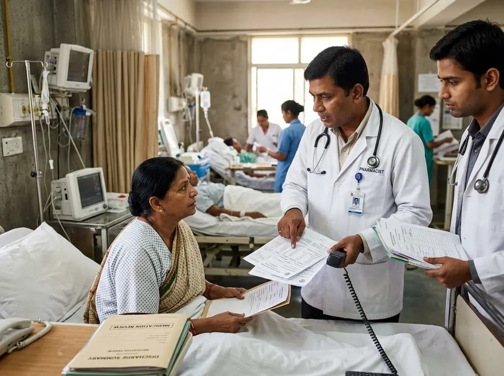
A retrospective cohort study demonstrates that comprehensive pharmacy transitions of care—including admission medication history, discharge review, and 72-hour telephone follow-up—significantly reduced 30-day readmission rates in acute care patients, highlighting the value of pharmacist-led clinical interventions.
Today

Meghalaya recorded a historic 55% reduction in maternal deaths, dropping from 244 to 107 over five years. Through the MOTHER app, dedicated transit homes, and expanded infrastructure like the 150-bed Tura Maternity Hospital with its first public NICU, the state demonstrates an effective public health model.
Today

Explore modern perioperative management protocols for clinical xenotransplantation. Learn how costimulation blockade, anti-inflammatory perfusion, and coagulopathy rescue overcome interspecies hurdles.
Today
A systematic review explores basivertebral nerve anatomy and its pivotal role in vertebrogenic pain. Learn how endplate inflammation and Modic changes inform targeted radiofrequency ablation.
Today

At Homa CME 2026 at Hyderabad's T-Hub, healthcare leaders emphasized moving beyond routine metrics through digital calculators and precision stratification. Discover why characterizing individual insulin secretion, resistance patterns, and vascular risks transforms chronic metabolic care in India.
Today
