.webp&w=384&q=75)

Loading, please wait...
Loading, please wait...
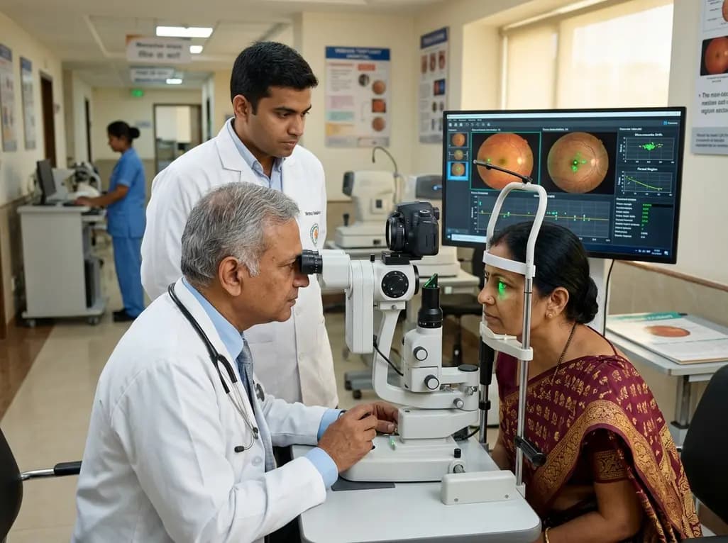
When individuals attempt to hold a steady gaze on a fine target, their eyes are never truly stationary. Instead, the visual system engages in continuous, microscopic shifts known as fixational eye movements. These fine adjustments comprise slow ocular drifts and rapid microsaccadic flicks that shift the incoming image across the photoreceptor mosaic. Recent groundbreaking research using adaptive optics imaging demonstrates that these involuntary motor dynamics are far from random noise. Rather, the oculomotor system operates with machine-like temporal and spatial precision to maintain optimal visual resolution. By steering visual inputs directly onto the preferred retinal locus, the visual apparatus ensures that high-demand discrimination tasks occur without perceptual blurring or target fading.
Fixational eye movements prevent neural adaptation in the retina while simultaneously stabilizing gaze on fine visual details. If an image remains completely motionless on the photoreceptor lattice, neural signaling rapidly habituates, causing the visual percept to fade entirely. To counteract this adaptation, the human visual pathway coordinates slow drift trajectories and rapid microsaccades. Ocular drift allows smooth scanning across adjacent cone photoreceptors, generating continuous changes in luminance that refresh retinal ganglion cell firing. However, uncorrected drift could eventually displace the target away from high-acuity foveal cones. Consequently, the brain executes corrective microsaccades to reposition the visual stimulus back toward the central locus. Adaptive optics tracking reveals that these combined dynamics are remarkably repeatable across repeated visual presentations. Therefore, human fixation operates as an active, highly regulated sensory-motor feedback loop rather than a passive mechanical state.
The central fovea represents the anatomical region of highest cone photoreceptor packing, but functional gaze targeting centers specifically on the preferred retinal locus (PRL). High-resolution Vernier acuity testing confirms that fine spatial resolution peaks directly at this functional center and within its immediate surrounding micro-zone. When visual stimuli land directly on the PRL or within a tiny radius, observers achieve optimal spatial discrimination. In contrast, visual performance declines consistently when target landing shifts more than 5 arcminutes away from this optimal locus. Fortunately, the oculomotor control system tightly restricts fixational variance. As a result, the landing location of an anticipated stimulus exhibits minimal spatial spread during the critical temporal window of stimulus presentation. This targeted confinement ensures that fine spatial details land precisely on the retinal zone best equipped to process high spatial frequencies.
Spatial precision alone does not guarantee superior visual performance; temporal coordination of ocular motion is equally vital. Recent physiological findings demonstrate that visual acuity is heavily dependent on the timing of preceding microsaccades. When a microsaccade occurs immediately prior to or during stimulus presentation, saccadic suppression and motion blur temporarily impair visual processing. Conversely, spatial discrimination reaches peak efficacy when the last microsaccade occurs sufficiently in advance of target presentation. During this quiescent post-saccadic interval, ocular drift stabilizes, and neural responsiveness across the primary visual cortex remains uninhibited. Consequently, the oculomotor apparatus times corrective movements to precede anticipated visual events. This precise coordination ensures that retinal motion remains minimal during critical discrimination phases, thereby safeguarding visual acuity from motion-induced degradation.
The implementation of adaptive optics scanning laser ophthalmoscopy has transformed visual neuroscience by overcoming optical aberrations of the ocular media. This technology allows researchers to project microscopic stimuli directly onto individual photoreceptors while recording retinal motion with sub-arcminute resolution. Through concurrent real-time tracking, investigators can determine the exact spatiotemporal coordinates of every visual presentation relative to individual cone photoreceptors. These precise measurements reveal that the oculomotor system dynamically adapts its motor program based on task demands. During demanding Vernier discrimination, microsaccadic frequency and drift trajectories adjust to constrain the retinal image within an exceptionally tight radius. Thus, modern optical instrumentation confirms that human gaze control maintains near-optimal spatial confinement under natural physiological conditions, effectively bridging the gap between cellular anatomy and functional perception.
Understanding the physiological boundaries of fixational stability provides critical insights for managing diverse ocular and neurological disorders. Conditions such as macular degeneration, amblyopia, and central serous chorioretinopathy frequently alter the anatomical architecture of the fovea, forcing patients to establish eccentric preferred retinal loci. By examining how fixational eye movements adapt to retinal pathology, clinicians can better assess functional visual impairment and guide targeted visual rehabilitation programs. Furthermore, subtle disruptions in microsaccadic dynamics, fixational drift, or ocular stability serve as early biomarkers for neurodegenerative disorders, including Parkinson disease and progressive supranuclear palsy. Integrating high-resolution gaze tracking into clinical evaluations will refine diagnostic sensitivity and support personalized rehabilitation for patients suffering from unstable ocular fixation.
Fixational eye movements are minute, involuntary ocular motions consisting of microsaccades, ocular drifts, and microtremors that occur during sustained visual gaze. They are essential because stationary images cause retinal photoreceptors to habituate, leading to visual fading. In addition, these movements dynamically shift images across high-density foveal cones, maintaining neural responsiveness and optimizing spatial resolution for high-acuity tasks.
The preferred retinal locus is the precise retinal sub-region that the visual system uses for steady fixation and fine visual discrimination. Because cone photoreceptor density and neural connectivity are maximized at and around this locus, visual resolution is highest when images land within a few arcminutes of it, declining sharply if targets shift beyond this zone.
Microsaccade timing directly affects visual acuity due to transient saccadic suppression and motion-induced blur. Visual resolution is highest when the preceding microsaccade occurs well before target presentation, allowing retinal image velocity to stabilize. If a microsaccade occurs immediately before or during presentation, visual sensitivity and spatial discrimination performance drop noticeably.
Disclaimer: This content is for informational and educational purposes only and should not be construed as medical advice. Healthcare professionals should evaluate clinical findings in the context of individual patient scenarios and refer to current clinical guidelines and institutional protocols.
References
Gautier J et al. How Gaze Direction and Dynamics Affect Visual Resolution. eNeuro. 2026 Aug 25. doi: undefined. PMID: 42642326.
Bowers NR, Gautier J, Lin S, Roorda A. Fixational eye movements in passive versus active sustained fixation tasks. J Vis. 2021;21(11):18.
Nghiem TAE, Witten JL, Dufour O, Harmening WM, da Silveira RA. Fixational eye movements as active sensation for high visual acuity. Proc Natl Acad Sci U S A. 2025;122(6):e2416266122.

Read summarized clinical updates, watch expert medical content, and earn CME certifications right from your smartphone.
Open your smartphone camera to scan and install.





New adaptive optics research shows how microscopic ocular drifts and microsaccades tightly confine visual targets to the preferred retinal locus. This machine-like oculomotor precision preserves visual resolution and prevents perceptual degradation during high-acuity tasks.
last month

Discover how to identify superimposed entrapment neuropathy in diabetic patients. This review details clinical evaluation, surgical decompression evidence, and a structured algorithm for primary care referrals to prevent ulcers and preserve limb function.
Today
A retrospective cohort study evaluated 1,000 low-risk singleton births to assess maternal and neonatal outcomes during office-hours versus off-hours. Off-hours deliveries were significantly associated with higher odds of intrapartum cesarean sections and episiotomies, highlighting critical obstetric staffing considerations.
Today
A comprehensive systematic review reveals that CT-based epicardial adipose tissue radiomics combined with machine learning holds significant promise for predicting atrial fibrillation occurrence and recurrence. However, technical heterogeneity and validation gaps currently limit immediate clinical adoption.
Today

Researchers have engineered an artificial sensory-pain integrated receptor using a multi-threshold organic synaptic transistor. The system distinguishes benign touch from noxious stimuli, enabling safer, more responsive prosthetic limbs with built-in memory and simplified hardware architecture.
Today

Medical robotics is transitioning beyond operative suites into diagnostics, cardiology, and endocrinology. Explore how innovations in tele-robotics, AI integration, and nanomedicine are overcoming geographical barriers and transforming precision healthcare delivery across India's evolving disease landscape.
Today
