.webp&w=384&q=75)

Loading, please wait...
Loading, please wait...

Epilepsy clinicians frequently rely on subtle ictal motor behaviors to pinpoint seizure onset zones within complex cortical networks. Recently, medical researchers identified a novel motor manifestation termed the Five Sign in patients presenting with drug-resistant focal epilepsy. This distinct semiological posture features complete extension along with prominent spreading of all five fingers during the ictal phase. Clinicians often encounter diagnostic challenges when evaluating deep hemispheric seizures, particularly those originating within the insular cortex or adjacent opercular structures. Consequently, identifying reliable surface motor markers remains an essential objective in modern epileptology. The clinical emergence of Five Sign focal seizures provides a non-invasive diagnostic clue that significantly enhances presurgical localization accuracy. By carefully observing hand motor patterns, neurologists can distinguish deep insulo-opercular discharges from superficial frontal or temporal onset events. Furthermore, integrating this motor posture into routine video-EEG monitoring protocols improves the speed and precision of diagnostic workflows. This breakthrough offers crucial insights into the functional connectivity bridging cortical sensory motor networks with subcortical structures.
Ictal motor semiology serves as a crucial diagnostic pillar during the presurgical evaluation of patients with refractory focal epilepsy. Historically, clinicians recognized classical motor postures such as asymmetric tonic limb posturing, dystonic limb turning, and rhythmic hand motions. However, deep structural seizures originating in the insulo-opercular region frequently present with subtle, misleading, or rapidly propagating clinical manifestations. The discovery of Five Sign focal seizures addresses this critical diagnostic gap by identifying a reproducible motor posture tied to specific deep networks. Specifically, this motor manifestation involves sustained, tonic extension of the digits combined with prominent abductive finger spreading. Patients typically exhibit this characteristic hand posture either at seizure onset or during early ictal evolution. Consequently, video-EEG monitoring teams can readily identify this sign during routine clinical review. In addition, neurophysiologists emphasize that recognizing this unique hand posture prevents clinical misattribution to primary motor or premotor cortices. Because insulo-opercular networks connect extensively with adjacent parietal and frontal opercula, early motor signs provide indispensable spatial guidance. Therefore, systematic documentation of digital extension and spreading during seizure onset allows clinical teams to formulate highly accurate anatomical hypotheses prior to invasive monitoring.
To evaluate the clinical accuracy of this motor phenomenon, researchers conducted a comprehensive retrospective study examining 510 patients with drug-resistant focal epilepsy. Among this cohort, 34 individual patients demonstrated Five Sign positive seizures during prolonged video-EEG monitoring sessions. Quantitative statistical analysis demonstrated a remarkably strong preferential association between this hand posture and insulo-opercular seizure localization. Specifically, the Five Sign exhibited its highest clinical prevalence among insulo-opercular cases, reaching 23.7% in this patient subgroup. Furthermore, the presence of this posture yielded an impressive positive predictive value of 64.7% for insulo-opercular epileptogenic zone localization. Statistical calculations confirmed that observing this distinct digital posture significantly increased the diagnostic likelihood of insulo-opercular involvement. In contrast, patients lacking this sign demonstrated a substantially lower probability of deep opercular involvement. Consequently, these robust clinical metrics highlight the high specificity of the sign in distinguishing insular seizures from temporal or neocortical frontal onset seizures. Moreover, hierarchical cluster analysis revealed that the Five Sign frequently co-occurs with distinct somatosensory and autonomic semiological features. Therefore, combining this motor sign with classical insular symptoms creates a highly reliable semiological triad for epilepsy specialists.
Understanding the precise neural circuitry generating this unique hand posture required comprehensive multimodal neuroimaging and electrophysiological investigations. Researchers utilized interictal Fluorine-18-fluorodeoxyglucose positron emission tomography group analysis alongside stereoelectroencephalography-based epileptogenicity mapping. These advanced diagnostic tools revealed that the Five Sign reflects intense functional engagement of a distinct cortico-subcortical network. Specifically, this epileptic network centers directly on the posterior insulo-opercular region and its downstream motor execution pathways. Electroencephalographic recordings confirmed high signal epileptogenicity within the posterior insula during active digital extension and spreading. Furthermore, metabolic PET imaging demonstrated marked focal hypometabolism localized to the posterior insula and contiguous opercular gyri. Consequently, these physiological findings confirm that the posture does not represent a simple local motor cortex discharge. Instead, it signifies rapid propagation through interconnected extrapyramidal and parietal-insular corticostriatal circuits. In addition, temporal analysis indicated that early onset of the sign correlates strongly with primary posterior insular involvement. Conversely, delayed appearance suggests secondary propagation from adjacent temporal or parietal structures. Therefore, mapping the temporal evolution of this hand posture provides clinicians with detailed spatial insights regarding seizure network dynamics.
The clinical identification of the Five Sign profoundly transforms presurgical evaluation strategies for patients facing surgery for drug-resistant epilepsy. Deep brain structures like the insular cortex present substantial anatomical challenges due to overlying dense vascular networks and complex opercular cover. Consequently, designing optimal stereoelectroencephalography trajectory plans demands highly precise non-invasive spatial hypotheses. Recognizing Five Sign focal seizures empowers multidisciplinary epilepsy teams to target invasive electrode coverage directly into the posterior insular gyri. Moreover, this focused sampling strategy minimizes unnecessary electrode implantations in non-epileptogenic neocortical regions. As a result, surgical teams can significantly reduce procedural risks while maximizing seizure focus localization yield. Furthermore, identifying this posture early in diagnostic evaluation prevents misdiagnoses that could lead to inappropriate resective surgical plans. When surgical interventions target the validated posterior insulo-opercular epileptogenic zone, post-operative seizure freedom rates improve dramatically. In addition, stereotactic radiofrequency thermocoagulation or laser interstitial thermal therapy can be deployed with greater spatial accuracy. Consequently, incorporating this semiological marker into preoperative decision-making protocols offers measurable therapeutic benefits for patients with complex focal epilepsy.
Integrating novel semiological markers into everyday neurological practice bridges critical gaps between clinical observation and advanced neuroimaging technology. The formal characterization of the Five Sign provides clinicians with a rapid, cost-effective bed-side tool during routine video-EEG telemetry. Furthermore, nursing staff and telemetry technicians can be trained to look for full finger extension and abduction during acute seizure events. Early identification by bedside personnel ensures immediate documentation and prompt clinical escalation. Consequently, patient diagnostic pathways become streamlined, reducing unnecessary diagnostic delays and redundant neuroimaging procedures. Additionally, this discovery underscores the ongoing value of detailed video-EEG semiological analysis in the modern era of high-resolution neuroimaging. Although sophisticated MRI and PET modalities offer crucial structural and functional data, semiology reflects active network dynamics during live seizure events. Therefore, semiology and advanced neuroimaging act as complementary pillars in comprehensive epilepsy care. Moving forward, incorporating automated video computer-vision algorithms may enable automated recognition of this hand posture in monitoring units. Ultimately, these clinical advances enhance diagnostic accuracy, refine surgical planning, and elevate quality of life for individuals suffering from drug-resistant focal epilepsy.
The Five Sign is an ictal hand posture characterized by full extension and broad spreading of all five fingers during a focal seizure. Clinical research demonstrates that this specific posture strongly correlates with epileptic discharges originating from or involving the insulo-opercular region of the brain. Recognizing this motor posture assists neurologists in differentiating deep insular seizure activity from superficial temporal or frontal lobe onset events during presurgical evaluations.
Observing the Five Sign provides a high positive predictive value for insulo-opercular epilepsy localization in patients undergoing presurgical evaluation. Because the insula lies deep within the lateral sulcus, surface electroencephalography often produces subtle or misleading findings. Identifying full digital extension allows clinical teams to design precise stereoelectroencephalography electrode trajectories, directly targeting the posterior insular network while avoiding unnecessary invasive sampling across unaffected brain regions.
Advanced multimodal mapping, combining positron emission tomography and invasive stereoelectroencephalography, demonstrates that the Five Sign involves a cortico-subcortical network centered on the posterior insulo-opercular region. Epileptic activity within this posterior network propagates through connected motor execution pathways, triggering involuntary tonic extension and finger abduction. Furthermore, tracking the temporal evolution of this hand posture helps clinicians differentiate primary insular onset from secondary propagation.
Disclaimer: This content is for informational and educational purposes only, intended for healthcare professionals, and does not constitute medical advice, diagnosis, or treatment. Refer to the latest local and national guidelines for clinical practice.
References

Read summarized clinical updates, watch expert medical content, and earn CME certifications right from your smartphone.
Open your smartphone camera to scan and install.





The Five Sign, characterized by full extension and spreading of the fingers during focal seizures, serves as a highly specific localizing marker for insulo-opercular epilepsy. Multimodal mapping demonstrates its reliance on a cortico-subcortical network centered on the posterior insulo-opercular region.
last month
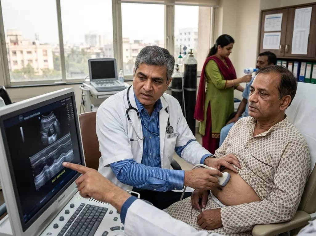
Diaphragmatic dysfunction affects over a third of chronic stroke patients, worsening sleep, mood, and functional mobility. Ultrasound assessment and inspiratory training improve outcomes.
Today

The ClinGen Prenatal Gene Curation Expert Panel evaluated 63 disease relationships across 61 genes, establishing clinical validity for severe fetal phenotypes like hydrops and stillbirth to enhance prenatal genomic interpretation and clinical care.
Yesterday

Managing refractory hypoxemia after cardiothoracic surgery becomes perilous when prolonged air leak limits positive-pressure ventilation. This case-based review details how high-flow nasal cannula, paired with awake rehabilitation, enabled successful extubation and avoided invasive re-intubation.
Today

Idiopathic axillary web syndrome (IAWS) is a rare cause of shoulder pain and mobility restriction characterized by palpable axillary cording without prior surgery or trauma. Early diagnosis, nonsteroidal anti-inflammatory therapy, and physical rehabilitation lead to complete resolution of symptoms.
Today

SWEDEPAD-1 trial insights show paclitaxel-coated devices do not improve long-term limb salvage in patients with chronic limb-threatening ischemia and tissue loss. While one-year reinterventions decreased, the devices were linked to a higher risk of major amputation at three months.
Today
