

Loading, please wait...
Loading, please wait...

Flow diverter devices have completely transformed endovascular treatment strategies for unruptured and complex intracranial aneurysms. However, delayed luminal narrowing remains an unsettling clinical obstacle for interventional teams. Specifically, flow diverter in-stent stenosis represents a significant complication that clinicians frequently encounter during post-procedural surveillance. While most stenotic lesions remain clinically silent, progressive neointimal hyperplasia can occasionally compromise cerebral perfusion or induce thromboembolic events. Consequently, neurointerventionalists urgently need reliable biochemical markers to predict vascular remodeling before deploying flow-diverting stents. Recent multicenter clinical evidence indicates that admission plasma fibrinogen plays an unexpected, nuanced role in vascular healing and intimal formation following device implantation. Understanding this biomarker allows clinicians to stratify patient risks more effectively and refine follow-up strategies.
A comprehensive multicenter retrospective cohort study across four high-volume stroke centers evaluated 394 consecutive patients who underwent endovascular reconstruction with flow diverters between March 2016 and October 2024. The investigative team tracked patients over an average angiographic follow-up period of 12.63 months to determine the true incidence of neointimal proliferation. Notably, 90 patients, representing 22.84% of the entire cohort, developed angiographically confirmed in-stent stenosis defined by luminal narrowing equal to or greater than 25%. Fortunately, all affected individuals presented with asymptomatic stenosis during their routine surveillance evaluations. Investigators collected extensive baseline clinical parameters, including patient demographics, cardiovascular risk factors, procedural nuances, and specific aneurysm dimensions. Most importantly, the researchers focused on baseline admission fibrinogen levels as the primary candidate predictor. Using rigorous multivariable Cox logistic regression modeling, the researchers adjusted for potential confounders, such as antiplatelet regimens, vessel diameters, and device types. The findings revealed that systemic coagulation profiles establish distinct biological environments that dictate how endoluminal stents integrate into parent intracranial vessels over subsequent months. Ultimately, these real-world data confirm that neointimal narrowing remains a frequent occurrence in neurovascular practice.
Interestingly, standard linear regression models initially failed to capture the true dynamic relationship between plasma fibrinogen and vascular patency. Therefore, the investigators executed a threshold effect analysis using sophisticated two-piecewise linear regression models. This mathematical approach uncovered a definitive nonlinear relationship, pinpointing an inflection point at exactly 2.71 g/L. Below this critical threshold of 2.71 g/L, each unit increase in admission fibrinogen conferred substantial protection against narrowing. In fact, patients demonstrated a 73% relative reduction in stenosis hazard per unit increase in fibrinogen (adjusted hazard ratio: 0.27; 95% confidence interval: 0.10 to 0.69; P = 0.006). Conversely, once fibrinogen levels rose above 2.71 g/L, this protective correlation disappeared completely, transitioning into a statistically neutral plateau. This notable inflection explains why previous smaller investigations reported conflicting conclusions regarding coagulation markers and neointimal proliferation. Coagulation dynamics do not behave in a simple monotonic fashion after intracranial stent deployment. Instead, physiological fibrinogen concentrations appear necessary to establish appropriate cellular stability without triggering excessive hypercoagulable thrombosis. Recognizing this biphasic pattern provides critical clarity for neurointerventionalists interpreting baseline laboratory panels.
Understanding why moderately higher fibrinogen levels below 2.71 g/L protect against stenosis requires examining vessel wall biology. Fibrinogen serves as an indispensable precursor for fibrin meshwork generation, which forms the temporary extracellular matrix needed for endothelial migration. Following device deployment, circulating endothelial progenitor cells must quickly adhere to stent struts to re-endothelialize the parent artery. When admission fibrinogen is critically low, impaired scaffold deposition delays rapid and uniform re-endothelialization. Consequently, the exposed metallic struts provoke sustained inflammatory signaling and excessive vascular smooth muscle cell proliferation. This disordered proliferative response ultimately drives exuberant neointimal hyperplasia, thereby narrowing the parent lumen. Furthermore, subnormal fibrinogen levels often reflect systemic nutritional deficits, hepatic synthetic impairment, or uncoordinated baseline hemostasis. When fibrinogen remains within the optimal physiological window near 2.70 g/L, healthy endothelialization occurs rapidly. This swift endothelization effectively caps the metallic foreign body, terminating the proliferative cascade before significant luminal compromise develops. Therefore, adequate substrate availability fosters constructive vascular remodeling rather than aggressive tissue hyperplasia.
These findings offer practical decision-making tools for interventional neuroradiologists and neurosurgeons managing intracranial aneurysms. Clinicians routinely evaluate baseline complete blood counts and basic coagulation panels prior to endovascular therapy. However, teams often overlook admission fibrinogen values unless overt coagulopathy threatens hemorrhagic safety. Identifying patients with admission fibrinogen levels well below 2.71 g/L enables operators to flag individuals who face heightened vulnerability to significant luminal narrowing. Moreover, this biomarker provides an objective parameter to calibrate postoperative imaging schedules. High-risk patients presenting with subthreshold fibrinogen may warrant early digital subtraction angiography or high-resolution magnetic resonance angiography at three to six months. In contrast, patients with balanced fibrinogen levels above the inflection point might safely follow standard noninvasive imaging intervals. Furthermore, neurointerventionalists can incorporate fibrinogen metrics into multidisciplinary perioperative discussions to weigh device selection, stent porosity, and parent vessel anatomical constraints. Proactive risk identification ensures that clinicians can address structural changes long before hemodynamic deficits manifest.
Managing flow diverter cases requires strict adherence to dual antiplatelet therapy to prevent acute thrombosis, yet medication regimens also influence vascular remodeling. When managing patients with low baseline fibrinogen, clinicians must exercise caution when selecting aggressive antiplatelet strategies. Excessive platelet inhibition coupled with deficient fibrin matrix formation might further impede vascular wall stabilization. Additionally, some centers utilize cilostazol as an adjunct antiplatelet agent because it selectively suppresses smooth muscle cell proliferation without increasing major hemorrhage risks. While the multicenter study did not recommend artificially infusing fibrinogen to prevent stenosis, identifying this threshold highlights novel therapeutic pathways. Future prospective trials should investigate whether correcting borderline nutritional deficits or modulating specific inflammatory pathways can normalize vascular healing. Moreover, interventional registries should record baseline inflammatory and coagulation biomarkers systematically to validate these thresholds across diverse global populations. Ultimately, integrating pre-procedural fibrinogen evaluation into institutional protocols bridges the gap between hematological status and endovascular device mechanics, fostering safer long-term outcomes for patients with intracranial aneurysms.
Flow diverter in-stent stenosis is a post-procedural complication characterized by progressive narrowing of the parent artery lumen within a deployed flow diverter. Most cases result from exuberant neointimal hyperplasia stimulated by stent struts. While frequently asymptomatic, severe stenosis can compromise cerebral hemodynamics and require careful angiographic monitoring.
Critically low fibrinogen levels impair the initial formation of a stable fibrin matrix over stent struts. This structural delay impedes swift endothelial cell migration and coverage. As a consequence, prolonged inflammatory signaling promotes abnormal vascular smooth muscle cell proliferation, which accelerates neointimal tissue growth and causes parent vessel narrowing.
Interventional teams typically manage asymptomatic in-stent stenosis conservatively with continued dual antiplatelet therapy and serial angiographic follow-up, as many lesions spontaneously regress over time. However, if patients develop ischemic symptoms or hemodynamic compromise, clinicians may introduce adjunctive agents like cilostazol or perform balloon angioplasty.
Disclaimer: This content is for informational and educational purposes only and should not be considered medical advice. Always consult a qualified healthcare professional regarding any medical condition or treatment. While we strive to present accurate and evidence-based information, healthcare practices evolve constantly. The author and publisher are not liable for any consequences resulting from the use of this material. Refer to the latest local and national guidelines for clinical practice.
References

Read summarized clinical updates, watch expert medical content, and earn CME certifications right from your smartphone.
Open your smartphone camera to scan and install.





A multicenter study reveals that admission fibrinogen levels below 2.71 g/L serve as a nonlinear predictor of in-stent stenosis following flow diverter placement for intracranial aneurysms, with higher levels below this inflection point conferring significant protection.
Today

A randomized crossover trial evaluated fifth-year medical students interacting with an AI standardized patient versus a human standardized patient. The study revealed key differences in Mini-CEX scores, active engagement, and interview dynamics.
Today

A comparative evaluation of INNO-LiPA, Cobas 4800, and Amplicor highlights significant assay-dependent variation in high-risk HPV detection, emphasizing the clinical distinction between analytical sensitivity and screening specificity.
Today

A Canadian nutrition study reveals that higher diet costs predict lower dietary energy density and higher fruit intake, but also increase sodium and processed meat consumption.
Today
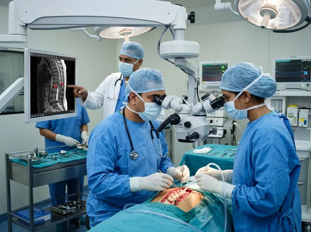
Idiopathic spinal cord herniation is a rare pathology presenting with progressive thoracic myelopathy. Early MRI identification and meticulous microsurgical reduction with dual bolstered dural defect repair prevent irreversible neurological deterioration.
Today

A novel smartphone-assisted biosensor utilizing molecularly imprinted polymers and catalytic nanozymes offers antibody-free, enzyme-free urinary microalbumin testing, enabling rapid, low-cost point-of-care screening for early chronic kidney disease.
Today
