

Loading, please wait...
Loading, please wait...

Early human development shapes lifelong immune competency long before birth. Recently, researchers recognized that fetal hematopoiesis unfolds across layered developmental waves rather than a single stem cell lineage. Consequently, transient embryonic progenitor cells establish permanent tissue-resident immune compartments across the body. In addition, maternal environmental perturbations fundamentally disrupt these fragile embryonic trajectories. Therefore, understanding early hematopoietic programming provides critical insights into postnatal immunopathology and pediatric disease.
Classical textbooks historically claimed that adult hematopoietic stem cells continuously regenerate all human blood cells. However, modern developmental biology demonstrates that hematopoiesis develops through several distinct, time-restricted waves. Initially, an extra-embryonic primitive wave emerges in the yolk sac to supply early red blood cells and macrophages. Subsequently, an erythro-myeloid progenitor wave colonizes the fetal liver without originating from definitive stem cells. Notably, these early progenitors seed vital organs and differentiate into self-renewing tissue-resident macrophages. Microglia in the brain, Kupffer cells in the liver, and alveolar macrophages originate directly from these embryonic waves. Furthermore, the fetal liver generates specialized innate-like lymphoid subsets, including distinct B-1 cells and early gamma-delta T cells. Because these resident cells persist throughout adult life, their initial developmental programming dictates long-term tissue homeostasis. Thus, layered embryonic hematopoiesis establishes an irreplaceable structural foundation for human immunity.
The intrauterine environment directly regulates the delicate progression of embryonic blood formation. When prenatal complications occur, systemic maternal inflammatory signals readily pass through the placenta. For instance, maternal infections, chorioamnionitis, and maternal obesity release substantial quantities of circulating inflammatory cytokines. Consequently, elevated interleukin-6 and interferon levels prompt embryonic progenitors to execute premature emergency myelopoiesis. This emergency response dramatically expands inflammatory myeloid cells while restricting normal lymphoid expansion. Moreover, sustained gestational distress alters histone marks and DNA methylation within developing progenitor populations. These persistent epigenetic alterations permanently impair regulatory T cell development and heighten neonatal monocyte reactivity. As a result, affected children face significantly heightened lifelong risks for allergic asthma, eczema, and chronic metabolic inflammation. Accordingly, managing maternal health during pregnancy protects nascent fetal immune systems from enduring structural dysregulation.
The distinct molecular signature of embryonic progenitors strongly influences susceptibility to childhood malignancies. Many childhood leukemias, including B-cell acute lymphoblastic leukemia, originate during fetal life inside the womb. Specifically, initiating oncogenic events like ETV6-RUNX1 fusions or KMT2A gene rearrangements arise in utero within primitive progenitors. However, these chromosomal translocations rarely cause acute leukemia before delivery without additional postnatal mutations. Instead, embryonic progenitors offer an exceptionally permissive transcriptional environment that protects these abnormal preleukemic clones. Fetal cells maintain unique chromatin accessibility, elevated tolerance for genomic stress, and rapid cell-cycle progression. In contrast, adult bone marrow stem cells rapidly undergo senescence or apoptosis when exposed to similar genetic lesions. Subsequently, common postnatal infections deliver a secondary proliferative trigger that converts preleukemic clones into overt leukemia. Therefore, investigating oncofetal reprogramming helps oncologists uncover unique vulnerabilities in aggressive pediatric blood cancers.
Translating laboratory findings on developmental hematopoiesis into human clinical applications remains technically challenging. Rodent models provide fundamental biological principles, yet significant immunological differences exist between mammalian species. For example, human infants reach different developmental immune milestones during gestation compared to newborn rodents. To resolve these discrepancies, modern investigators employ single-cell multi-omics to study authentic human fetal tissues. Researchers combine single-cell RNA sequencing with chromatin accessibility assays to map developmental trajectories at single-cell resolution. Furthermore, spatial transcriptomics clarifies how the surrounding fetal liver and bone marrow niches nurture embryonic progenitors. These innovative technologies effectively identify rare preleukemic clones long before physical symptoms develop. Consequently, multi-omic mapping bridges the longstanding gap between experimental discovery and pediatric medicine. Thus, high-resolution single-cell profiling establishes a dependable framework for identifying clinically meaningful biomarkers of immune disease.
Insights into fetal immune development generate practical diagnostic and therapeutic opportunities across perinatal care. First, obstetricians must aggressively detect and treat maternal metabolic imbalances, infections, and gestational inflammation. By stabilizing the intrauterine environment, clinicians safeguard developing stem cell niches against irreversible epigenetic damage. In addition, neonatologists caring for preterm infants must address how premature birth disrupts normal liver hematopoiesis. Preterm infants lack late gestational progenitor maturation, predisposing them to devastating neonatal sepsis and inflammatory bowel necrosis. Meanwhile, cellular engineers use fetal ontogeny programs to manufacture specialized, ontogeny-aware cell therapies. Unlike standard adult stem cell products, fetal-programmed cell therapies offer enhanced tissue regeneration and reduced alloreactivity. Furthermore, targeted therapies can selectively attack oncofetal antigens on pediatric leukemia cells without harming adult bone marrow. Ultimately, integrating developmental hematology into clinical practice will significantly advance personalized medicine for mothers and children.
Fetal hematopoiesis occurs in successive, distinct waves across anatomical compartments, beginning in the yolk sac before moving to the fetal liver and spleen. Unlike adult bone marrow hematopoiesis, which depends entirely on self-renewing adult hematopoietic stem cells, embryonic hematopoiesis includes stem cell-independent progenitor waves. These unique progenitors generate self-maintaining tissue-resident macrophages and specialized innate-like lymphoid cells that populate peripheral tissues permanently. Furthermore, fetal progenitors possess distinctive epigenetic programming, prioritizing rapid tissue growth and immunological tolerance over mature inflammatory responsiveness.
Fetal hematopoietic progenitors demonstrate rapid proliferative cycling, broad developmental plasticity, and unique chromatin accessibility patterns compared to adult stem cells. Consequently, initiating genetic events such as ETV6-RUNX1 or KMT2A translocations readily transform these embryonic precursors into stable preleukemic clones. While these genetic anomalies frequently induce senescence or apoptosis in mature adult marrow cells, fetal cells readily accommodate them. These dormant preleukemic clones persist through birth until secondary postnatal infections trigger subsequent oncogenic transformation into pediatric leukemia.
Maternal inflammation exposes fetal stem cells to systemic cytokines, altering fetal liver and marrow microenvironments during critical developmental windows. In response to inflammatory signals like interleukin-6, fetal progenitors undergo premature emergency myelopoiesis, depleting lymphoid pools and establishing durable epigenetic marks. Consequently, these persistent chromatin alterations permanently reprogram myeloid and lymphoid lineages. As the child matures, this prenatal immune priming manifests clinically as heightened susceptibility to severe asthma, atopic allergies, chronic autoimmune disorders, and recurrent systemic infections.
Disclaimer: This content is for informational and educational purposes only. It is not intended to provide medical advice, diagnosis, or treatment and should not replace professional medical judgment. Consult a qualified medical practitioner for specific health concerns. The information reflects available evidence at publication and is subject to change. While we strive to present accurate and up-to-date material, we make no guarantees regarding its completeness or accuracy. Mention of specific products, therapies, or clinical approaches does not imply endorsement. Refer to the latest local and national guidelines for clinical practice.
References

Read summarized clinical updates, watch expert medical content, and earn CME certifications right from your smartphone.
Open your smartphone camera to scan and install.





Recent advances in fetal hematopoiesis reveal that embryonic progenitor waves establish permanent immune compartments. Prenatal stressors like maternal inflammation alter developmental trajectories, predisposing offspring to immune dysfunction and preleukemic transformation.
Today
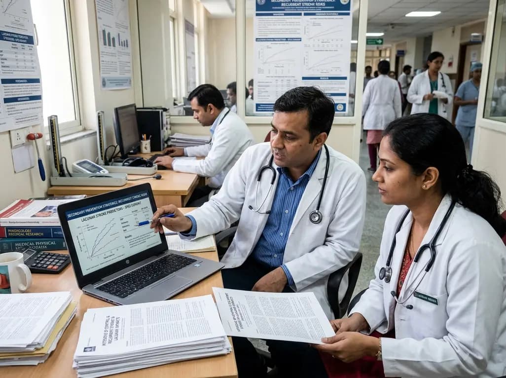
Explore vital recurrent stroke predictors in lacunar stroke patients managed with intensive blood pressure lowering. This analysis examines trial findings from SPS3 to uncover residual vascular risks, metabolic factors, and targeted secondary prevention strategies for high-risk neurological patients.
Today

The Directorate General of Health Services directs Indian states to halt the hazardous reuse and reprocessing of single-use medical devices. Discover regulatory implications under the Drugs and Cosmetics Act, critical infection risks, legal liabilities, and operational changes required across clinical specialties.
Today
Mitochondria-targeted nanoparticles self-assembled from EGCG and L-glutamic acid effectively restore mitochondrial homeostasis, suppress synovial inflammation, and attenuate chondrocyte senescence in osteoarthritis, offering a promising disease-modifying nanotherapy.
Today
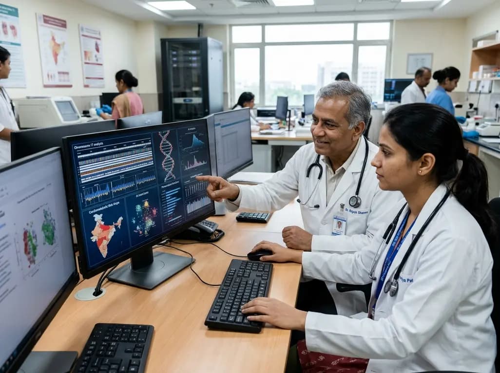
India is advancing preventive healthcare by integrating genomics, artificial intelligence, and quantum technologies. With the SANGAM programme sequencing one million Indian genomes, clinicians can unlock personalized therapies, detect cardiometabolic risks early, and curb the burden of diabetes and heart disease.
Today
