.webp&w=384&q=75)

Loading, please wait...
Loading, please wait...
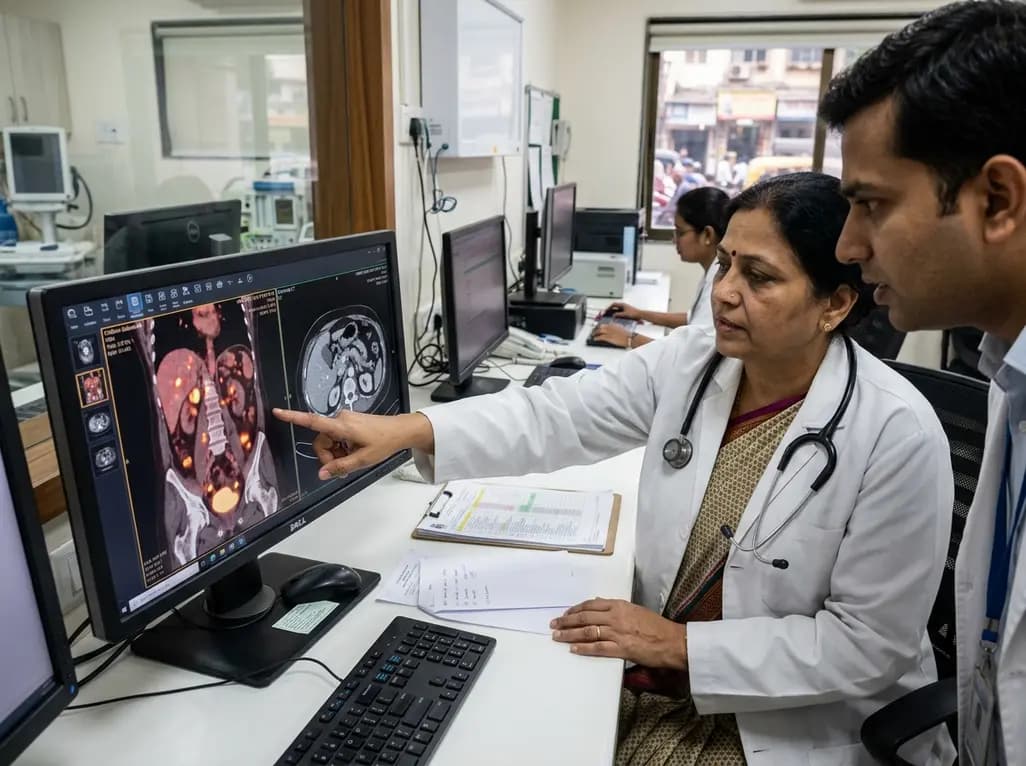
Managing advanced epithelial malignancies demands precise anatomical and functional assessment before extensive cytoreductive interventions. Recent clinical evidence demonstrates that FAPI PET ovarian cancer staging offers distinct diagnostic advantages over traditional cross-sectional imaging modalities. Consequently, this innovative molecular approach accurately maps subtle peritoneal tumor deposits that frequently escape conventional detection. Clinicians can therefore tailor individual therapy regimens with heightened confidence.
Peritoneal dissemination occurs in more than seventy percent of advanced epithelial ovarian carcinoma cases at presentation. Indeed, determining the exact disease extent dictates whether a patient receives upfront cytoreduction or neoadjuvant chemotherapy. However, conventional contrast-enhanced computed tomography often struggles to detect tiny, flat implants scattered across the abdominal cavity. Specifically, radiologic evaluation faces major hurdles when identifying tumor nodules on the small bowel serosa and the mesenteric root. Incomplete surgical cytoreduction significantly compromises survival outcomes when unanticipated extensive disease prevents total macroscopic clearance. Furthermore, unheralded bowel involvement can convert a planned curative resection into an incomplete procedure with elevated surgical morbidity. Diagnostic laparoscopy previously served as the standard invasive triage tool to avoid futile exploratory laparotomies. Nevertheless, laparoscopy carries procedural risks and may overlook isolated retroperitoneal or occult upper abdominal deposits. Therefore, oncology teams urgently require noninvasive imaging that accurately visualizes occult peritoneal carcinomatosis before entering the operating theater. Achieving high diagnostic sensitivity ultimately transforms preoperative strategic planning across multidisciplinary oncology boards. Consequently, enhanced detection prevents unnecessary surgical interventions in unresectable presentations.
A prospective trial in Bengaluru evaluated two hundred women presenting with primary epithelial ovarian cancer. Researchers compared preoperative gallium-68 FAPI-04 PET/CT against conventional contrast-enhanced computed tomography before cytoreductive surgery. Importantly, surgical inspection and histological verification served as the definitive reference standards. The trial demonstrated that FAPI PET achieved an impressive diagnostic sensitivity of 81.8% for peritoneal disease. In contrast, conventional computed tomography achieved a sensitivity of only 54.6%. Furthermore, FAPI PET attained an overall diagnostic accuracy of 86%, whereas computed tomography reached 78%. Standard computed tomography frequently underestimated the total volume and distribution of intra-abdominal malignancy. Conversely, the novel tracer identified subtle, millimetric implants that standard scans completely missed. As a result, the molecular scan demonstrated markedly superior diagnostic power across challenging anatomical territories. For example, the imaging modality successfully identified subtle tumor seeding on diaphragmatic surfaces and hepatic ligaments. Moreover, the scan accurately depicted small mesenteric deposits that dictate surgical resectability. Thus, these rigorous clinical findings establish the diagnostic superiority of fibroblast activation protein targeting over anatomical imaging. Consequently, clinicians gain an exceptional noninvasive roadmap for navigating complex abdominal pathology. This reliable precision provides oncologists with dependable clarity during therapy stratification.
Traditional metabolic imaging with fluorodeoxyglucose PET/CT relies on cellular glucose consumption to visualize active tumors. However, this metabolic mechanism introduces major diagnostic hurdles within the peritoneal cavity. Normal physiological peristalsis, high background bowel uptake, and regional inflammatory reactions frequently obscure subtle malignant lesions. In contrast, fibroblast activation protein inhibitor tracers bypass these limitations by targeting a completely different biological pathway. Specifically, the radiotracer binds with high affinity to fibroblast activation protein expressed on cancer-associated fibroblasts. These specialized stromal cells surround malignant epithelial cells in abundance throughout the tumor microenvironment. Because normal abdominal organs exhibit minimal tracer uptake, the scan creates an exceptionally high tumor-to-background contrast ratio. Consequently, tiny peritoneal deposits appear as distinct focal hot spots rather than indistinct shadows. In addition, the tracer clears rapidly from circulation without requiring dietary restriction or glucose stabilization. Therefore, nuclear medicine specialists can interpret peritoneal scans with far greater anatomical clarity. Ultimately, this targeted molecular approach resolves diagnostic ambiguities that routinely plague conventional metabolic imaging. Furthermore, the tracer delivers consistent tumor visualization even in treated or inflamed tissues.
Surgeons utilize the Peritoneal Cancer Index score to quantify disease dissemination across thirteen distinct abdominopelvic regions. Indeed, this standardized scoring system correlates directly with surgical resectability, post-operative morbidity, and overall patient survival. In the prospective Bengaluru trial, intraoperative assessment established a mean surgical score of 15.7. Remarkably, preoperative FAPI PET closely mirrored these operative findings with an average calculated score of 14.1. Conversely, conventional computed tomography substantially underestimated disease spread, yielding a mean score of only 10.8. Such underestimation often exposes patients to aborted cytoreductions or suboptimal surgical debulking. Furthermore, the molecular scan demonstrated superior correlation in critical anatomical sites, including the mesenteric root and jejunoileal surfaces. Because extensive tumor burden in these delicate areas precludes complete resection, accurate preoperative staging remains vital. Consequently, clinicians can identify patients who cannot achieve complete macroscopic clearance during upfront surgery. Therefore, this imaging modality prevents unnecessary laparotomies while improving clinical resource allocation. In addition, accurate preoperative scoring establishes reliable baselines for monitoring systemic therapeutic responses over time. Oncologists can thus design individualized surgical roadmaps tailored to each patient's specific disease burden. Ultimately, precise disease scoring aligns preoperative expectations with actual intraoperative realities.
Accurate preoperative staging directly influences fundamental clinical choices in gynecologic oncology. In the reported cohort, findings from the molecular scan altered definitive treatment pathways in 18% to 21% of patients. For instance, several women scheduled for immediate surgery instead received neoadjuvant chemotherapy after scans revealed occult unresectable disease. Conversely, other patients deemed unsuitable on conventional imaging safely proceeded to primary surgery when scans excluded diffuse mesenteric involvement. Consequently, this diagnostic precision shields patients from invasive surgical complications and futile operative procedures. In addition, the modality may diminish the need for routine diagnostic laparoscopy, lowering healthcare expenditures and operative delays. Currently, this advanced radiotracer is accessible only at select tertiary oncology centers across India. However, domestic radiopharmaceutical production is expanding rapidly to meet rising clinical demand nationwide. As validation studies continue across global institutions, clinical guidelines will likely incorporate these targeted scans into standard protocols. Therefore, multidisciplinary tumor boards can anticipate broader clinical adoption in the near future. Ultimately, integrating advanced molecular diagnostics into standard care pathways elevates therapeutic precision for women battling ovarian cancer. This technological progress marks a transformative leap forward in personalized oncologic care.
Q1: Why is FAPI PET superior to contrast CT for detecting ovarian cancer spread?
FAPI PET targets fibroblast activation protein on cancer-associated fibroblasts rather than relying on tissue density or non-specific glucose metabolism. Because normal bowel and surrounding tissues show negligible tracer uptake, the scan creates high tumor contrast. Consequently, it detects small, flat peritoneal deposits on the bowel and upper abdomen that contrast CT routinely misses. This molecular specificity provides an accurate assessment of tumor burden before major surgery.
Q2: How does FAPI PET influence clinical treatment decisions in ovarian cancer?
In clinical studies, FAPI PET altered management plans in approximately 18% to 21% of patients. By providing an accurate Peritoneal Cancer Index score, the scan reveals whether complete surgical debulking is achievable. If widespread mesenteric or upper abdominal deposits preclude complete resection, oncologists can switch to neoadjuvant chemotherapy. Conversely, confirming localized disease prevents unnecessary delays, directing suitable candidates straight to curative cytoreductive surgery without invasive laparoscopy.
Q3: How does FAPI PET compare to standard FDG PET scans in ovarian cancer?
Standard FDG PET relies on glucose metabolism, which causes high physiological uptake in the gut and inflammatory regions. This background activity often masks subtle peritoneal metastases. In contrast, FAPI PET targets stromal fibroblast activation protein, producing minimal non-specific bowel uptake and high tumor-to-background contrast. In addition, FAPI scans do not require prolonged fasting or blood sugar management. Therefore, they offer superior diagnostic clarity for evaluating complex peritoneal carcinomatosis.
Disclaimer: This content is for informational and educational purposes only. It does not constitute medical advice or replace professional judgment. Refer to the latest local and national guidelines for clinical practice.
References

Read summarized clinical updates, watch expert medical content, and earn CME certifications right from your smartphone.
Open your smartphone camera to scan and install.





A landmark prospective study reveals gallium-68 FAPI PET/CT achieves 81.8% sensitivity in mapping peritoneal carcinomatosis in epithelial ovarian cancer, surpassing contrast CT. By resolving small bowel and mesenteric deposits, this molecular imaging alters treatment decisions in up to 21% of surgical candidates.
Today

A review highlights how N-acetyl-L-cysteine influences pancreatic beta cells differently across metabolic states. While it protects islets during glucolipotoxicity and diabetes, excessive antioxidant exposure during physiological or aging states can induce reductive stress and impair insulin release.
Today

Atherosclerotic cardiovascular disease guidelines often rely on European-derived lipoprotein(a) cutoffs. However, baseline Lp(a) levels vary widely across ancestries, including South Asians and Africans. Clinicians must interpret Lp(a) values within overall absolute cardiovascular risk.
Today

A longitudinal study of 1,055 chronic low back pain patients reveals that higher patient activation fosters constructive pain medication attitudes, reduces disability, and decreases short- and long-term opioid use through collaborative, multimodal care.
Yesterday

A landmark cross-sectional study in Asian adults with type 2 diabetes identifies novel urinary metabolites linked to mild cognitive impairment. Elevated urinary sucrose and altered metabolites highlight non-invasive pathways for early cognitive risk stratification and targeted metabolic management.
Yesterday
